Molecular Insights into Human Monoamine Oxidase B Inhibition by the Glitazone Antidiabetes Drugs (original) (raw)
Abstract
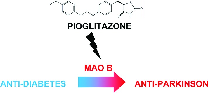
The widely employed antidiabetic drug pioglitazone (Actos) is shown to be a specific and reversible inhibitor of human monoamine oxidase B (MAO B). The crystal structure of the enzyme–inhibitor complex shows that the _R_-enantiomer is bound with the thiazolidinedione ring near the flavin. The molecule occupies both substrate and entrance cavities of the active site, establishing noncovalent interactions with the surrounding amino acids. These binding properties differentiate pioglitazone from the clinically used MAO inhibitors, which act through covalent inhibition mechanisms and do not exhibit a high degree of MAO A versus B selectivity. Rosiglitazone (Avandia) and troglitazone, other members of the glitazone class, are less selective in that they are weaker inhibitors of both MAO A and MAO B. These results suggest that pioglitazone may have utility as a “repurposed” neuroprotectant drug in retarding the progression of disease in Parkinson's patients. They also provide new insights for the development of reversible isoenzyme-specific MAO inhibitors.
Keywords: Antidiabetes drug, drug design, monoamine oxidase, neurodegeneration, Parkinson's disease, pioglitazone
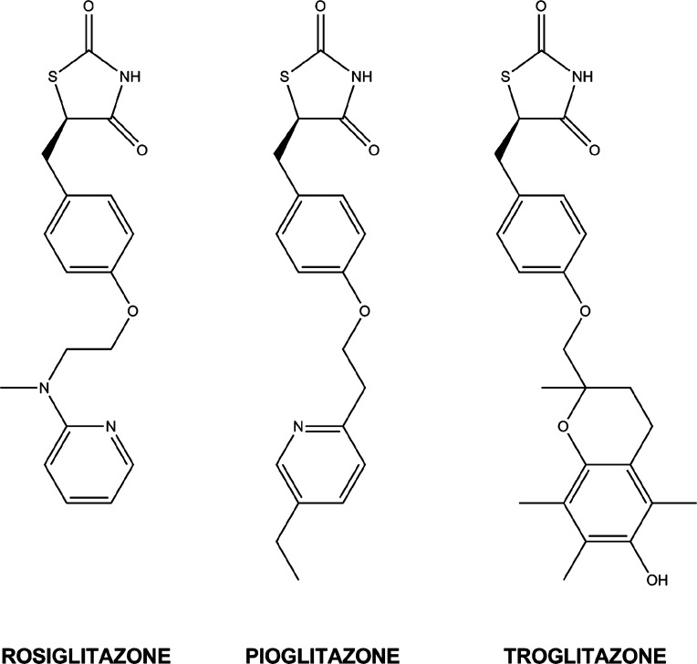
The incidence of type II diabetes has reached nearly epidemic proportions in western countries and constitutes a major health risk (current estimate of 19.7 million diagnosed individuals in the United States).1−3 The antidiabetes drugs belonging to the glitazone class act as peroxisome proliferator-activated receptor-δ agonists and counter cellular insulin resistance. Pioglitazone (Actos) accounted for $2.5 billion in sales in the United States in 2009, being one of the nine FDA-approved antidiabetes medications (Figure 1). Rosiglitazone (Avandia) is another member of this class; however, its cardiotoxic side effects have resulted in restrictions in its clinical use.4,5
Figure 1.
Structures of the glitazone (_R_-enantiomers) drugs used in this study.
Pioglitazone was shown to function as an effective neuroprotectant in the MPTP-mouse model for Parkinson's disease (PD).6 It was suggested to be an inhibitor of monoamine oxidases (MAOs), which are flavin-dependent mitochondrial enzymes that catalyze the oxidative degradation of various aromatic amine-containing neurotransmitters.7 The two human isoenzymes (MAO A and B) share considerable sequence identity (73%)8 but exhibit only partly overlapping substrate specificities.9 For instance, MAO A (but not MAO B) is able to oxidize serotonin or epinephrine, whereas MAO B preferentially acts on phenethylamine. On these bases, MAO A has classically been considered a target for antidepressant drugs, whereas MAO B inhibitors are currently used to support treatment of PD.10 A further point of interest is that MAOs are major sources of hydrogen peroxide in the cells, and their role in pathological oxidative stress is under active investigation.11,12
Presently, there are several clinically used MAO inhibitors (MAO-Is), including the recently developed rasagiline.13 A property common to all of these compounds is that their molecular mechanisms of inhibition feature the formation of a covalent adducts between the flavin cofactor and the inhibitor molecule.14 In this context, it was of interest to evaluate the inhibitory activity of glitazones against MAOs, especially since a number of clinical trials are now initiated to test the effectiveness of pioglitazone in the treatment of Parkinson's patients (http://parkinsontrial.ninds.nih.gov/). Here, we demonstrate that pioglitazone is a highly selective MAO B inhibitor, highlighting potential additional pharmacological actions of this widely used drug.
The main questions addressed in our study were as follows: (i) Are these glitazone drugs effective MAO-Is? (ii) Do they function as reversible or as irreversible inhibitors? (iii) Do they discriminate between the isoenzymes? (iv) Can they become lead compounds for further drug-design studies?
To quantitatively establish the inhibition activities of glitazone compounds, we used recombinant human MAO A and B heterologously expressed in Pichia pastoris.15,16 These experiments were also performed using the recombinant rat enzymes since the rat model is often employed in drug development studies. We found that (R,S)-pioglitazone (see the structures in Figure 1) competitively inhibits MAO B with a submicromolar _K_i value for the human enzyme. Of interest, the drug does not exhibit any inhibitory activity in the accessible concentration range (∼100 μM) against either human or rat MAO A (Table 1). In contrast, (R,S)-rosiglitazone is nonspecific and competitively inhibits both A and B isoenzymes, although slightly more weakly MAO A. Studies with troglitazone show complex inhibition behavior, which appears as noncompetitive with both MAO A and MAO B with _K_i values ∼10 μM. Thus, these glitazone molecules are MAO-Is with clearly different properties. In particular, pioglitazone exhibits high specificity and submicromolar affinity to human MAO B, and the others exhibit nonselectivity and weaker inhibition with both isozymes.
Table 1. Inhibition Properties of Glitazone Compounds with Purified Human and Rat MAOs.
| | MAO A (_K_i μM) | MAO B (_K_i μM) | | | | | ----------------------- | --------------- | --------------- | --------- | ---------- | | | human | rat | human | rat | | | pioglitazone | nonea | nonea | 0.5 ± 0.1 | 2.1 ± 0.4 | | rosiglitazone | 27.6 ± 3.0 | 32.6 ± 5.1 | 4.2 ± 0.8 | 5.8 ± 1.0 | | troglitazoneb | 10.5 ± 1.2 | 4.8 ± 0.6 | 9.5 ± 0.7 | 10.9 ± 1.0 |
We also tested the human histone demethylase LSD1 for any inhibition by pioglitazone and by rosiglitazone.17,18 This histone-modifying amine oxidase is weakly homologous to MAO A and B and is emerging as potential target for anticancer drugs. Of relevance to this study, LSD1 is inhibited by several MAO-I compounds such as tranylcypromine, raising the question of whether any glitazone inhibition may occur. No inhibition of LSD1 is observed by these glitazones at concentrations up to 100 μM, further highlighting their specificities for MAOs.
The high-resolution crystal structures of human MAO B in complex with both pioglitazone and rosiglitazone show no evidence for any covalent attachment of the inhibitor thiazolidinedione ring to the flavin or to active site residues (Figure 2 and Table S1 in the Supporting Information).14,19 The noncovalent nature of the binding was further confirmed by the absence of any change in the absorption spectrum of the protein-bound flavin cofactor. The benzyl and pyridyl rings of pioglitazone extend through the substrate cavity into the entrance cavity of the bipartite active site of MAO B (Figure 3), similar to safinamide, a compound in phase III clinical trials for the treatment of PD (Figure 4).20 A detailed structural analysis of pioglitazone in the active site of MAO B shows H-bonds of oxygen and nitrogen in the thiazolidinedione ring with conserved active site water molecules within the aromatic cage of Tyr398 and Tyr435 (Figure 3b). Phe103, located in the loop guarding the active site cavity, is displaced slightly in conformation to avoid steric clashes with the ethyl side chain on the pyridine ring (Figure 3b). These specific interactions and, more generally, the bipartite structure of the MAO B active site and its hydrophobic environment account for the specificity of pioglitazone binding to MAO B and not to MAO A.
Figure 2.
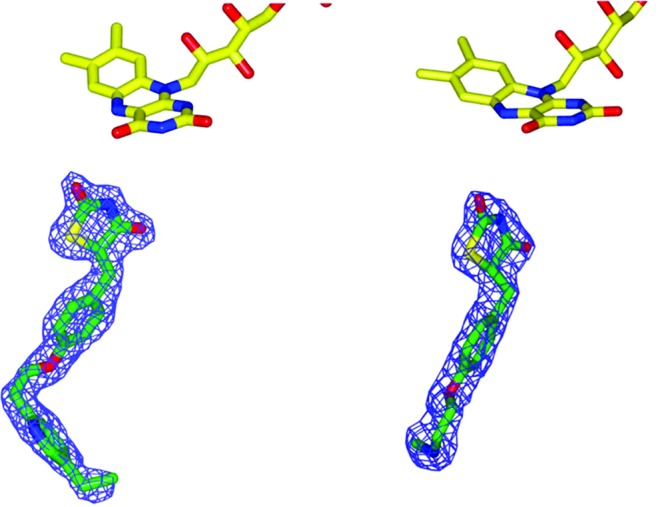
Unbiased electron density maps and stick models of pioglitazone (left) and rosiglitazone (right; the pyridyl moiety of rosiglitazone is not visible in the electron density; see the text) in complex with human MAO B. A stick model of the flavin is shown at the top of each structure.
Figure 3.
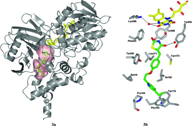
Structural studies on pioglitazone binding to MAO B. (a) Overall structure of the MAO B–pioglitazione complex. The binding cavity is shown as a semitransparent red surface. (b) Detailed structure of pioglitazone bound to the active site of human MAO B. Hydrogen bonds are denoted by dashed lines. The carbon skeleton of the flavin ring is in yellow, and that of pioglitazone is in green. Interacting amino acid residues are in gray. Oxygen atoms are red, nitrogens are in blue, and sulfurs are in yellow. Water molecules are depicted as red spheres.
Figure 4.
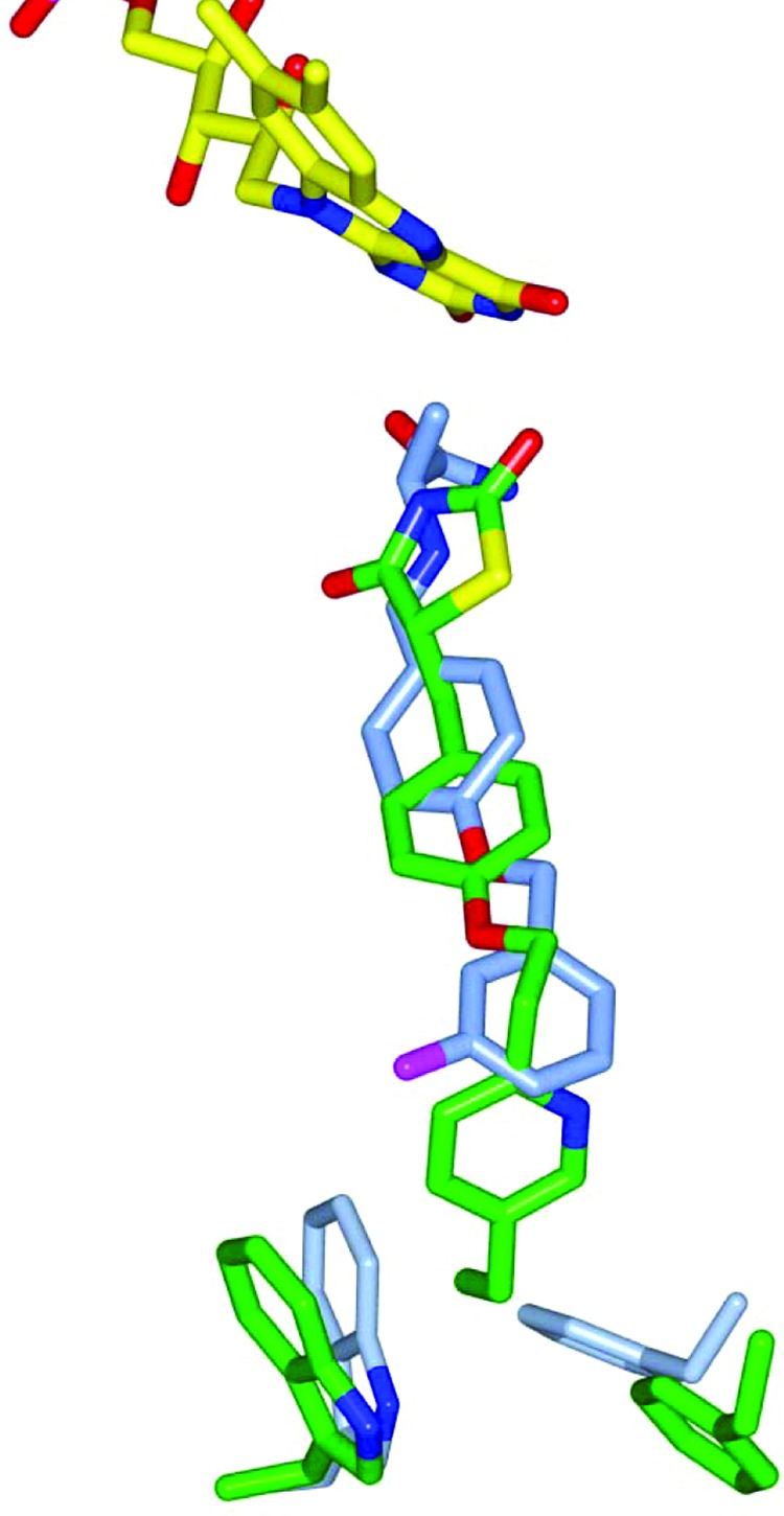
Overlay of the pioglitazone structure (green carbon chain) with that of safinamide (blue chain) (PDB code: 2V5Z) in the binding site of human MAO B. In both complexes, Ile199, the gating residue between the substrate and the entrance cavities (Figure 3), is in its open conformation. The conformations of Phe103 and Trp119 for the two bound inhibitors are shown. The flavin rings are in yellow. Atom designation is the same as in Figure 3.
The crystallographic investigation shows that rosiglitazone binds in the same position and conformation as does pioglitazone (Figure 2). The electron density does not include the pyridyl ring of the inhibitor. We have found that this inhibitor is also a very slow substrate of both MAO A and MAO B (in a time course of hours). The time required for crystal formation may allow for the oxidative cleavage of the bond adjacent to the tertiary anilino nitrogen with consequent loss of the pyridyl ring. On the basis of model fitting to electron density, the _R_-enantiomer of the racemic mixture of either pioglitazone or rosiglitazone appears to preferentially bind to the enzyme. Racemization of these compounds occurs reasonably rapidly,21 which precludes the administration of either enantiomer to enhance therapeutic specificity as well as a more thorough analysis of their differences in binding affinities.
Attempts to determine the structure of the troglitazone–MAO B complex were unsuccessful. No definitive electron density of the bound ligand could be observed, which suggests heterogeneity of binding modes to the oxidized enzyme or a low affinity and no covalent binding. The inhibition behavior observed suggests a higher affinity of this glitazone for the enzyme–substrate/product intermediates in catalysis.
The fundamental conclusion of our study is that pioglitazone is a reasonable MAO B inhibitor with two key features: its high selectivity and its noncovalent binding mode. These properties set it aside with respect to currently used MAO-I drugs, which act through covalent inhibition mechanisms and do not exhibit such a MAO A versus B selectivity.8 Pioglitazone is a widely and routinely used drug that is known to reach plasma concentrations in monkeys with induced symptoms characteristic of PD22 (∼4.9 μM), which are ∼10- fold higher than the _K_i value determined here for MAO B. Moreover, pioglitazone can cross the blood–brain barrier to reach therapeutic neuroprotective concentrations22 (∼0.14 μM), which are in a concentration range comparable to the _K_i value (0.5 μM). A knowledge of its inhibitory activity against MAO B should be helpful in the evaluation of pioglitazone's clinical benefits and possible side effects. These data provide a molecular basis for “repurposing” pioglitazone as a neuroprotectant in the treatment of patients at early stages of PD. Additional applications may also be considered in pathological conditions known to involve abnormal production of reactive oxygen species, as indicated by preliminary studies on the beneficial effects of MAO-Is in heart failure and muscular dystrophy.12 In this regard, glitazones may represent clinically validated scaffolds to open new avenues for the design of isoenzyme-selective noncovalent MAO-Is. These findings support the recently emerging trends in current drug development to probe existing drugs for therapeutic activities beyond those currently exploited in clinical applications.23 Furthermore, they are fully consistent with NIH guidelines to retrieve abandoned drugs for new uses.24
Experimental Procedures
Rosiglitazone was purchased from Cayman Chemical, and pioglitazone and troglitazone were from Sigma-Aldrich. MAO A and B recombinant proteins were overexpressed in Pichia pastoris and purified following previously published procedures.15,16 Enzymatic activities were measured spectrophotometrically using the horseradish peroxidase coupled Amplex red assay (Δϵ = 54 000 M–1 cm–1, λ = 560 nm) with _p_-CF3-benzylamine and benzylamine or phenethylamine as substrates for MAO A and MAO B, respectively. Inhibitor _K_i values were determined by measuring the initial rates of substrate oxidation (six different concentrations) in the presence of varying concentrations of inhibitor (a minimum of four different concentrations). _K_i values were determined using global fit analysis of the hyperbolic fits of enzyme activity with inhibitor concentrations using Graphpad Prism 5.0 software. Crystals of MAO B complexes were grown under conditions described previously14 in the presence of ∼500 μM inhibitors. X-ray diffraction data were collected at the ESRF (Grenoble, France) and at the SLS (Villigen, Switzerland) synchrotrons. Data processing and structure refinement were performed using programs of the CCP4 package following standard protocols (Table S1 in the Supporting Information).25 Structural representations were generated with CCP4 mg.26 Purification of recombinant human LSD1/CoREST complex and inhibition assays against this enzyme were carried out as described.27
Glossary
Abbreviations
MAO
monoamine oxidase
PD
Parkinson's disease
MAO-I
MAO inhibitor
Supporting Information Available
X-ray structural data collection and refinement statistics for the human MAO B–glitazone complexes. This material is available free of charge via the Internet at http://pubs.acs.org.
Author Contributions
C.B., M.A., and M.T. performed the experiments. C.B., W.J.G., A.M., and D.E.E. analyzed the data and wrote the manuscript.
Supported by NIH Grant GM29433, Cariplo Foundation (2008.3148 and 2010-0778), MIUR-PRIN09, and AIRC.
The authors declare no competing financial interest.
Funding Statement
National Institutes of Health, United States
Supplementary Material
References
- Breidert T.; Callebert J.; Heneka M. T.; Landreth G.; Launay J. M.; Hirsch E. C. Protective action of the peroxisome proliferator-activated receptor-gamma agonist pioglitazone in a mouse model of Parkinson's disease. J. Neurochem. 2002, 82, 615–624. [DOI] [PubMed] [Google Scholar]
- Schintu N.; Frau L.; Ibba M.; Caboni P.; Garau A.; Carboni E.; Carta A. R. PPAR-gamma-mediated neuroprotection in a chronic mouse model of Parkinson’s disease. Eur. J. Neurosci. 2009, 29, 954–963. [DOI] [PubMed] [Google Scholar]
- DeFronzo R. A.; Tripathy D.; Schwenke D. C.; Banerji M.; Bray G. A.; Buchanan T. D.; Clement S. C.; Henry R. R.; Hodis H. N.; Kitabchi A. E.; Mack W. J.; Mudaliar S.; Ratner R. E.; Williams K.; Stentz F. B.; Musi N.; Reaven P. D. Pioglitazone for Diabetes Prevention in Impaired Glucose Tolerance. N. Engl. J. Med. 2011, 364, 1104–1115. [DOI] [PubMed] [Google Scholar]
- Nathan D. M. Rosiglitazone and cardiotoxicity—Weighing the evidence. N. Engl. J. Med. 2007, 357, 64–66. [DOI] [PubMed] [Google Scholar]
- Nissen S. K.; Wolski K. Effect of rosiglitazone on the risk of myocardial infarction and death from cardiovascular causes. N. Engl. J. Med. 2007, 356, 2457–2471. [DOI] [PubMed] [Google Scholar]
- Quin L. L. P.; Crook B.; Hows M. E.; Vidgeon-Hart M.; Chapman H.; Upton N.; Medhurst A. D.; Virley D. J. The PPARδ agonist pioglitazone is effective in the MPTP mouse model of Parkinson’s disease through inhibition of monoamine oxidase B. Br. J. Pharmacol. 2008, 154, 226–233. [DOI] [PMC free article] [PubMed] [Google Scholar]
- Gedenhuys W. J.; Darvesh A. S.; Franl M. O.; Van der Schyf C. J.; Carroll R. T. Identification of novel monoamine oxidase B inhibitors by structure-based virtual screening. Bioorg. Med. Chem. Lett. 2010, 20, 5295–5298. [DOI] [PubMed] [Google Scholar]
- Bach A. W.; Lan N. C.; Johnson D. L.; Abell C. W.; Bembenek M. E.; Kwan S. W.; Seeburg P. H.; Shih J. C. cDNA cloning of human liver monoamine oxidases A and B: Molecular basis of differences in enzymatic properties. Proc. Natl. Acad. Sci. U.S.A. 1988, 85, 4934–4938. [DOI] [PMC free article] [PubMed] [Google Scholar]
- Edmondson D. E.; Binda C.; Wang J.; Upadhyay A. K.; Mattevi A. Molecular and mechanistic properties of the membrane-bound mitochondrial monoamine oxidases. Biochemistry 2009, 48, 4220–4230. [DOI] [PMC free article] [PubMed] [Google Scholar]
- Youdim M. B.; Edmondson D.; Tipton K. F. The therapeutic potential of monoamine oxidase inhibitors. Nat. Rev. Neurosci. 2006, 7, 295–309. [DOI] [PubMed] [Google Scholar]
- Trouche E.; Mias C.; Seguelas M. H.; Ordener C.; Cussac D.; Parini A. Characterization of monoamine oxidases in mesenchymal stem cells: role in hydrogen peroxide generation and serotonin-dependent apoptosis. Stem Cells Dev. 2010, 19, 1571–1578. [DOI] [PubMed] [Google Scholar]
- Menazza S.; Blaauw B.; Tiepolo T.; Toniolo L.; Braghetta P.; Spolaore B.; Reggiani C.; Di Lisa F.; Bonaldo P.; Canton M. Oxidative stress by monoamine oxidases is causally involved in myofiber damage in muscular dystrophy. Hum. Mol. Genet. 2010, 19, 4207–4215. [DOI] [PubMed] [Google Scholar]
- Weinreb O.; Amit T.; Bar-Am O.; Youdim M. B. Rasagiline: A novel anti-Parkinsonian monoamine oxidase-B inhibitor with neuroprotective activity. Prog. Neurobiol. 2010, 92, 330–344. [DOI] [PubMed] [Google Scholar]
- Binda C.; Li M.; Hubálek F.; Restelli N.; Edmondson D. E.; Mattevi A. Insights into the mode of inhibition of human mitochondrial monoamine oxidase B from high resolution crystal structures. Proc. Natl. Acad. Sci. U.S.A. 2003, 100, 9750–9755. [DOI] [PMC free article] [PubMed] [Google Scholar]
- Newton-Vinson P.; Hubálek F.; Edmondson D. E. High-Level Expression of Human Liver Monoamine Oxidase B in Pichia pastoris. Protein Expression Purif. 2000, 20, 334–345. [DOI] [PubMed] [Google Scholar]
- Wang J.; Edmondson D. E. High-level expression and purification of rat monoamine oxidase A (MAO A) in Pichia pastoris: Comparison with human MAO A. Protein Expression Purif. 2010, 70, 211–217. [DOI] [PMC free article] [PubMed] [Google Scholar]
- Culhane J. C.; Cole P. A. LSD1 and the chemistry of histone demethylation. Curr. Opin. Chem. Biol. 2007, 11, 561–568. [DOI] [PMC free article] [PubMed] [Google Scholar]
- Forneris F.; Binda C.; Battaglioli E.; Mattevi A. LSD1: Oxidative chemistry for multifaceted functions in chromatin regulation. Trends Biochem. Sci. 2008, 33, 181–189. [DOI] [PubMed] [Google Scholar]
- Reddy V. V. B. G.; Karanam B. V.; Gruber W. L.; Wallace M. A.; Vincent S. H.; Franklin R. B.; Baille T. A. Mechanistic Studies on the Metabolic Scission of Thiazolidinedione Derivatives to Acyclic Thiols. Chem. Res. Toxicol. 2005, 18, 880–888. [DOI] [PubMed] [Google Scholar]
- Binda C.; Wang J.; Pisani L.; Caccia C.; Carotti A.; Salvati P.; Edmondson D. E.; Mattevi A. Structures of human monoamine oxidase B complexes with selective noncovalent inhibitors: safinamide and coumarin analogs. J. Med. Chem. 2007, 50, 5848–5852. [DOI] [PubMed] [Google Scholar]
- Jamali B.; Bjørnsdottir I.; Nordfang O.; Honoré Hansen S. Investigation of racemisation of the enantiomers of glitazone drug compounds at different pH using chiral HPLC and chiral CE. J. Pharm. Biomed. Anal. 2008, 46, 82–87. [DOI] [PubMed] [Google Scholar]
- Swanson C. R.; Bonarenko J. V.; Brunner V.; Simmons H. A.; Zeigler t.e.; Kemnitz J. W.; Johnson J. A.; Emborg M. E. The PPAR-gamma agonist pioglitazone modulates inflammation and induces neuroprotection in parkinsonian monkeys. J. Neuroinflammation 2011, 8, 91–104. [DOI] [PMC free article] [PubMed] [Google Scholar]
- Campillos M.; Kuhn M.; Gavin A. C.; Jensen L. J.; Bork P. Drug target identification using side-effect similarity. Science 2008, 321, 263–266. [DOI] [PubMed] [Google Scholar]
- Kaiser J. Biomedicine. NIH's secondhand shop for tried-and-tested drugs. Science 2011, 332, 1492. [DOI] [PubMed] [Google Scholar]
- Winn M. D.; Ballard C. C.; Cowtan K. D.; Dodson E. J.; Emsley P.; Evans P. R.; Keegan R. M.; Krissinel E. B.; Leslie A. G.; McCoy A.; McNicholas S. J.; Murshudov G. N.; Pannu N. S.; Potterton E. A.; Powell H. R.; Read R. J.; Vagin A.; Wilson K. S. Overview of the CCP4 suite and current developments. Acta Crystallogr., Sect. D: Biol. Crystallogr. 2011, 67, 235–242. [DOI] [PMC free article] [PubMed] [Google Scholar]
- McNicholas S.; Potterton E.; Wilson K. S.; Noble M. E. Presenting your structures: The CCP4mg molecular-graphics software. Acta Crystallogr., Sect. D: Biol. Crystallogr. 2011, 67, 386–394. [DOI] [PMC free article] [PubMed] [Google Scholar]
- Binda C.; Valente S.; Romanenghi M.; Pilotto S.; Cirilli R.; Karytinos A.; Ciossani G.; Botrugno O. A.; Forneris F.; Tardugno M.; Edmondson D. E.; Minucci S.; Mattevi A.; Mai A. Biochemical, structural, and biological evaluation of tranylcypromine derivatives as inhibitors of histone demethylases LSD1 and LSD2. J. Am. Chem. Soc. 2010, 132, 6827–6833. [DOI] [PubMed] [Google Scholar]
Associated Data
This section collects any data citations, data availability statements, or supplementary materials included in this article.
