A Non-Mouse-Adapted Enterovirus 71 (EV71) Strain Exhibits Neurotropism, Causing Neurological Manifestations in a Novel Mouse Model of EV71 Infection (original) (raw)
Abstract
Enterovirus 71 (EV71) is a neurotropic pathogen that has been consistently associated with the severe neurological forms of hand, foot, and mouth disease. The lack of a relevant animal model has hampered our understanding of EV71 pathogenesis, in particular the route and mode of viral dissemination. It has also hindered the development of effective prophylactic and therapeutic approaches, making EV71 one of the most pressing public health concerns in Southeast Asia. Here we report a novel mouse model of EV71 infection. We demonstrate that 2-week-old and younger immunodeficient AG129 mice, which lack type I and II interferon receptors, are susceptible to infection with a non-mouse-adapted EV71 strain via both the intraperitoneal (i.p.) and oral routes of inoculation. The infected mice displayed progressive limb paralysis prior to death. The dissemination of the virus was dependent on the route of inoculation but eventually resulted in virus accumulation in the central nervous systems of both animal groups, indicating a clear neurotropism of the virus. Histopathological examination revealed massive damage in the limb muscles, brainstem, and anterior horn areas. However, the minute amount of infectious viral particles in the limbs from orally infected animals argues against a direct viral cytopathic effect in this tissue and suggests that limb paralysis is a consequence of EV71 neuroinvasion. Together, our observations support that young AG129 mice display polio-like neuropathogenesis upon infection with a non-mouse-adapted EV71 strain, making this mouse model relevant for EV71 pathogenesis studies and an attractive platform for EV71 vaccine and drug testing.
INTRODUCTION
Enterovirus 71 (EV71) is responsible for hand, foot, and mouth disease (HFMD), mainly in infants and young children. It has been consistently associated with the most severe complications of the disease, including aseptic meningitis, brain stem encephalitis, and acute flaccid paralysis (AFP), a polio-like syndrome (7). Neuronal degeneration, severe central nervous system (CNS) inflammation, and pulmonary edema have also been reported in fatal cases (1). Over the past 12 years, EV71 has become the cause of major and regular epidemics across the Asia-Pacific region (28) and is now believed to be the most significant pathogen globally among the enteroviruses, now that poliovirus has been largely eradicated in most parts of the world through vaccination. With no effective vaccine or antiviral treatment available thus far, EV71 certainly poses a pressing economical and public health threat in Asia.
Despite much speculation about possible viral and immunopathological mechanisms, the pathogenesis and the transmission route of EV71 remain ill defined (40). The ability to further our knowledge of EV71 and to develop effective prophylactic and therapeutic approaches has been severely hampered by the lack of a relevant animal model, a deficit resulting from the limited species tropism of EV71. Experimental infection of rhesus and cynomolgus monkeys has been reported, but their use is limited for ethical and economical reasons (6, 15, 17, 41). Furthermore, EV71-infected monkeys do not develop any typical neurological complications, including flaccid paralysis and ataxia, while only 10% of infected animals develop clinical symptoms, which is in agreement with reports of human patients but impractical at the experimental level.
The small-animal (mouse) model has also been explored, and 1-day-old mouse neonates were found to be susceptible to high infectious doses of EV71 (8, 25). However, the immaturity of their immune system has greatly limited the investigations and calls for a cautious interpretation of any data generated with neonate mice. In addition, this mouse model is not suitable for anti-EV71 drug or vaccine testing, with a susceptibility window of a couple of days after birth only (12). Antibody-mediated protection could, however, be assayed indirectly through the passive transfer of immune serum or by immunizing the mothers (38). Lastly, the necessity to administer, and thus produce in vitro, high viral doses combined with the fragility and extremely small size of neonatal mice make this model technically challenging and overall unsatisfactory.
Alternatively, the mouse adaptation of EV71 strains has been reported. A mouse brain-adapted EV71 strain successfully infected mice of up to 14 days of age (4), while a murine muscle cell line-adapted EV71 strain has also been described for the neonate model (35). Host adaptation is a well-known strategy to improve the infectivity of a virus in a nonnatural host. However, it represents the disadvantage of biasing the natural tropism of the pathogen. In particular, viruses from the family Picornaviridae are known to exhibit diverse tissue tropisms (18), and adapting the virus through serial passages in the mouse brain (19) or muscle cells (35) inevitably selects for neurotropic and myotropic variants, respectively. Consequently, the adaptation process may account for some of the observations made with these mouse-adapted virus strains, particularly the tropism-related features, rendering them less relevant in the clinical context. Thus, an adult mouse model that is susceptible to infection with non-mouse-adapted EV71 strains is highly desirable. This would allow the unbiased study of (some aspects of) EV71 pathogenesis as well as the evaluation of EV71 vaccine and drug candidates.
AG129 mice lack alpha/beta interferon (IFN-α/β) and IFN-γ receptor genes and were initially generated to study the in vivo antiviral effects of IFN-α/β and IFN-γ (30). Without a functional interferon system, AG129 mice support both the spread from the primary infection site and the persistence in vivo of a number of viruses, including dengue virus (29), Sindbis virus (24), and rhesus rotavirus (10). IFNs have also been shown to play an important role in the antiviral defense against EV71 infection. The induction of type I interferon was shown previously to improve the survival rate of EV71-challenged mice by decreasing tissue viral titers (16). In addition, treatment with a neutralizing antibody to type I IFN exacerbated disease progression and worsened the survival outcome of EV71-infected mice (16). We thus reasoned that AG129 mice, which lack both type I and type II IFNs, may be permissive to EV71 infection.
Here we report the successful infection of AG129 mice with a non-mouse-adapted strain of EV71 (27). In this model, the virus exhibited neurotropism and caused neurological damage that was likely responsible for the limb paralysis observed for the infected animals.
MATERIALS AND METHODS
Virus and growth conditions.
Non-mouse-adapted EV71 virus strain 41 (5865/SIN/00009) was used for this study. It belongs to subgenogroup B4 and was originally isolated from a fatal human case of encephalitis during an HFMD outbreak in Singapore in 2000 (27). Virus growth was performed by using rhabdomyosarcoma (RD) cells (ATCC CCL-136), and virus purifications were carried out as reported previously (8).
Virus quantitation by plaque assay.
RD cell monolayers in 24-well plates (Nunc, NY) were infected with 10-fold serially diluted viral suspensions in Dulbecco's modified Eagle's medium (DMEM) (Invitrogen). After incubation at 37°C in a 5% CO2 atmosphere for 2 h with rocking at 15-min intervals, the medium was decanted, and 1 ml of 1.2% Avicel (FMC BioPolymer, Germany) in DMEM supplemented with 2% fetal bovine serum (FBS) was added. After 4 days of incubation at 37°C with 5% CO2, the cells were fixed with 4% paraformaldehyde and stained for 30 min with 1% crystal violet. The plates were dried, and the numbers of PFU were scored visually.
Mouse infection.
AG129 mice were obtained from B&K Universal (United Kingdom). They were housed under specific-pathogen-free conditions in individual ventilated cages. The institutional guidelines for animal care and use were strictly followed. Mice were administered with 103 to 107 PFU of EV71 strain 41 via the intraperitoneal (i.p.) route (0.4 ml in sterile phosphate-buffered saline [PBS]). For oral infection via gavage, a 24-gauge feeding tube was used to inoculate the mice with the virus (400 μl) after 6 h of fasting. Total limb paralysis was used as a criterion for early euthanasia.
For passive transfer studies, heat-treated (56°C at 30 min) pooled sera from naïve or EV71-immunized adult mice with a neutralizing titer of 1:3.2 × 10⁵ were injected i.p. into the mice 24 h before, 24 h after, or 48 h after lethal challenge with 107 PFU of EV71 via the i.p. or oral route. For ribavirin studies, i.p. infected mice (107 PFU) received PBS i.p. or ribavirin (100 mg/kg of body weight) daily for 9 days, starting 2 h after infection.
Histology.
Organs and tissues were harvested from euthanized mice and immediately incubated in a Formica-4 fixative decalcifier (Decal Chemical Corporation, Tallman, NY) for 72 h at room temperature. Fixed tissues were paraffin embedded, sectioned, and stained with hematoxylin and eosin (H&E) or a mouse anti-EV71 monoclonal antibody (Chemicon International).
IHC.
Immunohistochemistry (IHC) was performed on 4-mm-thick array sections by using a BenchMark XT automated slide stainer (Ventana Medical Systems, Roche Diagnostics, AZ), and an ultraView DAB detection kit (Ventana Medical System) according to the manufacturer's recommendations. The primary antibody used was a mouse anti-EV71 monoclonal antibody (Chemicon International).
Determination of virus titers in infected mice.
Euthanized animals were perfused systemically with 50 ml sterile PBS prior to organ harvesting. The tissues and organs were harvested, weighed, and stored at −80°C. They were homogenized by using a mechanical homogenizer (Omni) in DMEM on ice. The virus titers in the supernatants of clarified homogenates and in serum were determined by a plaque assay as described above and are expressed as PFU per gram or per ml, with a limit of sensitivity set at 20 PFU.
Viral RNA quantification.
Intestines, limb muscles, and brains from mice were aseptically harvested in DMEM and homogenized for total RNA extraction by using an RNeasy extraction kit (Qiagen, Hilden, Germany) according to the manufacturer's instructions. The extracted RNA was then analyzed for the presence of the EV71 RNA genome by using a real-time hybridization probe and reverse transcription-PCR (RT-PCR) as described previously (8). Each assay was carried out in duplicates.
Cytokine quantification.
The levels of cytokines in the sera from infected or uninfected mice were measured by using individual detection kits (eBioscience, San Diego, CA) according to the manufacturer's instructions.
Flow cytometry analysis.
Total splenocyte suspensions were prepared as described previously (8) and stained with the following antibodies for 45 min on ice: Pacific Blue-conjugated anti-CD4 (RM4-5; eBioscience), fluorescein isothiocyanate (FITC)-conjugated anti-CD8α (53-7.7; eBioscience), Cy-5-conjugated anti-CD19 (6D5; Beckman Coulter, Brea, CA), phycoerythrin-conjugated anti-CD69 (H1.2F3; eBioscience), and allophycocyanin (APC)-conjugated anti-CD138 (281-2; BD Biosciences) antibodies. The stained cells were then analyzed by use of a Cyan flow cytometer using Summit software (Beckman Coulter).
Determination of antibody titers.
Briefly, 96-well plates (Costar; Corning, NY) were coated overnight at 4°C with 10 μg/ml of heat-inactivated (65°C for 30 min) purified EV71 in 0.1 M carbonate buffer (pH 9.6). After blocking, plates were incubated with 50 μl of serum at 37°C for 1 h and washed with PBS containing 0.1% Tween 20. Detection was performed by using secondary horseradish peroxidase (HRP)-conjugated goat anti-mouse IgM (Chemicon) or IgG(H+L) (Bio-Rad Laboratories) antibody at 37°C for 1 h. The reaction was revealed by the addition of _o_-phenylenediamine dihydrochloride substrate (Sigma-Aldrich), and the absorbance at 450 nm was measured by an enzyme-linked immunosorbent assay (ELISA) plate reader (model 680; Bio-Rad).
Statistics.
All statistical analyses were done with GraphPad Prism, version 5.0 (GraphPad 4 Software, San Diego, CA), for Mac. Kaplan-Meier survival curves were analyzed by a log rank test. Clinical score curves were analyzed by the Wilcoxon test. Other experiments were analyzed by Student's t test. A two-tailed P value of <0.05 was considered significant.
RESULTS
Two-week-old or younger AG129 mice develop fatal EV71 infection.
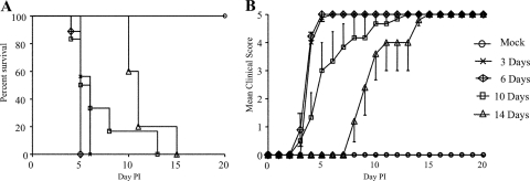
Groups of 3-, 6-, 10-, 14-, and 22-day-old mice were infected i.p. with 107 PFU of non-mouse-adapted EV71 strain 41. Three- and six-day-old infected animals died by day 6 postinfection (p.i.) (Fig. 1A). Ten- and fourteen-day-old infected mice died by day 15 p.i., with 10-day-old animals showing a greater susceptibility to EV71 infection, as evidenced by the death of some animals as early as 4 days p.i. (Fig. 1A).
Fig 1.
Age-dependent mortality of AG129 mice infected intraperitoneally with EV71. Three-day-old to two-week-old AG129 mice were inoculated i.p. with 107 PFU of EV71 strain 41. Survival (A) and clinical scores (B) were monitored daily after infection. Clinical scores were defined as follows: 0, healthy; 1, ruffled hair and hunchbacked; 2, limb weakness; 3, paralysis in 1 limb; 4, paralysis in both limbs; 5, death. Control mice received PBS instead. Each group consisted of 5 to 8 mice. Results are representative of 2 independent experiments.
Fourteen-day-old or younger mice consistently displayed initial clinical signs including hunchback and limb weakness, which further progressed to rear-limb paralysis. Paralysis was initially mild, with decreased limb movements, and severe paralysis was then observed for all limbs 1 or 2 days prior to death (Fig. 1B). Interestingly, none of the animals experienced significant body weight loss throughout the course of infection, indicating that body weight loss does not constitute a valid criterion for early euthanasia (see Fig. S1 in the supplemental material). Therefore, in subsequent experiments, the infected animals were euthanized when they displayed the characteristic 2-limb paralysis observed a couple of days prior to death. Mice of 22 days of age were not found to be susceptible to EV71 infection, with 100% survival and no clinical manifestations (data not shown).
AG129 mice are susceptible to EV71 infection via the i.p. and oral routes in a dose-dependent manner.
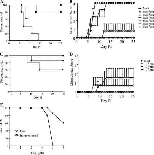
Groups of 2-week-old mice were infected i.p. with 10-fold serially diluted viral suspensions of EV71 strain 41 ranging from 103 to 107 PFU per mouse. A dose-dependent mortality was observed, with the highest viral dose resulting in the earliest time of death. Upon infection with 106 PFU and higher, all mice died by 14 days p.i., whereas all mice survived at infectious doses of 105 PFU and lower (Fig. 2A). Mice infected with lethal doses consistently displayed hunchback, limb weakness, and limb paralysis, while mice infected with a nonlethal dose remained healthy throughout the experiment (Fig. 2B).
Fig 2.
Survival rates of AG129 mice infected with a dose range of EV71. Two-week-old AG129 mice were infected i.p. (A and B) or orally (C and D) with serially diluted doses of EV71 ranging from 103 to 107 PFU/mouse. Survival (A and C) and clinical scores (B and D) were monitored on a daily basis. Clinical scores were graded as described in the legend of Fig. 1. Control mice received PBS (Mock). (E) Comparative dose dependency of EV71-induced death in 2-week-old AG129 mice infected via the i.p. or oral route. Each group consisted 5 to 7 mice. Results are representative of 2 independent experiments.
Since EV71 is an enteric virus, disease manifestations and outcomes upon oral infection of 2-week-old AG129 mice with 107 PFU of EV71 strain 41 were also investigated. Approximately 40% of the mice died by 15 days p.i. (Fig. 2C), and importantly, they displayed relevant clinical symptoms similar to those observed for i.p. infected animals (Fig. 2D). The survivors appeared healthy throughout the course of the experiment. The oral administration of 106 PFU led to only 15% mortality (Fig. 2C). Table 1 summarizes the mean day of death as a function of the route of infection and the infectious dose.
Table 1.
Mean day of death_a_
| Dose (PFU) | Intraperitoneal infection | Oral infection | ||
|---|---|---|---|---|
| Mean day of death p.i. | No. of mice | Mean day of death p.i. | No. of mice | |
| Mock | No death | 5 | No death | 5 |
| 103 | No death | 5 | Not tested | |
| 104 | No death | 5 | Not tested | |
| 105 | No death | 5 | No death | 5 |
| 5 × 10⁵ | 13 | 5 | Not tested | |
| 106 | 9 | 5 | 10 | 7 |
| 107 | 8.62 | 8 | 11 | 5 |
EV71 strain 41 displays neurotropism in AG129 mice.
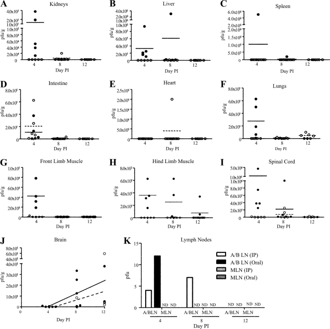
The viral titers in the blood, pooled lymph nodes, kidneys, spleen, intestines, limb muscles, liver, lungs, heart, spinal cord, and brain from animals i.p. or orally infected with 107 PFU of EV71 strain 41 were monitored by plaque assay. No infectious viral particles were detected in the blood at all time points tested and regardless of the route of infection (data not shown). At an early stage of infection (day 4 p.i.), a substantial number of viral particles were recovered in all the organs assayed, except for the heart from the i.p. infected animals (Fig. 3A to J). In contrast, the majority of the infectious viral particles were confined to the intestines of the orally infected mice, with few infectious viral particles being recovered from the brain and axillary and bronchial lymph nodes (Fig. 3D, J, and K). Interestingly, viable infectious particles were absent from the limb muscles of all the orally infected animals but one, for which a low level of viable virus particles was detected in the front limb muscle at day 4 p.i. (Fig. 3G). However, real-time PCR analysis clearly revealed the presence of viral RNA in the muscle tissues from all the orally infected mice and at all the time points tested (see Fig. S2 in the supplemental material). Together, these data suggest that the virus does travel from the gut to the intestines in orally infected mice, but the amount of infectious virus particles that reach the limb muscles is likely to be insufficient to be detectable by plaque assay.
Fig 3.
Virus titers in organs from AG129 mice infected with EV71 via the i.p. and oral routes. Two-week-old mice were inoculated i.p. (closed circles and solid lines) or orally (open circles and dashed lines) with EV71 at 107 PFU/mouse. At days 4, 8, and 12 postinfection, animals (n = 5) were euthanized, and virus titers in the kidneys (A), liver (B), spleen (C), intestine (D), heart (E), lungs (F), front-limb muscle (G), hind-limb muscle (H), spinal cord (I), brain (J), and axillary and bronchial lymph nodes (A/B LN) and mesenteric lymph nodes (MLN) (K) were determined by a plaque assay. Results are expressed as PFU per gram of tissue and means for panels A to I. Results are expressed as PFU per gram of tissue and curve fits for panel J. Results are representative of 2 independent experiments.
At later time points postinfection (days 8 and 12 p.i.), infectious virus was detected mostly in the skeletal muscles from i.p. infected mice only and in the CNS (spinal cord and brain) from both animal groups. Whereas the number of viral particles decreased over time in the limb muscles from the i.p. infected mice (Fig. 3G and H), high and sustained PFU numbers were measured in the brains of both i.p. and orally infected animals (Fig. 3I and J).
Histopathological examination of EV71-infected mice.
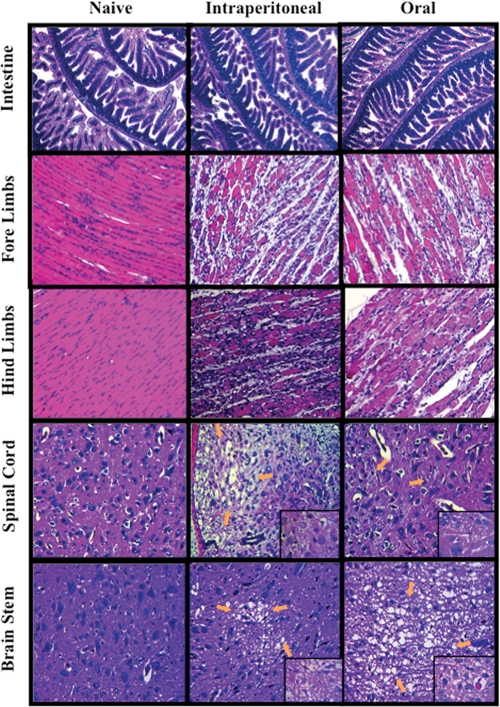
A histopathological examination of the infected mice at the moribund state was carried out. No marked lesions and/or obvious signs of inflammation were observed for the spleen, heart, lungs, and liver of the infected animals (data not shown). Interestingly, the histological analysis of the intestines did not reveal any obvious damage, even in animals infected orally, indicating that EV71 did not cause any cytopathic damage at its primary site of infection (Fig. 4). In contrast, massive damage was observed for the limb muscles, characterized by the presence of foci of myositis and myonecrosis associated with neutrophilic infiltration (Fig. 4).
Fig 4.
Histological examination of EV71-infected mice. Two-week-old AG129 mice were inoculated i.p. or orally with EV71 at 107 PFU/mouse. The animals were sacrificed at the moribund state, and paraffin sections of the organs were stained with H&E. Arrows indicate neuropil vacuolation and neuronal loss in the anterior horn area (spinal cord) and brainstem reticular formation (brainstem). The specimens shown are representative of 3 mice in each group with similar histologies. Observations were made at a magnification of ×20. Insets at the right bottom corners are observations made at a ×100 magnification.
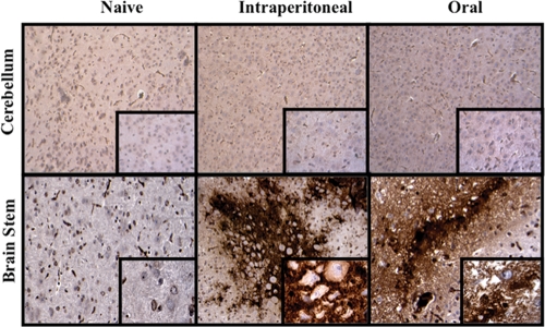
Hyperintense lesions in the anterior horn and ventral root of the spinal cord are believed to cause EV71-induced acute flaccid paralysis in patients (2). Similarly, the spinal cord and brainstem of EV71-infected AG129 mice also appeared to be severely damaged, in a pattern strikingly similar to that reported for patients. Neuropil vacuolation and neuronal degeneration or loss with no evidence of neutrophilic infiltration were observed in the anterior horn area and brainstem reticular formation (Fig. 4). No obvious pathological changes were identified in other parts of the CNS (data not shown). Immunohistochemistry (IHC) staining using an anti-EV71 monoclonal antibody further confirmed that most of the virus particles accumulated in the brainstem area, in which the cell bodies of neurons were stained strongly, but not in other areas of the brain, such as the cerebellum (Fig. 5).
Fig 5.
Detection of EV71 particles in the brain by immunohistochemistry. Two-week-old AG129 mice were inoculated i.p. or orally with EV71 at 107 PFU/mouse. The animals (n = 2) were sacrificed at the moribund state, and paraffin sections of the brains were stained with monoclonal antibody against EV71. Observations were made at a magnification of ×20. Insets at the right bottom corners are observations made at a ×100 magnification.
Proinflammatory cytokines are upregulated in EV71-infected mice.
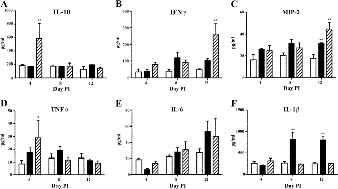
Enhanced cytokine production has been proposed to contribute to EV71 pathogenesis in both humans and mice (8, 14, 33). Consistently, the levels of several proinflammatory cytokines, namely, interleukin-1β (IL-1β), IL-10, macrophage inflammatory protein 2 (MIP-2), tumor necrosis factor alpha (TNF-α), and IFN-γ, were significantly elevated in EV71-infected AG129 mice. IL-10 and TNF-α were identified as early proinflammatory cytokines, as their levels were significantly increased in the i.p. infected mice at day 4 p.i. and declined to basal expression levels by day 8 p.i. (Fig. 6A and D). Instead, the IL-6, IFN-γ, and MIP-2 levels increased progressively in conjunction with disease advancement, reaching the highest levels at the time of death in mice infected via both the i.p. and oral routes (Fig. 6B, C, and E). Conversely, the level of IL-1β was found to be elevated only in mice infected orally with EV71 at the later stage of infection (days 8 and 12 p.i.) but not in i.p. infected animals (Fig. 6F). Of note, peak values of the systemic levels of these cytokines were significantly higher in i.p. infected animals than in the orally infected group, with the exception of IL-1β and IL-6.
Fig 6.
Systemic cytokine profile for EV71-infected AG129 mice. Two-week-old mice were infected via the i.p. (hatched bars) or oral (black bars) route with EV71 at 107 PFU/mouse. Mice from the mock-infected group (open bars) were administered an equal amount of PBS. At days 4, 8, and 12 postinfection, mice (n = 5) were bled and euthanized. Serum levels of IL-10 (A), IFN-γ (B), MIP-2 (C), TNF-α (D), IL-6 (E), and IL-1β (F) were quantified by ELISA. Serum samples were diluted 1:8. The data are expressed as the means ± standard errors of the means (SEM) of duplicates. *, P < 0.05; **, P < 0.01 (relative to the naïve group).
Adaptive immune response in EV71-infected AG129 mice.
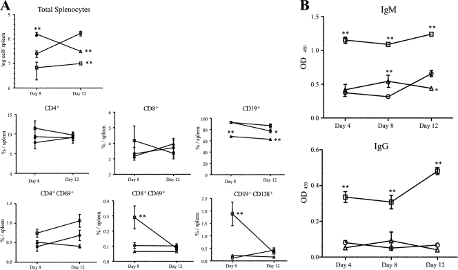
Children with EV71-associated neurological complications were reported previously to have depleted lymphocyte populations but intact humoral responses (39). Consistently, the number of total splenocytes measured by flow cytometry was significantly lower in mice i.p. (9.97 × 10⁶ cells/spleen) and orally (3.10 × 10⁷ cell/spleen) infected with EV71 at the moribund state than in uninfected controls (1.69 × 10⁸ cells/spleen) (Fig. 7A). However, the proportions of CD4+ and CD8+ T cell populations were comparable to those measured in the uninfected controls (Fig. 7A). In contrast, a decrease in the CD19+ B cell population was seen for the animals infected orally (Fig. 7A). Strikingly, whereas elevated percentages of activated CD8+ T and CD19+ B cells were measured in i.p. infected mice at day 8 p.i., such an increase was not seen with orally infected animals (Fig. 7A). Consistently, high levels of systemic EV71-specific IgM antibodies were detected at as early as day 4 p.i. in i.p. infected mice and were sustained throughout the course of infection (Fig. 7B). A significant systemic anti-EV71 IgG antibody response was also detected in the i.p. infected animals, which increased progressively to reach its peak at the time of death. In contrast, no significant levels of anti-EV71 IgG antibodies were detected in the serum of mice orally infected with EV71.
Fig 7.
Adaptive immune responses in EV71-infected AG129 mice. (A) Spleen composition of EV71-infected AG129 mice. Two-week-old mice were infected via the i.p. (squares) or oral (triangles) route with EV71 at 107 PFU/mouse. At days 4, 8, and 12 postinfection, mice (n = 5) were bled and euthanized, and their spleens were harvested individually. Total splenocytes were counted or stained with monoclonal antibodies specific to CD4, CD8, CD19, CD69, and CD138 and analyzed by flow cytometry. The data show the absolute numbers of total splenocytes and percentages of the respective cellular subsets, as indicated. (B) Specific anti-EV71 IgM and IgG titers were determined by ELISA for each individual serum sample. The data show the means ± SEM of duplicates. Mice from the mock-infected group (circle) were administered equal amount of PBS. Results are representative of 2 independent experiments. OD450, optical density at 450 nm. *, P < 0.05; **, P < 0.01 (in relation to the mock-infected group).
Model validation.
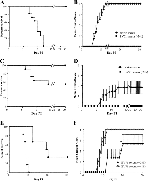
To validate this novel mouse model of EV71 infection, a passive transfer experiment was carried out, whereby a hyperimmune serum (neutralizing titer of 1:3.2 × 10⁵) generated in adult AG129 mice injected with heat-inactivated EV71 was prophylactically administered i.p. to 2-week-old AG129 naïve mice 24 h prior to a lethal challenge with 107 PFU of EV71 strain 41. A control group received the serum from naïve adult AG129 mice prior to EV71 infection. While the control animals developed severe hind-limb paralysis upon EV71 infection, mice that were injected with the anti-EV71 hyperimmune serum did not display any of the disease manifestations and were healthy throughout the experiment (Fig. 8A to D).
Fig 8.
Passive protection of EV71-infected AG129 mice. (A to D) Heat-treated anti-EV71 hyperimmune serum (neutralizing titer of 1:3.2 × 10⁵) was administered i.p. to 2-week-old AG129 mice (n = 10) 24 h prior to (with EV71 serum, −24 h) lethal challenge with EV71 at 107 PFU per mouse via the i.p. (A and B) or oral (C and D) route. The control animals were given serum from naïve mice. (E and F) Heat-treated anti-EV71 hyperimmune serum (neutralizing titer of 1:3.2 × 10⁵) was administered i.p. to 2-week-old AG129 mice (n = 10) 24 h (with EV71 serum, +24 h) or 48 h (with EV71 serum, +48 h) after lethal challenge with EV71 at 107 PFU per mouse via the i.p. route. Survival (A, C, and E) and clinical scores (B, D, and F) were then monitored daily after infection. Clinical scores were graded as described in the legend of Fig. 1.
To assess the therapeutic efficacy of the polyclonal antiserum, 2-week-old AG129 mice were challenged lethally with 107 PFU of EV71 strain 41 and subsequently injected i.p. with the hyperimmune serum 24 h or 48 h after virus challenge. Similar to the control group, all mice treated with the antiserum at 48 h postchallenge developed limb paralysis and were euthanized by day 10 p.i. (Fig. 8E and F). Instead, anti-EV71 serum treatment at an earlier time point (24 h postchallenge) was able to delay symptom manifestations significantly while conferring protection to approximately 30% of the challenged animals.
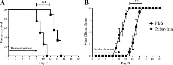
In a second set of experiments, we assessed the protective efficacy of a known EV71 drug, namely, ribavirin. Ribavirin can inhibit EV71 replication and was shown previously to reduce mortality in EV71-infected mice (13). AG129 mice were infected i.p. with 107 PFU of EV71 strain 41 and were treated with ribavirin (100 mg/kg) once daily for 9 days starting from 2 h p.i. All infected mice given PBS (control group) developed limb paralysis and were euthanized by day 11 p.i. (Fig. 9). Instead, the infected mice treated with ribavirin showed a marked delay in disease manifestation, with none of them showing disease symptoms until day 10 p.i. (Fig. 9B). Consequently, the ribavirin-treated animals experienced significantly delayed death compared to the controls (Fig. 9A). Thus, in this mouse model of EV71 infection, ribavirin treatment significantly decreased morbidity and delayed the time to death of the infected animals.
Fig 9.
Effect of ribavirin treatment on EV71-infected AG129 mice. Two-week-old AG129 mice were lethally infected i.p. with EV71 at 107 PFU/mouse. The infected mice were treated daily with PBS or ribavirin (100 mg/kg) for 9 days starting at 2 h postinfection. The survival rates (A) and clinical scores (B) were recorded. Clinical scores were graded as described in the legend of Fig. 1. **, P ≤ 0.01 by log rank test (A) and by Wilcoxon test (B).
Together, the data indicate that to be effective, antiviral treatment against EV71 must be provided very early postinfection in order to observe any beneficial impact on the disease outcome.
DISCUSSION
This work describes a novel mouse model of EV71 infection using mouse strain AG129, a mouse knockout strain that lacks IFN-α/β and IFN-γ receptors. The virus used for the study is non-mouse-adapted EV71 strain 41 (5865/SIN/00009), belonging to subgenogroup B4, which was originally isolated from a fatal human case of encephalitis during an HFMD outbreak in Singapore in 2000 (27). Subsequently, the virus was amplified in human RD cells for no more than 5 consecutive passages.
Our results indicated that 2-week-old or younger AG129 mice are susceptible to EV71 infection and develop CNS infection, as observed for humans. Upon infection via the peritoneal or oral route, AG129 mice consistently displayed hunchback, limb weakness, and limb paralysis prior to death. Similar to human manifestations of EV71 encephalomyelitis (7), the virus exhibited a strong tropism for the CNS of AG129 mice, with virus accumulation in the spinal cord and brain coinciding with the death of the animals. In addition, all sick mice exhibited massive neuronal damage, increased levels of cytokines, and splenic atrophy, as reported previously for severe cases of human EV71 disease (20). Furthermore, while not practical, the age-dependent susceptibility of AG129 mice to EV71 infection actually reflects the human situation, where children younger than 5 years of age display clinical manifestations upon EV71 infection, with a possibility of severe complications (22).
While i.p. infection with a lethal dose of EV71 strain 41 led to 100% mortality, the proportion of orally infected AG129 mice that developed CNS complications corresponded to the percentages (10 to 30%) of EV71-infected patients that present with encephalomyelitis (21, 22). The lower infectivity observed for the orally infected mice correlated with the less efficient spread of the virus from the gastrointestinal tract to the CNS. Consistently, a previous study suggested the existence of specific oral bottlenecks represented by physical barriers (colonic epithelium) that limit the virus trafficking from the oral gut to other body sites, including the CNS, upon oral infection with human poliovirus (HPV), a virus closely related to EV71 (11). Furthermore, the orally infected animals developed milder immune activation than did i.p. infected animals, likely a consequence of the impaired gut mucosal immunity previously reported for AG129 mice (9). Nevertheless, all the sick mice displayed comparable clinical manifestations and neurotropism regardless of the route of infection.
Viral dissemination was previously suggested to occur via retrograde axonal transmission along peripheral nerves (3, 19, 37). This suggestion was based on studies using a mouse brain-adapted EV71 strain that possibly predisposed the virus to a neuronal preference. Using a non-mouse-adapted EV71 strain, our present work supports the neurotropism of EV71 regardless of the route of inoculation. Upon i.p. administration to AG129 mice, the virus initially disseminated effectively in various organs and tissues, while it was confined largely to the intestines when administered orally. However, for both animal groups, the virus eventually reached and persisted within the CNS. This result is in sharp contrast with our previous observations made with 1-day-old neonates infected i.p. with the same virus strain, where viral RNA could be detected only in the intestines but not in the brain or limb muscles (8). The immaturity of the immune and neural systems in mouse neonates may account for the discrepancy in the observations, underscoring the need to develop a better mouse model of EV71 infection.
In humans, neuronal destruction during infection has been reported and was proposed to cause EV71-induced clinical symptoms such as acute flaccid paralysis (AFP) and brainstem encephalitis (36). Observations from magnetic resonance imaging (MRI) revealed that high-intensity lesions involving anterior horn cells may account for EV71-induced AFP in patients (2). In the case of EV71-associated encephalomyelitis, widespread lesions and damage of white and gray matter were observed for various regions of the CNS, including brainstem (26, 31). Similarly, the infected AG129 mice presented neuropathological features that closely resemble those observed for human disease. Severe pathological lesions were localized to the anterior horn and brainstem of the infected animals. At the time of histological analysis (moribund stage), an extensive amount of infectious viral particles was detected in the spinal cord and brain of the infected animals by both plaque assay and IHC. This finding supports that the apparent limb paralysis in the EV71-infected mice is of neurogenic origin, much akin to the effects of EV71-induced encephalomyelitis on patients.
Skeletal muscle has been proposed to support persistent enterovirus infection and to represent a viral source of entry into the CNS during poliovirus infection (5, 23). Consistently, EV71 replication in the limb muscles from i.p. infected AG129 mice was followed by rapid viral spread to the brain via the spinal cord. The apparent relationship between limb paralysis and the presence of infectious particles in the skeletal muscle from i.p. infected mice was not supported by our observations of orally infected mice. Although virus-specific RNA was present in the skeletal muscles from these mice, only minute amounts of viable infectious particles were detected by plaque assay. Virus replication was unlikely to occur in these tissues, as we did not measure any significant increase in the levels of viral RNA over time postinfection, despite the apparent myositis and limb paralysis observed for the moribund animals. Instead, virus replication was observed in the spinal cord and then ascending to the brain of the orally infected animals. Taken together, these observations seem to suggest that the paralytic poliomyelitis-like manifestations may not be associated directly with the viral cytopathic effect on the skeletal muscles but may rather be a consequence of EV71 neuronal destruction. We propose that for both orally and i.p. infected animals, the skeletal muscle represents a viral source of entry into the CNS and, ultimately, the brain. However, due to the existence of oral bottlenecks, the amount of viral particles that reach the muscles and eventually the brain is significantly lower in orally infected mice than that measured in i.p. infected animals. Subsequently, EV71 enters the CNS via peripheral somatic motor nerves that innervate the skeletal muscle, followed by anterior horn motor neurons of the spinal cord, before it spreads to the brainstem. We hypothesize that the neurotropic spread of the virus results in motor function impairment, leading to disease manifestations such as flaccid paralysis in the infected animals. However, we cannot exclude the possibility that paralysis may also be partially caused by severe myositis itself.
Several studies of humans and mice have suggested the possibility of EV71-induced immunopathogenesis (8, 34). Elevated levels of several cytokines and chemokines have been reported for children with brainstem encephalitis and pulmonary edema (14, 33, 34). Likewise, the levels of proinflammatory cytokines implicated in EV71 infection, namely, IL-1β, IL-10, MIP-2, TNF-α, and IFN-γ, were found to be significantly elevated in infected AG129 mice. However, the actual roles of the above-mentioned cytokines in the AG129 mouse model remain to be elucidated. A depletion of lymphocyte populations was also reported previously for children with edema, especially CD4, CD8, and NK cells, but with normal humoral responses (39). In the EV71-infected mice, there was a decrease in the absolute number of splenocytes, implying splenic atrophy. Furthermore, AG129 mice have been shown to produce normal antiviral antibody responses, although the IgG subclass distribution is heavily biased toward IgG1 (30). Consistently, the levels of systemic anti-EV71 antibody responses were elevated in the i.p. infected AG129 mice. In contrast, the oral inoculation of the virus did not trigger a substantial systemic antibody response against the virus, a defect that may be attributed to the impaired gut mucosal immunity in this mouse strain (9).
High levels of IFN-γ in blood and cerebrospinal fluid have been strongly associated with neurological complications in EV71 patients (14, 32). It was also suggested that type I IFNs represent an essential innate defense mechanism for controlling EV71 in mice (16). Thus, it was not surprising to find that the defect in IFN responses in AG129 mice rendered them susceptible to a lethal EV71 infection. However, the defect in IFN signaling may lead to some compensatory changes in the pattern of immune responses, which implies that the immunopathogenesis findings derived from this model may not accurately reflect the immunopathogenesis seen in immunocompetent patients, nor may it be of good predictive value for any IFN-related antiviral therapeutic approaches. Nevertheless, this mouse model represents an important step forward toward the development of a relevant animal model of EV71 infection and an improved platform for vaccine and drug testing.
Supplementary Material
Supplemental material
ACKNOWLEDGMENTS
This work was supported by the Ministry of Health/National Medical Research Council, Singapore (EDG09nov044).
We are grateful to FMC Biopolymer (United States) for giving away samples of Avicel RC-591.
Footnotes
Published ahead of print 30 November 2011
REFERENCES
- 1.Chang LY, Huang YC, Lin TY. 1998. Fulminant neurogenic pulmonary oedema with hand, foot, and mouth disease. Lancet 352:367–368 [DOI] [PubMed] [Google Scholar]
- 2.Chen CY, et al. 2001. Acute flaccid paralysis in infants and young children with enterovirus 71 infection: MR imaging findings and clinical correlates. Am. J. Neuroradiol. 22:200–205 [PMC free article] [PubMed] [Google Scholar]
- 3.Chen C-S, et al. 2007. Retrograde axonal transport: a major transmission route of enterovirus 71 in mice. J. Virol. 81:8996–9003 [DOI] [PMC free article] [PubMed] [Google Scholar]
- 4.Chen Y-C, et al. 2004. A murine oral enterovirus 71 infection model with central nervous system involvement. J. Gen. Virol. 85:69–77 [DOI] [PubMed] [Google Scholar]
- 5.Douche-Aourik F, et al. 2003. Detection of enterovirus in human skeletal muscle from patients with chronic inflammatory muscle disease or fibromyalgia and healthy subjects. J. Med. Virol. 71:540–547 [DOI] [PubMed] [Google Scholar]
- 6.Hashimoto I, Hagiwara A. 1983. Comparative studies on the neurovirulence of temperature-sensitive and temperature-resistant viruses of enterovirus 71 in monkeys. Acta Neuropathol. 60:266–270 [DOI] [PubMed] [Google Scholar]
- 7.Huang CC, et al. 1999. Neurologic complications in children with enterovirus 71 infection. N. Engl. J. Med. 341:936–942 [DOI] [PubMed] [Google Scholar]
- 8.Khong WX, Foo DGW, Trasti SL, Tan EL, Alonso S. 2011. Sustained high levels of interleukin-6 contribute to the pathogenesis of enterovirus 71 in a neonate mouse model. J. Virol. 85:3067–3076 [DOI] [PMC free article] [PubMed] [Google Scholar]
- 9.Kjerrulf M, et al. 1997. Interferon-gamma receptor-deficient mice exhibit impaired gut mucosal immune responses but intact oral tolerance. Immunology 92:60–68 [DOI] [PMC free article] [PubMed] [Google Scholar]
- 10.Kuebler JF, Czech-Schmidt G, Leonhardt J, Ure BM, Petersen C. 2006. Type-I but not type-II interferon receptor knockout mice are susceptible to biliary atresia. Pediatr. Res. 59:790–794 [DOI] [PubMed] [Google Scholar]
- 11.Kuss SK, Etheredge CA, Pfeiffer JK. 2008. Multiple host barriers restrict poliovirus trafficking in mice. PLoS Pathog. 4:e1000082. [DOI] [PMC free article] [PubMed] [Google Scholar]
- 12.Lee M-S, Chang L-Y. 2010. Development of enterovirus 71 vaccines. Expert Rev. Vaccines 9:149–156 [DOI] [PubMed] [Google Scholar]
- 13.Li Z-H, et al. 2008. Ribavirin reduces mortality in enterovirus 71-infected mice by decreasing viral replication. J. Infect. Dis. 197:854–857 [DOI] [PMC free article] [PubMed] [Google Scholar]
- 14.Lin T-Y, Hsia S-H, Huang Y-C, Wu C-T, Chang L-Y. 2003. Proinflammatory cytokine reactions in enterovirus 71 infections of the central nervous system. Clin. Infect. Dis. 36:269–274 [DOI] [PubMed] [Google Scholar]
- 15.Liu L, et al. 2011. Neonatal rhesus monkey is a potential animal model for studying pathogenesis of EV71 infection. Virology 412:91–100 [DOI] [PubMed] [Google Scholar]
- 16.Liu M-L, et al. 2005. Type I interferons protect mice against enterovirus 71 infection. J. Gen. Virol. 86:3263–3269 [DOI] [PubMed] [Google Scholar]
- 17.Nagata N, et al. 2004. Differential localization of neurons susceptible to enterovirus 71 and poliovirus type 1 in the central nervous system of cynomolgus monkeys after intravenous inoculation. J. Gen. Virol. 85:2981–2989 [DOI] [PubMed] [Google Scholar]
- 18.Nathanson N. 2008. The pathogenesis of poliomyelitis: what we don't know. Adv. Virus Res. 71:1–50 [DOI] [PubMed] [Google Scholar]
- 19.Ong K, et al. 2008. Pathologic characterization of a murine model of human enterovirus 71 encephalomyelitis. J. Neuropathol. Exp. Neurol. 67:532. [DOI] [PubMed] [Google Scholar]
- 20.Ooi MH, Wong SC, Lewthwaite P, Cardosa MJ, Solomon T. 2010. Clinical features, diagnosis, and management of enterovirus 71. Lancet Neurol. 9:1097–1105 [DOI] [PubMed] [Google Scholar]
- 21.Ooi MH, et al. 2009. Identification and validation of clinical predictors for the risk of neurological involvement in children with hand, foot, and mouth disease in Sarawak. BMC Infect. Dis. 9:3. [DOI] [PMC free article] [PubMed] [Google Scholar]
- 22.Ooi MH, et al. 2007. Human enterovirus 71 disease in Sarawak, Malaysia: a prospective clinical, virological, and molecular epidemiological study. Clin. Infect. Dis. 44:646–656 [DOI] [PubMed] [Google Scholar]
- 23.Ren R, Racaniello VR. 1992. Poliovirus spreads from muscle to the central nervous system by neural pathways. J. Infect. Dis. 166:747–752 [DOI] [PubMed] [Google Scholar]
- 24.Ryman KD, Meier KC, Gardner CL, Adegboyega PA, Klimstra WB. 2007. Non-pathogenic Sindbis virus causes hemorrhagic fever in the absence of alpha/beta and gamma interferons. Virology 368:273–285 [DOI] [PubMed] [Google Scholar]
- 25.Sasaki O, Karaki T, Imanishi J. 1986. Protective effect of interferon on infections with hand, foot, and mouth disease virus in newborn mice. J. Infect. Dis. 153:498–502 [DOI] [PubMed] [Google Scholar]
- 26.Shen WC, Chiu HH, Chow KC, Tsai CH. 1999. MR imaging findings of enteroviral encephaloymelitis: an outbreak in Taiwan. Am. J. Neuroradiol. 20:1889–1895 [PMC free article] [PubMed] [Google Scholar]
- 27.Singh S, Poh CL, Chow VTK. 2002. Complete sequence analyses of enterovirus 71 strains from fatal and non-fatal cases of the hand, foot and mouth disease outbreak in Singapore (2000). Microbiol. Immunol. 46:801–808 [DOI] [PubMed] [Google Scholar]
- 28.Solomon T, et al. 2010. Virology, epidemiology, pathogenesis, and control of enterovirus 71. Lancet Infect. Dis. 10:778–790 [DOI] [PubMed] [Google Scholar]
- 29.Tan GK, et al. 2010. A non mouse-adapted dengue virus strain as a new model of severe dengue infection in AG129 mice. PLoS Negl. Trop. Dis. 4:672. [DOI] [PMC free article] [PubMed] [Google Scholar]
- 30.van den Broek MF, Müller U, Huang S, Aguet M, Zinkernagel RM. 1995. Antiviral defense in mice lacking both alpha/beta and gamma interferon receptors. J. Virol. 69:4792–4796 [DOI] [PMC free article] [PubMed] [Google Scholar]
- 31.Verboon-Maciolek MA, et al. 2008. White matter damage in neonatal enterovirus meningoencephalitis. Neurology 71:536. [DOI] [PubMed] [Google Scholar]
- 32.Wang S-M, et al. 2007. Cerebrospinal fluid cytokines in enterovirus 71 brain stem encephalitis and echovirus meningitis infections of varying severity. Clin. Microbiol. Infect. 13:677–682 [DOI] [PubMed] [Google Scholar]
- 33.Wang S-M, et al. 2003. Pathogenesis of enterovirus 71 brainstem encephalitis in pediatric patients: roles of cytokines and cellular immune activation in patients with pulmonary edema. J. Infect. Dis. 188:564–570 [DOI] [PubMed] [Google Scholar]
- 34.Wang S-M, et al. 2008. Acute chemokine response in the blood and cerebrospinal fluid of children with enterovirus 71-associated brainstem encephalitis. J. Infect. Dis. 198:1002–1006 [DOI] [PubMed] [Google Scholar]
- 35.Wang W, et al. 2011. A mouse muscle-adapted enterovirus 71 strain with increased virulence in mice. Microbes Infect. 13:862–870 [DOI] [PubMed] [Google Scholar]
- 36.Weng K-F, Chen L-L, Huang P-N, Shih S-R. 2010. Neural pathogenesis of enterovirus 71 infection. Microbes Infect. 12:505–510 [DOI] [PubMed] [Google Scholar]
- 37.Wong KT, et al. 2008. The distribution of inflammation and virus in human enterovirus 71 encephalomyelitis suggests possible viral spread by neural pathways. J. Neuropathol. Exp. Neurol. 67:162–169 [DOI] [PubMed] [Google Scholar]
- 38.Wu CN, et al. 2001. Protection against lethal enterovirus 71 infection in newborn mice by passive immunization with subunit VP1 vaccines and inactivated virus. Vaccine 20:895–904 [DOI] [PubMed] [Google Scholar]
- 39.Yang K, Yang M, Li C, Lin S. 2001. Altered cellular but not humoral reactions in children with complicated enterovirus 71 infections in Taiwan. J. Infect. Dis. 183:85–86 [DOI] [PubMed] [Google Scholar]
- 40.Yi L, Lu J, Kung H-F, He M-L. 9 June 2011, posting date The virology and developments toward control of human enterovirus 71. Crit. Rev. Microbiol. doi:10.3109/1040841X.2011.580723 [DOI] [PubMed] [Google Scholar]
- 41.Zhang Y, et al. 9 May 2011, posting date Pathogenesis study of enterovirus 71 infection in rhesus monkeys. Lab. Invest. doi:10.1038/labinvest.2011.82 [DOI] [PubMed] [Google Scholar]
Associated Data
This section collects any data citations, data availability statements, or supplementary materials included in this article.
Supplementary Materials
Supplemental material
