In vivo Genome Editing Using High Efficiency TALENs (original) (raw)
. Author manuscript; available in PMC: 2013 May 1.
Published in final edited form as: Nature. 2012 Sep 23;491(7422):114–118. doi: 10.1038/nature11537
Abstract
The zebrafish (Danio rerio) is increasingly being used to study basic vertebrate biology and human disease using a rich array of in vivo genetic and molecular tools. However, the inability to readily modify the genome in a targeted fashion has been a bottleneck in the field. Here we show that improvements in artificial transcription activator-like effector nucleases (TALENs) provide a powerful new approach for targeted zebrafish genome editing and functional genomic applications1–5. Using the GoldyTALEN modified scaffold and zebrafish delivery system, we show this enhanced TALEN toolkit demonstrates a high efficiency in inducing locus-specific DNA breaks in somatic and germline tissues. At some loci, this efficacy approaches 100%, including biallelic conversion in somatic tissues that mimics phenotypes seen using morpholino (MO)-based targeted gene knockdowns6. With this updated TALEN system, we successfully used single-stranded DNA (ssDNA) oligonucleotides (oligos) to precisely modify sequences at predefined locations in the zebrafish genome through homology-directed repair (HDR), including the introduction of a custom-designed EcoRV site and a modified loxP (mloxP) sequence into somatic tissue in vivo. We further show successful germline transmission of both EcoRV and mloxP engineered chromosomes. This combined approach offers the potential to model genetic variation as well as to generate targeted conditional alleles.
Keywords: zebrafish, TALEN, genome engineering, loxP
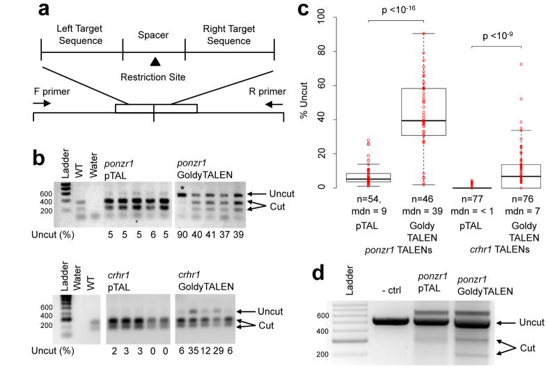
Custom zinc finger nucleases (ZFNs) 7–9 and TALENs1–5 have been used introduce locus-specific double-stranded breaks in the zebrafish genome, generating dozens of mutant alleles10. Recent work has been facilitated by the relatively straightforward DNA base recognition cipher underlying TALEN technology11,12. However, the efficacy of previously described custom sequence-specific nucleases was limiting in some applications1–5,7–9. For example, standard TALENs using the pTAL scaffold13 (Supplementary Fig. 1) targeting exon 2 of the zebrafish ponzr1 locus14 resulted in a measurable level of locus modification in somatic tissue (median value of 5%; Fig. 1b–c). This pTAL-ponzr1 pair yielded 4 germline-transmitting founder animals carrying a mutation in ponzr1 out of the 24 tested (Supplementary Fig. 3d). TALENs against a second locus (crhr1) using the pTAL scaffold yielded a modest rate of locus modification (<1%; Fig. 1b–c). These results are characteristic of the standard TALEN efficacy range demonstrating room for improvement.
Figure 1. Second-generation GoldyTALEN scaffold improves genome-editing efficacy.
a, A schematic showing the layout of TALEN target sites. TALENs were targeted to flanking sequences surrounding a restriction enzyme site for easy screening through introduction of a restriction fragment length polymorphism. b, Relative activity of the GoldyTALEN and pTAL scaffolds at two loci, ponzr1 and crhr1. Under each lane is the percent uncut DNA of a single larva, illustrating the increased activity of GoldyTALEN. c, Whisker plots of the percent uncut DNA demonstrates TALEN cutting efficiency at two loci. ponzr1 TALENs demonstrate a significant (p < 10−16), 6-fold increase in activity using GoldyTALEN. crhr1 TALENs also demonstrate a significant (p < 10−9), 15-fold increase in activity. n = number of embryos screened, mdn = the median percent cut. d, The ponzr1 GoldyTALENs were more active in a cell-free restriction enzyme digestion assay. ponzr1 DNA is labeled in both uncut and cut forms. − ctrl = negative control.
Multiple TALEN scaffold designs have been described13,15,16, including those with different N- and C-terminal truncations, diverse FokI nuclease linkers, and various nuclear localization sequences. To improve in vivo efficacy, we tested the GoldyTALEN scaffold (Supplementary Fig. 1; Supplementary Fig. 2) in an mRNA expression vector backbone (pT3TS17) using DNA analysis that measures the loss of a restriction enzyme recognition sequence at the TALEN cut site (Fig. 1a). Using the same recognition domains in the GoldyTALEN scaffold, there is a 6-fold increase in somatic gene modification at the ponzr1 locus (Fig. 1b–c; Supplementary Fig. 3b) over the pTAL scaffold. The germline modification rate was similarly increased when switching scaffolds, from 17% (4/24; pTAL-ponzr1; Supplementary Fig. 3d) to 71% (10/14; GoldyTALEN-ponzr1; Supplementary Fig. 3e). We also detected improved efficacy using a cell-free assay system with in vitro translated TALEN protein and purified ponzr1 PCR DNA (Fig. 1d). The GoldyTALENs against crhr1 showed an increase in the genome modification rate, improving from <1% to 7% median cutting efficacy (Fig. 1b–c; Supplementary Fig. 3c). Sequence comparisons of pTAL and GoldyTALEN scaffolds in both loci demonstrate similar indels at the cut site, which is diagnostic of NHEJ repair (Supplementary Fig. 3).
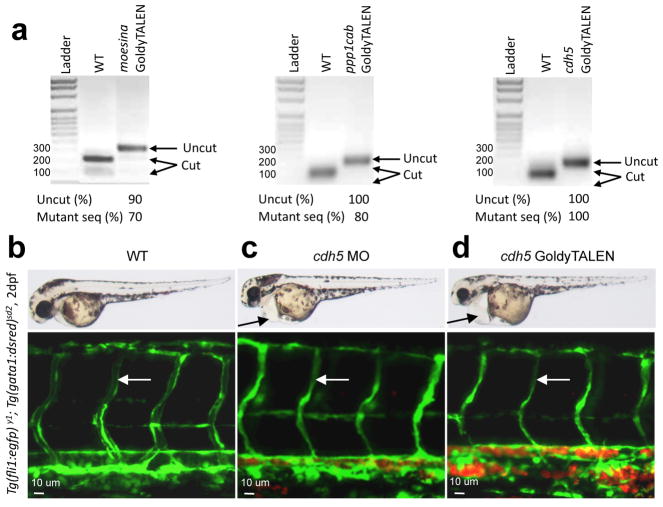
To further test the efficacy of the GoldyTALEN scaffold, we generated TALENs against three additional loci (moesina, ppp1cabb and cdh5; Supplementary Fig. 4a). We observed efficient gene modification at each locus (5 out of 5 loci total; Fig. 1 and Fig. 2a). In three instances, the mutagenesis efficiency ranged from 70 to 100% as demonstrated by loss of the restriction enzyme recognition sequence at the TALEN cut sites (Fig. 2a) and DNA sequence analyses (Supplementary Fig. 4b–d) of amplicons from pooled injected embryos. To determine the time-course of the GoldyTALEN-induced changes, we examined restriction enzyme nuclease activity at 256-cell, 28 hours post fertilization (hpf) and 50 hpf stages. A majority of the DNA was modified by the 256-cell stage (Supplementary Fig. 5). Together, these results indicate early, efficient gene targeting in somatic tissues including biallelic conversion in some animals. Somatic targeting efficacy using the GoldyTALEN scaffold compares favorably with previous TALEN scaffolds in zebrafish, with 3 of 5 GoldyTALENs demonstrating as high or higher mutation frequency as any of the previously reported loci using the first generation TALEN systems1–5.
Figure 2. Increased TALEN efficiency results in biallelic gene targeting.
a, GoldyTALENs were designed against the moesina, ppp1cab and cdh5 genes. All three gene targets contained a restriction enzyme site within the spacer region between the TALEN binding sites. Injection of GoldyTALEN mRNAs demonstrated a nearly complete loss of the restriction enzyme site in the amplicons of somatic tissue. Each lane is the amplification product from a group of 10 embryos. Mutant seq (%) = percentage of amplicons that carry mutant sequences as determined by sequencing 10 clones (Supplementary Fig. 4). b–d, Injection of cdh5 GoldyTALENs (d) phenocopies the MO-based loss-of-function phenotype (c). Brightfield images (top panels) show pronounced cardiac edema (arrows) in both GoldyTALEN (d)- and MO (c)-injected larvae at 2 days post fertilization. Using the Tg(_fli1_-egfp)y1 line, the intersomitic vessels were visualized (bottom panels) and show a loss of lumen formation (white arrow) in both the MO (c)- and GoldyTALEN (d)-injected larvae. The Tg(gata1:dsred)sd2 line revealed reduced circulation in GoldyTALEN- and MO-injected larvae, demonstrated by the increase in red fluorescence in the confocal images (see Movies 1–3).
In response to the increased efficacy of the GoldyTALENs, we asked whether injection of TALENs could recapitulate a known MO6 loss of function phenotype. We conducted a dose-response curve of the moesina, ppp1cab and cdh5 GoldyTALEN pairs, optimizing GoldyTALEN concentration to the number of embryos with biallelic changes, and percent dead or malformed embryos (Supplementary Fig. 6). Embryos injected with either cdh5 GoldyTALENs (Fig. 2d) or MOs18 (Fig. 2c) displayed similar vascular phenotypes: pronounced cardiac edema (Fig. 2b, top panels), loss of patent lumens in the Tg(fli1-egfp)y1 vasculature19 (Fig. 2c, d, bottom panels), and loss of circulating Tg(gata1:dsred)sd2 red blood cells20 (Fig. 2c, d, bottom panels; Supplementary Movies 1–3). A similar pericardial edema phenotype was observed in F1 offspring from F0 cdh5 founder incrosses (data not shown), suggesting specificity of the phenotype described in F0s to cdh5 loss of function. Furthermore, cdh5 GoldyTALEN-injected embryos display little or no Cdh5 protein (Supplementary Fig. 7). Together, these results indicate that the GoldyTALEN platform can achieve efficient biallelic targeting recapitulating known loss-of-function phenotypes. Furthermore, these data demonstrate that GoldyTALENs have the potential to be a complementary, but distinct, approach to MO-based somatic phenotype assessment.
The biallelic GoldyTALEN-injected fish were raised to assess germline mutation transmission. The moesina, ppp1cab or cdh5 F0 founders were outcrossed. Ten pooled F1 embryos were screened and displayed a 9 to 55% locus mutation frequency (Supplementary Fig. 8a–c). From two founder F0 outcrosses per locus, 10 individual F1 embryos were sequenced with mutant alleles identified in 20% to 100% of the F1 offspring (Supplementary Fig. 8). Furthermore, in two out of three of these loci we detected germline mosaicism, suggesting several independent repair events. These data indicate that the efficient somatic TALEN targeting is effectively passed through the germline.
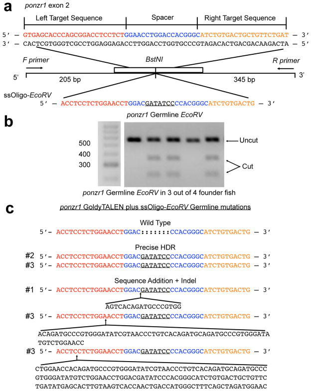
Recent in vitro work demonstrates that ssDNA can be an effective donor for HDR-based genome editing at a ZFN-induced double-stranded break21,22. With the highly efficient genome modification success of GoldyTALENs, we hypothesized that synthetic oligos designed to span the predicted TALEN cut site could serve as a template for HDR in vivo (Fig. 3). Using ponzr1 as a test locus, we introduced an EcoRV restriction site by co-injection of ponzr1 GoldyTALENs and a ssDNA oligo (Fig. 3a). In these experiments, 42 of 74 injected embryos displayed a detectable level of chromosomes containing the introduced EcoRV sequence with an estimated 9% ratio of converted chromosomes in these animals (Supplementary Fig 9a). Sequence analysis indicated two precisely modified chromosome events from different larvae (Supplementary Fig. 9b) demonstrating successful somatic HDR at the ponzr1 locus. Other events show precise addition at the 3′ end while small indels were noted at the 5′ side of the modification site (Supplementary Fig. 9c). Several homology arm lengths were tested for the highest HDR signal. In this experimental approach, an increase in homology arm length that spanned the TALEN binding site decreased the frequency of HDR events (Supplementary Table 1).
Figure 3. Targeted genome editing using GoldyTALENs.
a, A schematic of the ponzr1 locus with the ssDNA sequence used to introduce a targeted exogenous EcoRV sequence into the genome in vivo. The left and right TALEN binding sites are shown in red and orange, respectively, and the spacer region is in blue. b, A representative gel demonstrating germline transmission of the HDR-based EcoRV sequence in 3 out of 4 fin tissue-positive fish. c, Sequence analysis of the three germline-transmitting lines. The first fish transmitting HDR-based genome changes through the germline (#1) yielded 7 out of 96 embryos with an incorporated EcoRV site. The genomes of all 7 embryos showed the same modified sequence. The second founder fish (#2) yielded 7 out of 46 embryos with EcoRV incorporation. All 7 embryos showed precise HDR-based addition of the EcoRV sequence. The third fish with germline transmission (#3) yielded 5 out of 18 embryos with an incorporated EcoRV site, and showed a mosaic germline as demonstrated by offspring with three different modified sequences. One embryo included precise HDR-based EcoRV addition. The other 4 embryos contained sequence insertions on the 5′ end with two embryos each harboring the specific sequences changes.
To test whether the HDR sequence modification was stably maintained in zebrafish somatic tissue, fin biopsies from two month-old fish were assayed for addition of the EcoRV sequence at the ponzr1 locus. Out of 186 fish, 8 showed a visible incorporation of EcoRV (Supplementary Fig. 9c). To determine whether a lack of somatic EcoRV incorporation also indicated a lack of germline incorporation, 13 randomly selected fish with _EcoRV_-negative fin biopsies were outcrossed. The offspring from all 13 adults were negative for EcoRV incorporation at the ponzr1 locus (clutch sizes ranged from 16–96 embryos). Therefore, fin biopsy positive fish were prioritized for determining germline transmission. Outcross embryos from three out of four fin tissue positive fish yielded clutches with introduction of the EcoRV site at the ponzr1 locus (Fig. 3b). Two out of three of these germline fish demonstrated precise EcoRV addition (Fig. 3c).
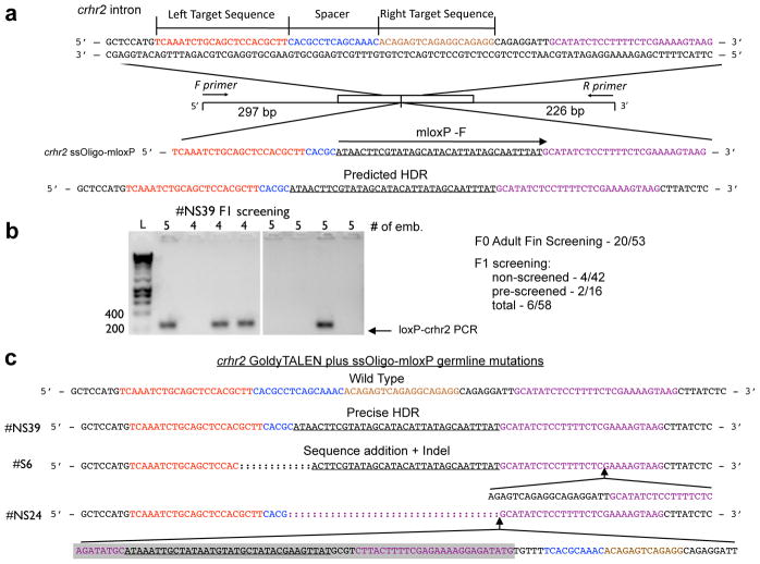
We next asked whether TALEN/oligo co-injection could introduce larger sequences such as a loxP site, an essential step in making Cre-dependent conditional genetic alleles. We used TALENs against an intron in the crhr2 gene and a ssDNA oligo were used to add a modified loxPJTZ17 (mloxP)23 site at this location (Fig. 4a). PCR analysis demonstrates somatic introduction of the mloxP sequence at the crhr2 TALEN cut site (Supplementary Fig. 10a). Sequence characterization confirmed integration of the mloxP site in 3 assayed somatic chromosomes (Supplementary Fig. 10b). A similar method was used to introduce an mloxP sequence at the ponzr1 locus (Supplementary Fig. 11a). Sequencing confirmed precise somatic addition at this locus (Supplementary Fig. 11b–c).
Figure 4. Germline mloxP integration into the crhr2 locus.
a, A diagram of the TALEN target sites with the mloxP ssDNA oligo. The left and right TALEN target sequences are red and orange, respectively, the spacer region is blue, and the oligo’s right homology arm is in purple. The mloxP sequence is underlined. b, Germline screening of the crhr2 locus. 53 adult fish were prescreened via fin biopsy. Of those prescreened, 20 demonstrated mloxP maintenance. 16 F0s were outcrossed with 2 showing germline transmission. 42 unscreened F0s were outcrossed and 4 demonstrated germline transmission. c, Sequence confirmation of three mloxP germline fish. One fish demonstrated precise germline HDR while two showed indels. In #NS24, we saw the reverse complement of the mloxP was noted (shaded in grey).
Maintenance of somatic mloxP-modified crhr2 chromosomes by fin bioposy was used to identify germline transmission of the mloxP sequence. Positive chromosomes were detected by quantitative PCR in 20 of 53 animals (Supplementary Fig. 10a). Embryos were obtained from 16 of the somatic-positive fish as well as 42 fish that had not been pre-screened by PCR. Both groups transmitted HDR events through the germline (Fig. 4b). However, no significant enrichment for likely germline transmitting animals was noted, perhaps due to the less stringent PCR assay than used for ponzr1. In total, 6 out of 58 injected animals transmitted mloxP-modified chromosomes through the germline at the crhr2 locus (Fig. 4b). Sequence confirmation of three of these fish demonstrated a precise HDR event as well as other, non-precise events (Fig. 4c).
Here, we focused on local genome editing changes induced by TALENs, especially those induced by HDR. However, more complete analyses will be required to assess any, off-target effects of TALENs or ssDNA-based HDR. Whole genome sequencing on germline-transmitting fish from different parental lines would be particularly instructive. Should this analysis demonstrate off-target mutations, TALENs using obligate heterodimer-based nuclease fusions have recently been reported as an alternative approach3,5,24. Using obligate heterdimers in the GoldyTALEN scaffold is one future method for potentially optimizing HDR-directed gene editing specificity.
To our knowledge, these results represent the first description of successful HDR in zebrafish and the first demonstration of HDR using ssDNA as a donor template in vivo. This approach complements the error-prone NHEJ toolkit for model organisms (Fig. 5). The use of ssDNA facilitates an array of genome changes, including the introduction of single nucleotide polymorphisms for vertebrate genetic applications. The asymmetry in precise editing suggests an additional mechanism for genome editing that incorporates both HDR and NHEJ (Fig. 5). For example, the donor ssDNA may serve as a primer for new strand synthesis at the TALEN break. Extension from the 3′ end of the oligo would create long regions of homology for recombination. However, the 5′ end of the oligo limits the extent of strand invasion and a limited opportunity for HDR. This leads to 5′ end resolution by either HDR or NHEJ. For applications where new sequences are introduced into non-coding genomic regions, such as the introduction of loxP sites into intronic sequences, either event will likely be of high utility.
Figure 5. in vivo TALEN-induced genome editing outcomes.
TALENs efficiently create double-stranded breaks in chromosomal DNA and catalyze three major outcome classes. First, error-prone NHEJ produces an indel in and near the spacer region of the TALEN binding site. If a complementary ssDNA oligonucleotide is also added, two different outcomes are noted. First, HDR precisely uses the exogenous sequence information in the ssDNA to add sequence at the cut site. Alternatively, ssDNA acts as a primer for 3′ integration of the oligonucleotide but the 5′ end undergoes error-prone NHEJ22.
Using the zebrafish, we report an updated TALEN system for use in genome modification and functional genomic applications. The high efficacy enables new approaches, including somatic gene targeting for reverse genetics applications. Furthermore, we show that synthetic ssDNA oligos can be used with this TALEN system for genome editing including the precise introduction of exogenous DNA sequence at a specific locus. Although deployed here in zebrafish, this approach has the potential to be effective for in vivo applications in a wide array of model organisms.
Methods Summary
TALENs were assembled via the GoldenGate method13. For ease of analysis, TALENs recognition sequences flanked a unique restriction site within the targeted gene. TALEN RVDs were cloned into a pT3TS17-driven TALEN scaffold, and mRNA was injected into single-cell zebrafish embryos. The injected larvae were either molecularly tested or raised for germline mutation analysis. Somatic and germline TALEN-induced mutations were evaluated via PCR and restriction fragment length polymorphisms. To induce HDR events, singe-stranded DNA oligonucleotides with either an EcoRV or mloxP site were designed with short homology arms around a TALEN target site and were injected into one-cell zebrafish embryos. PCR analysis of modified loci was used to detect the resulting somatic and germline HDR events. Detailed methods can be found in the Supplementary Information.
Full Methods
TALEN Design
The software developed by the Bogdanove laboratory (https://boglab.plp.iastate.edu/node/add/talen) was initially used to find candidate binding sites as described13. Three criteria were used for TALEN design. First, TALEN binding sites were selected that ranged from 15–25 bases in length. Second, the spacer length was initially selected to be 14 to 18 base pairs (bps), but subsequent GoldyTALEN designs were restricted to 15–16 bps. Additionally, when possible TALEN cut sequences were selected around a restriction enzyme centrally located within the spacer. To simplify the TALEN design process, a free, open access software (Mojo Hand) was created and made available online (www.talendesign.org). Mojo Hand downloads sequence from NCBI and uses an exhaustive database of commercially available restriction enzymes to identify TALEN binding sites with a restriction enzyme site in the spacer region to simplify downstream analysis (Neff et al (unpublished)). Mojo Hand also features a BLAST interface that will search genomes for potential second site effects.
TALEN Binding Sites and Spacer Regions
The ponzr1 TALEN recognition sequences are: left TALEN 5′-GTGAGCACCCAGCGGACCTCCTCT-3′ and right TALEN 5′-ATCAGAACAACAGTCAGAGAT-3′. Between the two binding sites is an 18 bp spacer with a _BstN_I sequence (GGAACCTGGACCACGGGC, _BstN_I underlined). The crhr1 TALEN recognition sequences are: left TALEN 5′-TGCAACACTGAGCTCTGTAAACCT-3′ and right TALEN 5′-CTGCTGCCGACTGGACCCTGAGGT-3′. Between the two binding sites is a 15 bp spacer with a _BstU_I site (GTCCGCGTGTGGCGA, _BstU_I underlined). The moesina TALEN recognition sequences are: left TALEN 5′-ACCCAGAAGACGTTT-3′ and right TALEN 5′-CTTTGAGTGGCCTCCT-3′. Between the two binding sites is a 15 bp spacer with an _Xmn_I site (CTGAGGAACTGATTC, _Xmn_I underlined). The ppp1cab TALEN recognition sequences are: left TALEN 5′-CCACCAGAGAGTAACT-3′ and right TALEN 5′-GCCTCTGTCAACATAGT-3′. Between the two binding sites is a 15 bp spacer with a _Bs_II site (ACCTATTTCTGGGAG, _Bs_II underlined). The cdh5 TALEN recognition sequences are: left TALEN 5′-CTCCTCAACATACATACT-3′ and right TALEN 5′-ACAAATGATTCATCTT-3′. Between the two binding sites is a 16 bp spacer with a _Hinc_II site (GGAGAGTTAGTTGACA, _Hinc_II underlined). The crhr2 binding sites are: left TALEN 5′-GTCAAATCTGCAGCTCCACGCTT-3′ and right TALEN 5′-CCTCTGCCTCTGACTCTGT-3′. Between the two binding sites is a 15 bp spacer (CACGCCTCAGCAAAC).
TALEN Constructs
TALEN assembly of the RVD-containing repeats was conducted using the Golden Gate approach13. Once assembled, the RVDs were cloned into a pT3TS destination vector with the appropriate TALEN backbone to generate mRNA expression plasmids – pT3TS-TAL (pTAL) and pT3TS-GoldyTALEN (GoldyTALEN). In vitro transcription of TALEN mRNA was conducted by linearizing the expression plasmids with _Sac_I endonuclease at 37°C for 2–3 hours, transcribing the linearized DNA (T3 mMessage Machine kit, Ambion) and purifying the mRNA by phenol/chloroform extraction (T3 mMessage Machine kit user manual protocol) for injection.
TALEN Mutation Screening
One-cell embryos were microinjected with 50–400 pg of TALEN mRNA. The dose of each pair of TALENs injected was empirically determined, with up to a 3-fold difference noted between different TALEN pairs. In each case, conditions were used where over 50% embryos survived post-injection. Genomic DNA for Figures 1, 3 and 4 were collected at 2–4 dpf from 24–32 individual larvae by incubating in 50mM NaOH at 95°C, followed by cooling to 4°C and adding 1/10 volume 1M Tris-HCL pH 8.025. Genomic DNA for Figure 2 was isolated from groups of 10 larval zebrafish using DNAeasy Blood and Tissue kit (Qiagen). Genotyping was conducted using PCR followed by restriction enzyme digest. For ponzr1, the primers were 5′-GTTCACACAAAATGTCTCTCAAGTCTCTAAATC -3′ and 5′-AGTGGCCAGTGAGTGTATGTTACCT -3′. For crhr1 the primers were 5′-CGTGAAAGAGACAGCGAAGGGATTG -3′ and 5′-AGAAACTACCATTGTCACACTGAGCGAAG -3′. The primers for moesina were 5′-GTTACGGCTCAAGACGTC-3′ and 5′-CAGGATGCCCTCTTTAAC-3′. The primers for ppp1cab were 5′-GATGTTCATGGTCAGTAC-3′ and 5′-TGATTGAGGCACATTCATGG-3′. The primers for cdh5 were 5′-TTGTTGTCCTTGCAAAGCTG-3′ and 5′-TCTAGAGGATTCGCTGAT-3′. The primers for crhr2 were 5′-CCCTGATTGTGGAACTTTTCAGAACGTA-3′ and 5′-TGGTTTGGAATTAGTGCAGCATGAGTA-3′. Mutations were assessed by loss of restriction enzyme digestion. To sequence-verify mutations, the gel-purified, uncut PCR products were cloned into the TOPO® TA Cloning® Kit (Invitrogen).
Analysis of cdh5
A cdh5 morpholino18 was injected at the 1–4 cell stage into Tg(fli1:efgp)y1 embryos19. The vascular phenotype of the MO and the GoldyTALEN-injected embryos were assessed using a confocal microscope. Antibody staining using the cdh5 antibody26 was performed as described18.
Genome Editing
For the ponzr1 locus, a ssDNA oligo was designed to target the spacer sequence between the TALEN cut sites. The oligo extends to half the length of the TALEN recognition site. An _EcoR_V site (5′-GATATC-3′) or a modified loxP (mloxP) site (5′-ATAACTTCGTATAGCATACATTATAGCAATTTAT-3′) was introduced near the center of the oligo resulting in a 20-base homology arm on the 5′ end and an 18-base homology arm on the 3′ end. For the crhr2 locus, the crhr2 mloxP oligo (5′ –TCAAATCTGCAGCTCCACGCTTCACGCATAACTTCGTATAGCATACATTATAGCAAT TTATGCATATCTCCTTTTCTCGAAAAGTAAG – 3′) was designed to replace the 3′ TALEN binding site with an mloxP site while providing 27 bases of homology at both 5′ and 3′ end. The oligos were ordered from Integrated DNA Technologies (IDT) and purified using the Nucleotide Removal Kit (Qiagen).
One-cell embryos were microinjected with both the GoldyTALEN mRNA and ssDNA donor. The ssDNA oligo dose was varied to improve the rate of HDR without impacting toxicity beyond 50% embryonic death post injection. For the ponzr1 locus, 50–75 pg of ponzr1 GoldyTALEN mRNA and 50–75 pg of the ssDNA donor. For the crhr2 locus, 50 pg of crhr2 GoldyTALEN mRNA was injected with either 25 pg or 50 pg of crhr2 mloxP oligo. Genomic DNA was isolated as described above. If the embryos were injected with the _EcoR_V oligo, PCR was conducted using the same primers as listed above and the product was digested using EcoRV. The full-length amplicon from _EcoRV_-positive larvae was cloned into a TOPO® TA Cloning® Kit (Invitrogen). Colony PCR was used to identify plasmids with _EcoRV_-modified inserts. Those plasmids were subsequently sequenced to confirm _EcoR_V integration and determine details of sequence changes due to HDR. If the embryos were injected with the mloxP oligo, the genomic DNA was amplified using the same forward primer as listed above and a mloxP reverse primer, 5′-ATAAATTGCTATAATGTATGCTATACGAAGT-3′, or the same reverse primer as listed above and a mloxP forward primer, 5′-ACTTCGTATAGCATACATTATAGCAATTTAT-3′. For sequence analysis, the complete amplicon was produced using the gene-specific primers listed above and cloned (TOPO® TA Cloning® Kit, Invitrogen). Colony PCR was used to find mloxP-positive plasmids. The positive plasmids were sequenced for confirmation of mloxP integration.
Injected fish from the same batch of somatically screened embryos were raised. When the fish were at least two months old, fin tissue was obtained using standard protocols pre-approved by Institutional Animal Care and Use Committee guidelines. The fish were anesthetized using Tricaine (approximately 200 μg/ml). The tail fins were trimmed with a fresh razor blade for each fish to prevent contamination. The most caudal 2–3mm of fin was biopsied and placed on ice until all fin biopsies were collected. 150 μl of 50mM NaOH was added to the fin clips prior to DNA isolation (above). Those fish that maintained somatic modifications were outcrossed to wild type fish and the embryos were screened for germline mutations. Somatic mutations were determined by RFLP analysis for EcoRV integration into ponzr1. Quantitative PCR of mloxP integrations into the crhr2 locus were compared to a reference gene, RPS6Kb1. Twenty of 53 fish included >0.2% of their DNA containing mloxP integrations into crhr2 (CT of ≤10) and were prioritized for screening. For mloxP integration into crhr2, 42 fish that were not screened by quantitative PCR were also tested for germline transmission and no appreciable difference in germline transmission between these two methods was noted.
The PCR product for germline HDR events were cloned and sequenced. In one clone that contained a sequence insertion along with integration of the EcoRV site, the sequencing was more difficult presumably because the insertion tended to form a hairpin and disrupted the sequencing reaction. To obtain the full sequence, the PCR product was digested with EcoRV and each half sequenced separately. Similar cloning difficulties were observed in some crhr2 lineages, but not for precise HDR or limited sequence addition.
The sequence addition process using ssDNA oligos is inherently less efficient than the relatively simpler NHEJ events seen in the GoldyTALEN-alone injected embryos. Therefore, to identify a precise HDR event, more fish will need to be raised and screened. Fin clipping the fish for maintenance of the somatic insertion may be a good indicator of germline transmission. Continued investigation into the mechanism of HDR incorporation in zebrafish will likely increase the efficiency of this technique.
Zebrafish Work
The zebrafish work was conducted under full animal care and use guidelines with prior approval by the local institutional animal care committee’s approval. Danio rerio transgenic lines were described previously: Tg(fli1:efgp)y1 vasculature19 and Tg(gata1:dsred)sd2 red blood cells20.
Data Analysis and Statistics
ImageJ was used to quantify the percent GoldyTALEN-modified chromosomes by measuring the intensity of bands post-digestion. For each gel, the background was subtracted and each lane isolated to generate individual intensity plot profiles. A straight line was drawn across the bottom of each plot to eliminate inconsistencies caused by baseline skew. The intensity measurement for each band was added together to determine total intensity. To calculate percent cutting, the intensity of the top band was divided by the total intensity. A student’s T-test was used to compare TALEN scaffold cutting efficiencies. To measure the differences between pTAL and GoldyTALEN at two different loci, several whisker plots were constructed (Figure 1C). The interquartile range (IQR; Q3-Q1) is shown as a box, with the median value (Q2) being the horizontal line within the box. The upper and lower whiskers are the highest and lowest data point within 1.5 times the IQR added or subtracted from Q3 or Q1, respectively.
A similar approach was used to calculate the percent of HDR-converted chromosomes. The intensity of the digested products were added together and divided by the total intensity. The percent of embryos with an HDR signal was determined by dividing the number of embryos with signal by the total number of screened embryos.
Cell-free TALEN Restriction Endonuclease Assay
In vitro translation of 2 μg of each TALEN mRNA was conducted using the TNT® Quick Coupled Transcription and Translation System (Promega). 5 μg of the ponzr1 PCR product was included in the assay mix during in vitro translation of different TALEN combinations, allowing the translation and in vitro nuclease digestion to occur simultaneously. The highest signal was obtained when translation and digestion steps were conducted simultaneously presumably because the TALEN protein is unstable using these in vitro conditions. Translation was conducted for 2 hours at 30°C. To further facilitate TALEN in vitro nuclease activity, the assay mix was diluted five fold in in vitro digestion buffer (20mM Tris-HCl pH 7.5, 5mM MgCl2, 50 mM KCl, 5% Glycerol, and 0.5mg/ml BSA)27. The assay mix was incubated at 30°C for 4 hours. The digested DNA was purified using the PCR Purification kit (Qiagen), concentrated via ethanol precipitation, and run on a 2% agarose gel. No TALEN mRNA was added to the negative control.
Supplementary Material
1
2
3
Acknowledgments
Grants: State of Minnesota grant H001274506 to SCE and DFV; NIH GM63904 to SCE; NIH grant P30DK084567 to SCE and KJC; NIH grant DK083219 to VMB; Mayo Foundation; NIH DA032194 to KJC; NIH grant R41HL108440 to DFC and SCF; NIH grant GM088424 to JJE; NSF grant DBI0923827 to DFV; General Research Fund (HKU771611, HKU771110, HKU 769809M) from the Research Grant Council, The University of Hong Kong and the Tang King Yin Research Fund to ACM and AYHL. We thank Dr. Greg Davis for discussion on ssDNA use with custom restriction enzymes, Hind J. Fadel for in vitro RNA synthesis, and Stephanie Westcot for comments on this manuscript. We thank Gary Moulder for help in DNA analyses and members of the Mayo Clinic Zebrafish Core Facility for excellent animal care.
Footnotes
Author contribution statements
WT designed and constructed full-length pT3TS-Tal vectors targeting Danio rerio crhr1 and crhr2 against exon sequences provided by KJC. DFC and SCF designed and produced the pT3TS-Tal and GoldyTALEN cloning vectors. DFC synthesized crhr1 and crhr2 TALEN mRNA from the pT3TS-Tal vectors. CGS designed and assembled ponzr1 TALENs and transferred TAL repeats from original crhr1 and crhr2 TALEN vectors into GoldyTALEN. TLP and KJC did initial crhr1 TALEN microinjections. TLP performed initial characterization of crhr1 mutagenesis efficiency by PCR and RFLP analysis. TLP and KJC microinjected crhr2 TALEN and loxP oligos. TLP performed initial characterization by PCR demonstrating loxP integration. RGK fully characterized efficiency and sequence of somatic loxP insertions in the crhr2 locus. KJC screened adult fin clips for mloxP integrations into crhr2. TLP screened F1 offspring for mloxP integrations into crhr2 and together with JMC cloned and sequenced integration events. KJC designed experiments associated for crhr1 and crhr2 modification. KJC selected loxP mutant JTZ17 for integration. ACM developed initial zebrafish genetic testing, TALEN cell-free assays, and ssDNA HDR protocols. SGP conducted the cell-free TALEN endonuclease assay. AYHL contributed to the design of initial TALEN and ssDNA HDR experiments. YW and JJE conducted biallelic conversion TALEN experiments in somatic and germline tests. DFV and SCE initiated the strategy to use custom restriction enzymes for genome editing in zebrafish. SCE developed the plan for HDR targeting using ssDNAs, conducted overall project design and data analysis, and wrote the initial manuscript text. All authors contributed to manuscript composition. VMB and JMC conducted ponzr1 and crhr1 TALEN scaffold comparison experiments. VMB and KJC designed ssDNA oligonucleotides for HDR experiments. VMB and JMC injected and screened the EcoRV and mloxP HDR experiments at the ponzr1 locus. JMC ran quantitative data assessments and statistical analyses. VMB and JMC made first drafts and legends of Figures 1, 3 and 5. VMB and JMC conducted fin biopsy analyses of the ponzr1 locus. VMB completed the analysis of ponzr1 germline transmission with assistance from JMC.
Declaration of Competing Financial Interests
SCF, JJE, KJC and DFV hold shares in Recombinetics, Inc., a company that utilizes TALENs for genome modification in large animals. DFV is a listed inventor on a patent application titled “TAL effector-mediated DNA modification” that is co-owned by Iowa State Univ. and the Univ. of Minnesota, and has been licensed to Cellectis, a European biotechnology company.
References
- 1.Huang P, et al. Heritable gene targeting in zebrafish using customized TALENs. Nat Biotechnol. 2011;29:699–700. doi: 10.1038/nbt.1939. [DOI] [PubMed] [Google Scholar]
- 2.Sander JD, et al. Targeted gene disruption in somatic zebrafish cells using engineered TALENs. Nat Biotechnol. 2011;29:697–698. doi: 10.1038/nbt.1934. [DOI] [PMC free article] [PubMed] [Google Scholar]
- 3.Cade L, et al. Highly efficient generation of heritable zebrafish gene mutations using homo- and heterodimeric TALENs. Nucleic Acids Research. 2012 doi: 10.1093/nar/gks518. [DOI] [PMC free article] [PubMed] [Google Scholar]
- 4.Moore FE, et al. Improved somatic mutagenesis in zebrafish using transcription activator-like effector nucleases (TALENs) PLoS ONE. 2012;7:e37877. doi: 10.1371/journal.pone.0037877. [DOI] [PMC free article] [PubMed] [Google Scholar]
- 5.Dahlem TJ, et al. Simple Methods for Generating and Detecting Locus-Specific Mutations Induced with TALENs in the Zebrafish Genome. PLoS Genet. 2012;8:e1002861. doi: 10.1371/journal.pgen.1002861. [DOI] [PMC free article] [PubMed] [Google Scholar]
- 6.Nasevicius A, Ekker SC. Effective targeted gene ‘knockdown’ in zebrafish. Nat Genet. 2000;26:216–220. doi: 10.1038/79951. [DOI] [PubMed] [Google Scholar]
- 7.Doyon Y, et al. Heritable targeted gene disruption in zebrafish using designed zinc-finger nucleases. Nat Biotechnol. 2008;26:702–708. doi: 10.1038/nbt1409. [DOI] [PMC free article] [PubMed] [Google Scholar]
- 8.Meng X, Noyes MB, Zhu LJ, Lawson ND, Wolfe SA. Targeted gene inactivation in zebrafish using engineered zinc-finger nucleases. Nat Biotechnol. 2008 doi: 10.1038/nbt1398. [DOI] [PMC free article] [PubMed] [Google Scholar]
- 9.Foley JE, et al. Rapid mutation of endogenous zebrafish genes using zinc finger nucleases made by Oligomerized Pool ENgineering (OPEN) PLoS ONE. 2009;4:e4348. doi: 10.1371/journal.pone.0004348. [DOI] [PMC free article] [PubMed] [Google Scholar]
- 10.Lawson ND, Wolfe SA. Forward and reverse genetic approaches for the analysis of vertebrate development in the zebrafish. Dev Cell. 2011;21:48–64. doi: 10.1016/j.devcel.2011.06.007. [DOI] [PubMed] [Google Scholar]
- 11.Moscou MJ, Bogdanove AJ. A simple cipher governs DNA recognition by TAL effectors. Science. 2009;326:1501. doi: 10.1126/science.1178817. [DOI] [PubMed] [Google Scholar]
- 12.Boch J, et al. Breaking the code of DNA binding specificity of TAL-type III effectors. Science. 2009;326:1509–1512. doi: 10.1126/science.1178811. [DOI] [PubMed] [Google Scholar]
- 13.Cermak T, et al. Efficient design and assembly of custom TALEN and other TAL effector-based constructs for DNA targeting. Nucleic Acids Research. 2011;39:e82. doi: 10.1093/nar/gkr218. [DOI] [PMC free article] [PubMed] [Google Scholar]
- 14.Bedell VM, et al. The lineage-specific gene ponzr1 is essential for zebrafish pronephric and pharyngeal arch development. Development. 2012;139:793–804. doi: 10.1242/dev.071720. [DOI] [PMC free article] [PubMed] [Google Scholar]
- 15.Miller JC, et al. A TALE nuclease architecture for efficient genome editing. Nat Biotechnol. 2011;29:143–148. doi: 10.1038/nbt.1755. [DOI] [PubMed] [Google Scholar]
- 16.Mussolino C, et al. A novel TALE nuclease scaffold enables high genome editing activity in combination with low toxicity. Nucleic Acids Research. 2011 doi: 10.1093/nar/gkr597. [DOI] [PMC free article] [PubMed] [Google Scholar]
- 17.Hyatt TM, Ekker SC. Vectors and techniques for ectopic gene expression in zebrafish. Methods Cell Biol. 1999;59:117–126. doi: 10.1016/s0091-679x(08)61823-3. [DOI] [PubMed] [Google Scholar]
- 18.Wang Y, et al. Moesin1 and Ve-cadherin are required in endothelial cells during in vivo tubulogenesis. Development. 2010;137:3119–3128. doi: 10.1242/dev.048785. [DOI] [PMC free article] [PubMed] [Google Scholar]
- 19.Lawson ND, Weinstein BM. In vivo imaging of embryonic vascular development using transgenic zebrafish. Developmental Biology. 2002;248:307–318. doi: 10.1006/dbio.2002.0711. [DOI] [PubMed] [Google Scholar]
- 20.Traver D, et al. Transplantation and in vivo imaging of multilineage engraftment in zebrafish bloodless mutants. Nat Immunol. 2003;4:1238–1246. doi: 10.1038/ni1007. [DOI] [PubMed] [Google Scholar]
- 21.Chen F, et al. High-frequency genome editing using ssDNA oligonucleotides with zinc-finger nucleases. Nat Meth. 2011;8:753–755. doi: 10.1038/nmeth.1653. [DOI] [PMC free article] [PubMed] [Google Scholar]
- 22.Radecke S, Radecke F, Cathomen T, Schwarz K. Zinc-finger nuclease-induced gene repair with oligodeoxynucleotides: wanted and unwanted target locus modifications. Mol Ther. 2010;18:743–753. doi: 10.1038/mt.2009.304. [DOI] [PMC free article] [PubMed] [Google Scholar]
- 23.Thomson JG, Rucker EB, 3, Piedrahita JA. Mutational analysis of loxP sites for efficient Cre-mediated insertion into genomic DNA. Genesis. 2003;36:162–167. doi: 10.1002/gene.10211. [DOI] [PubMed] [Google Scholar]
- 24.Doyon Y, et al. Enhancing zinc-finger-nuclease activity with improved obligate heterodimeric architectures. Nat Meth. 2011;8:74–79. doi: 10.1038/nmeth.1539. [DOI] [PubMed] [Google Scholar]
- 25.Meeker ND, Hutchinson SA, Ho L, Trede NS. Method for isolation of PCR-ready genomic DNA from zebrafish tissues. Biotechniques. 2007;43:610–612. 614. doi: 10.2144/000112619. [DOI] [PubMed] [Google Scholar]
- 26.Blum Y, et al. Complex cell rearrangements during intersegmental vessel sprouting and vessel fusion in the zebrafish embryo. Developmental Biology. 2008;316:312–322. doi: 10.1016/j.ydbio.2008.01.038. [DOI] [PubMed] [Google Scholar]
- 27.Mahfouz MM, et al. De novo-engineered transcription activator-like effector (TALE) hybrid nuclease with novel DNA binding specificity creates double-strand breaks. Proceedings of the National Academy of Sciences of the United States of America. 2011;108:2623–2628. doi: 10.1073/pnas.1019533108. [DOI] [PMC free article] [PubMed] [Google Scholar]
Associated Data
This section collects any data citations, data availability statements, or supplementary materials included in this article.
Supplementary Materials
1
2
3


