Adenoviral-mediated expression of Pcsk9 in mice results in a low-density lipoprotein receptor knockout phenotype (original) (raw)
Abstract
Proprotein convertase subtilisin kexin 9 (Pcsk9) is a subtilisin serine protease with a putative role in cholesterol metabolism. Pcsk9 expression is down-regulated by dietary cholesterol, and mutations in Pcsk9 have been associated with a form of autosomal dominant hypercholesterolemia. To study the function of Pcsk9 in mice, an adenovirus constitutively expressing murine Pcsk9 (Pcsk9-Ad) was used. Pcsk9 overexpression in wild-type mice caused a 2-fold increase in plasma total cholesterol and a 5-fold increase in non-high-density lipoprotein (HDL) cholesterol, with no increase in HDL cholesterol, as compared with mice infected with a control adenovirus. Fast protein liquid chromatography analysis showed that the increase in non-HDL cholesterol was due to an increase in low-density lipoprotein (LDL) cholesterol. This effect appeared to depend on the LDL receptor (LDLR) because LDLR knockout mice infected with Pcsk9-Ad had no change in plasma cholesterol levels as compared with knockout mice infected with a control adenovirus. Furthermore, whereas overexpression of Pcsk9 had no effect on LDLR mRNA levels, there was a near absence of LDLR protein in animals overexpressing Pcsk9. These results were confirmed in vitro by the demonstration that transfection of Pcsk9 in McA-RH7777 cells caused a reduction in LDLR protein and LDL binding. In summary, these results indicate that overexpression of Pcsk9 interferes with LDLR-mediated LDL cholesterol uptake. Because Pcsk9 and LDLR are coordinately regulated by cholesterol, Pcsk9 may be involved in a novel mechanism to modulate LDLR function by an alternative pathway than classic cholesterol inhibition of sterol regulatory element binding protein-mediated transcription.
Dietary cholesterol raises plasma low-density lipoprotein (LDL) cholesterol levels, but the response varies considerably among individuals, presumably on a genetic basis (1–3). In a study (4) designed to identify genes involved in dietary-cholesterol responsiveness, mice were fed low versus high-cholesterol diets and liver gene-expression profiling was performed. We found that 37 genes were consistently down-regulated by dietary cholesterol and that 32 genes were up-regulated. The down-regulated genes included many genes that were involved in cholesterol biosynthesis and transport as well as genes of unknown function. One of the latter genes was proprotein convertase subtilisin kexin 9 (Pcsk9), a member of the subtilisin serine protease family (5). This gene had also been cloned by Seidah et al. (6) as neural apoptosis-regulated convertase 1 (Narc-1) and was hypothesized originally to play a role in liver regeneration and neuronal differentiation.
The mammalian subtilisin family contains nine members, including Pcsk9. Seven members are proprotein convertases, whose protease activities activate a wide variety of serum proteins, prohormones, receptors, and zymogens (7, 8). These proteins have the highest similarity to the kexin subfamily of bacterial/yeast subtilisin proteases and cleave after basic residues in the consensus (K,R)(X)n(K,R)↓, where n = 0, 2, 4, or 6 (9). The eighth member of the subtilisin family is the site-1 protease (S1P), whose protease activity activates sterol regulatory element binding proteins (SREBPs) and brain-derived neurotrophic factor (10, 11). S1P shows highest similarity to the pyrolysin subfamily of bacterial subtilisin proteases (6) and cleaves after nonbasic residues in the consensus (K,R)(X)2(L,T)↓ (9). Pcsk9, whose function is unknown, is distinct from these family members because it is most similar to the proteinase K subfamily of bacterial subtilisin proteases. As for S1P, Pcsk9 does not cleave after basic residues (12), but its consensus cleavage site has not been defined. Pcsk9 requires activation by intramolecular self-cleavage (12), and two different intramolecular cleavage sites have been reported: (Y,I)VV(V,L)(L,M)↓ (6) and LVFAQ↓ (12). The natural substrates of Pcsk9, other than itself, are currently unknown.
Regulation of Pcsk9 by dietary cholesterol suggests that it may play an important role in cholesterol metabolism. This idea is supported by the observation that missense mutations in Pcsk9 are associated with a form of autosomal dominant hypercholesterolemia (ADH) referred to as Hchola3 (OMIM 603776; http://www.ncbi.nlm.nih.gov/omim) (13, 14). ADH is characterized clinically by highly elevated total and LDL cholesterol levels and is accompanied by cholesterol deposition in tissues and premature atherosclerosis (15). The most common form of ADH is familial hypercholesterolemia and is caused by mutations in the LDL receptor (LDLR) (16), which is responsible for clearing LDL from plasma. Another form of ADH is familial defective apolipoprotein B-100 (apoB), which is caused by structural mutations in apoB that preclude LDL binding to the LDLR (17). Hchola3 is a disease found in ADH families not linked to the LDLR or apoB loci (18, 19). Hchola3 has been linked to Pcsk9 missense mutations in two French families and one Utah kindred (S127R, F216L, and D374Y, respectively) (13, 14), and it has been suggested that they act as either gain-of-function or dominant-negative mutations. However, neither the precise mechanisms of these mutations nor the role of Pcsk9 in cholesterol metabolism is currently understood.
To begin to elucidate the role of Pcsk9 in cholesterol metabolism, we performed in vivo studies by using an adenovirus constitutively expressing the C57BL/6 mouse form of Pcsk9. These studies demonstrate an elevation of plasma LDL cholesterol levels coincident with the disappearance of hepatic LDLR protein, mimicking the phenotype observed in LDLR-deficient mice. Thus, Pcsk9 appears to play a major role in the regulation of LDLR-mediated LDL removal from plasma.
Methods
Materials. To create antibodies specific for Pcsk9, the murine Pcsk9 primary amino acid sequence was analyzed with protean software (DNAStar, Madison, WI) to identify regions with high antigenic index. The amino acid sequences of two positions, amino acids 500–520 and 583–603 of the C57BL/6 mouse form of Pcsk9 (GenBank accession no. AY273821), were submitted to Bethyl Laboratories (Montgomery, TX), where corresponding peptides were made and used to immunize two rabbits each. The resulting affinity purified antibodies were tested extensively in cells overexpressing Pcsk9 by transient transfection or infection. The antibodies consistently recognized two bands at ≈76 kDa and 62 kDa, corresponding to the precursor and processed forms of Pcsk9; binding could be blocked by coincubation of the antibodies with the immunogenic peptides (data not shown). Antibodies to the murine LDLR cytoplasmic tail (Ab 3143) were a generous gift from Joachim Herz (University of Texas Southwestern Medical Center, Dallas). Antibodies to γ-tubulin were obtained from Sigma. McA-RH7777 cells were obtained from the American Type Culture Collection (CRL-1601), and standard cell culture reagents were obtained from Gibco. Human LDL labeled with 1,1′-dioctadecyl-3,3,3′,3′-tetramethyl-indocarbocyanine perchlorate (DiI-LDL) was obtained from Biomedical Technologies (Stoughton, MA). Unlabeled human LDL was obtained from Calbiochem, and lipoprotein-deficient serum was made by ultracentrifugation by standard techniques.
Construction of Adenovirus and Plasmids Expressing Pcsk9. The Pcsk9 sequence used in this study (GenBank accession no. AY273821) was cloned by RT-PCR from C57BL/6 mouse liver as described (4) and aligns exactly to the public mouse genome. It contains the Asp-202–His-242–Ser-402 catalytic triad and the Asn-333 oxyanion hole characteristic of subtilisin proteases. The Pcsk9 ORF was inserted into the pAd5-CMV-NpA shuttling vector. Recombinant adenoviral particles containing the Pcsk9 ORF under the control of a constitutive CMV promoter were created by Viraquest (North Liberty, IA). These viruses are referred to as Pcsk9-Ad. Upon infection of multiple different cell lines, Pcsk9-Ad produced the expected 76-kDa precursor and 62-kDa processed forms by Western blotting (data not shown). A control adenovirus, referred to as “Ad empty,” was obtained from Viraquest. For transfection experiments, the Pcsk9 ORF was cloned into the expression vector pcDNA3 by standard techniques (Pcsk9-pcDNA) and the empty vector was used as a control. The Pcsk9 catalytic mutant, Pcsk9(S402A), was made by site-directed mutagenesis using the QuikChange kit (Stratagene). All constructs were sequence verified.
Animals and in Vivo Delivery of Recombinant Adenovirus. Wild-type C57BL/6 male mice (stock no. 00664) and LDLR knockout mice on the C57BL/6 background (B6.129S7-Ldlr tm1Her, stock number 002207) were purchased from The Jackson Laboratory. Animals were housed in a humidity- and temperature-controlled room with a 12:12 h dark/light cycle and maintained on standard laboratory chow. For adenovirus injections, animals were anesthetized with Nembutal and injected through the tail vein with 1 × 10⁸ plaque-forming units (pfu), 5 × 10⁸ pfu, 1 × 10⁹ pfu, or 3 × 10⁹ pfu of adenovirus in PBS per mouse. At 4 days after injection, animals were fasted for 5 h and anesthetized with ketamine and xylazine before blood and tissue collection. All animal protocols were approved by The Rockefeller University Animal Care and Use Committee.
Plasma, Liver, and Gallbladder Bile Analysis. Blood was collected by left ventricular puncture and plasma isolated by centrifugation. To separate high-density lipoprotein (HDL) and non-HDL plasma fractions, plasma was mixed 1:1 with KBr at a density of 1.12 g/ml and centrifuged at 40,000 rpm for 18 h. To obtain lipoprotein profiles, plasma was subjected to fast protein liquid chromatography (FPLC). Cholesterol in total plasma and fractions was measured by enzymatic assay (Roche). Triglycerides in total plasma and fractions were measured by enzymatic assay (Roche). Liver tissue was excised and frozen immediately in liquid nitrogen. Liver total, free, and esterified cholesterol (mg/g liver) were measured by gas chromatography with coprostanol as an internal standard as described (20). Gallbladder bile was isolated and analyzed for cholesterol, phospholipids, and bile acids by enzymatic assay (Roche, Sigma).
Quantitative RT-PCR. Quantitative RT-PCR was performed as described (4). Briefly, liver tissue in RNAlater was homogenized in TRIzol reagent (Invitrogen), and total RNA was isolated according to the manufacturer's instructions. Total RNA was then subjected to RNeasy clean-up (Qiagen, Valencia, CA) and treated with DNase I (Ambion, Austin, TX), and 5 μg of total RNA was reverse-transcribed by using Superscript II (Invitrogen) and a mixture of oligo(dT) and random hexamer primers. Each sample was amplified in duplicate for the genes of interest and a housekeeping gene, cyclophilin A, on an 7700 Sequence Detection System (Applied Biosystems) by using the quencher dye TAMRA as a passive reference. Primers and probes for quantitative RT-PCR were obtained from Applied Biosystems, and the sequences were as follows: LDLR, 5′GGATGGCTATACCTACCCCTCAA (forward) and 5′CGGCTCTCCCGGCTG (reverse); and probe, 6FAM-TCAGCCTGGAGGACGATGTGGCA-TAMRA. Pcsk9 and cyclophilin A sequences have been reported (4, 21). The threshold was set in the linear range of normalized fluorescence, and a threshold cycle (_C_t) was measured in each well; data were analyzed as in ref. 21.
Western Blotting. Proteins were isolated from cells or mouse liver by homogenization in RIPA buffer (50 mM Tris, pH 7.4/150 mM NaCl/1 mM EDTA/0.1% SDS/1% Triton X-100/1% deoxycholate). Crude extracts were centrifuged at 16,000 × g for 10 min to pellet cellular debris and nuclei. Protein concentration was measured by the BCA assay (Pierce), and 10–40 μg of protein was electrophoresed for Western blotting. The following dilutions were used: anti-Pcsk9–583, 1:5,000; anti-LDLR, 1:1,000; and anti-γ-tubulin, 1:10,000. Band intensities were measured by using image pro plus.
LDL Binding Studies. McA-RH7777 cells were plated at a density of 2 × 10⁵ cells and transfected 6–8 h later with 100 ng of plasmid DNA in Lipofectamine Plus (Invitrogen). After 36 h, cells were washed two times in ice-cold PBS and then treated with 10 μg/ml DiI-LDL without or with 200 μg/ml unlabeled LDL in medium containing lipoprotein-deficient serum for 2 h at 4°C. Cells were then washed three times in rapid succession in Tris-buffered saline (TBS)/2 mg/ml BSA, one time in TBS/BSA for 2 min, and finally two times in TBS. Cell lysates were made in RIPA buffer, and cell-associated fluorescence was measured in a microplate reader. DiI-LDL fluorescence was corrected for cellular protein. Specific binding was calculated by subtracting the normalized DiI-LDL fluorescence in the presence of excess unlabeled LDL from fluorescence in its absence.
Results
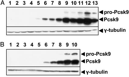
Cloning of Pcsk9 and Construction of Adenovirus. The mouse C57BL/6 Pcsk9 ORF sequence identical to that found in the public genome database was used to create adenovirus particles expressing Pcsk9. The Pcsk9 adenovirus (Pcsk9-Ad) was injected into C57BL/6 male mice through the tail vein at concentrations of 1 × 10⁸ to 3 × 10⁹ pfu per mouse and compared with mice injected with PBS alone or 1 × 10⁹ pfu of a control empty adenovirus (Ad empty). As shown in Fig. 1_A_, the livers of mice injected with PBS or Ad empty have no immunoreactive Pcsk9. However, injection of increasing concentrations of Pcsk9-Ad produced increasing amounts of both the pro and processed forms of Pcsk9. Because processing of Pcsk9 is an intramolecular event (12), the presence of the processed form in these mice indicates that the adenovirus expresses catalytically active Pcsk9.
Fig. 1.
Adenoviral-mediated expression of Pcsk9. (A) Male C57BL/6 mice of >10 weeks of age that were fed a chow diet were injected through the tail vein with PBS (lanes 1 and 2), Ad empty (lanes 3 and 4), or an adenovirus constitutively expressing the murine C57BL/6 Pcsk9 ORF under the control of a CMV promoter (Pcsk9-Ad, lanes 5–12). Pcsk9-Ad was injected at increasing concentrations from 1 × 10⁸ pfu per mouse to 3 × 10⁹ pfu per mouse (lanes 5–12). As a control, McA-RH7777 cells were infected with Pcsk9-Ad at an moi of 2,000 (lane 13). (B) Male LDLR knockout mice on a C57BL/6 background of >10 weeks of age that were fed a chow diet were injected through the tail vein with Ad empty (lanes 1–5) or increasing concentrations of Pcsk9-Ad (lanes 6–10). In both cases, liver homogenates were immunoblotted with an antibody to Pcsk9 and γ-tubulin.
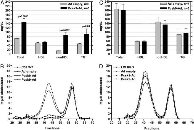
Adenoviral-Mediated Expression of Pcsk9 Increases Plasma LDL Cholesterol Levels. The effect of adenoviral-mediated Pcsk9 expression on plasma cholesterol levels was determined as shown in Fig. 2_A_. Injection of Pcsk9-Ad into C57BL/6 mice resulted in elevated plasma total cholesterol levels compared with injection of Ad empty (147 ± 17 mg/dl vs. 72 ± 6 mg/dl, P = 0.0003). This change was due to an increase in plasma non-HDL cholesterol levels (86 ± 14 mg/dl vs. 16 ± 3 mg/dl, P = 0.0002), with no change in HDL cholesterol (56 ± 2 mg/dl vs. 51 ± 2 mg/dl, no significant difference). There was also an increase in plasma triglycerides (91 ± 25 mg/dl vs. 47 ± 2 mg/dl, P = 0.03). The distribution of cholesterol in FPLC-separated lipoprotein fractions is shown in Fig. 2_B_. The increase in non-HDL cholesterol in Pcsk9-Ad-injected mice was shown to be due to a specific increase in LDL cholesterol, with little or no change in cholesterol in very-low-density lipoprotein or chylomicrons. The increase in triglycerides was also found to be in the LDL fractions (data not shown).
Fig. 2.
Overexpression of Pcsk9 increases LDL cholesterol levels in an LDLR-dependent manner. (A) Plasma was isolated from wild-type male C57BL/6 mice injected with Ad empty and Pcsk9-Ad, and HDL and non-HDL plasma fractions were separated by ultracentrifugation. Cholesterol levels in these fractions were measured by enzymatic assay. Overexpression of Pcsk9 caused an increase in total and non-HDL cholesterol with no change in HDL cholesterol levels. (B) Plasma was pooled from at least two wild-type animals (C57 WT), animals injected with Ad empty, and animals injected with Pcsk9-Ad; plasma was fractionated by FPLC; and cholesterol was measured by enzymatic assay. Two runs of Pcsk9-Ad-injected mice are shown to demonstrate reproducibility. Wild-type mice and mice injected with Ad empty had mainly HDL-derived cholesterol (fractions 52–62). Mice injected with Pcsk9-Ad had similar levels of HDL-derived and very-low-density lipoprotein-derived cholesterol, but they had high levels of LDL-derived cholesterol (fractions 35–50). (C) LDLR knockout mice were injected with Ad empty and Pcsk9-Ad. LDLR knockout mice injected with Ad empty had elevated total cholesterol and non-HDL cholesterol with no change in HDL cholesterol levels. Overexpression of Pcsk9 did not cause a further increase in total or non-HDL cholesterol levels in LDLR knockout mice. (D) FPLC analysis demonstrated that the lipoprotein profile of LDLR knockout mice injected with Pcsk9-Ad was indistinguishable from that of uninjected LDLR knockout mice (LDLRKO) and LDLR knockout mice injected with Ad empty.
The Pcsk9-Ad-Induced Increase in Plasma LDL Cholesterol Depends on the LDLR. Similar to what was observed in C57BL/6 mice, the livers of LDLR knockout mice injected with Ad empty had no immunoreactive Pcsk9, and injection of increasing concentrations of Pcsk9-Ad produced increasing amounts of both the pro and processed forms of Pcsk9 (Fig. 1_B_). Although LDLR knockout mice had higher levels of total cholesterol, non-HDL cholesterol and triglycerides than wild-type mice, no significant differences in these lipid levels were observed between Pcsk9-Ad and Ad empty-injected LDLR knockout mice (202 ± 26 mg/dl vs. 205 ± 27 mg/dl, no significant difference; 135 ± 19 mg/dl vs. 145 ± 23 mg/dl, no significant difference; 96 ± 20 mg/dl vs. 89 ± 25 mg/dl, no significant difference) (Fig. 2_C_). The distribution of cholesterol in FPLC separated lipoprotein fractions is shown in Fig. 2_D_. The increases in non-HDL cholesterol in Pcsk9-Ad-injected, Ad empty-injected, and uninjected LDLR knockout mice were all due to a specific increase in LDL cholesterol. The increases in triglycerides were also found to be in the LDL fractions (data not shown).
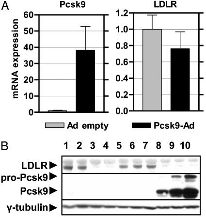
Pcsk9-Ad Injection Results in the Absence of Liver LDLR Protein with Normal mRNA Levels. The effects of Pcsk9 overexpression on liver LDLR mRNA and protein levels were examined. As shown in Fig. 3_A_, there was no significant change in LDLR mRNA levels despite a large increase in Pcsk9 mRNA levels in Pcsk9-Ad vs. Ad empty-injected mice. Effects on LDLR protein levels were determined by Western blotting with an antibody to the LDLR cytoplasmic tail as shown in Fig. 3_B_. Wildtype C57BL/6 mice have a band at ≈160 kDa, which is absent in LDLR knockout mice. This band is virtually absent in Pcsk9-Ad-injected mice, whereas it is unaffected in Ad empty-injected mice.
Fig. 3.
Overexpression of Pcsk9 results in an absence of LDLR protein with normal LDLR mRNA levels. (A) The levels of LDLR mRNA in livers of mice injected with Ad empty and Pcsk9-Ad was measured by quantitative RT-PCR. Overexpression of Pcsk9 did not change the levels of LDLR mRNA. (B) The levels of LDLR protein in livers of mice injected with Ad empty and Pcsk9-Ad were detected by immunoblotting with an antibody specific to the murine LDLR cytoplasmic tail. Wild-type C57BL/6 mice (lanes 1 and 2) had a predominant LDLR band at ≈160 kDa; this band was absent in LDLR knockout mice (lanes 3 and 4). Mice injected with Ad empty had normal levels of LDLR protein (lanes 5–7), whereas mice injected with Pcsk9-Ad had a complete absence of LDLR protein (lanes 8–10).
Pcsk9-Ad Injection Does Not Alter Hepatic Cholesterol Levels or Gallbladder Bile Composition. The effects of Pcsk9-Ad on other physiological measures related to cholesterol metabolism were studied. As shown in Table 1, there were no differences in hepatic total, free, or esterified cholesterol between Pcsk9-Ad and Ad empty-injected mice. There were also no differences in gallbladder bile concentrations of cholesterol, bile acids, or phospholipids.
Table 1. Liver cholesterol and gallbladder bile composition in mice overexpressing Pcsk9.
| Liver | Gallbladder bile | |||||
|---|---|---|---|---|---|---|
| Virus | Total cholesterol | Free cholesterol | Cholesterol ester | Cholesterol, mg/dl | Bile acids, mM | Phospholipids, mg/dl |
| Ad empty | 3.11 ± 0.72 | 2.49 ± 0.66 | 0.81 ± 0.31 | 62.3 ± 20.4 | 130.9 ± 54.1 | 1,430 ± 406 |
| Pcsk9-Ad | 3.12 ± 0.16 | 2.42 ± 0.19 | 0.74 ± 0.17 | 53.8 ± 10.9 | 140.7 ± 21.9 | 1,254 ± 182 |
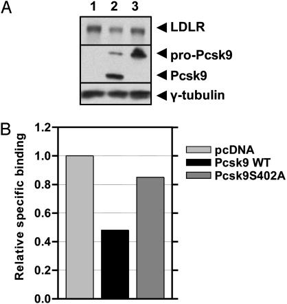
Overexpression of Pcsk9 in Rat Hepatoma Cells Leads to a Decrease in LDLR Protein and a Corresponding Decrease in LDL Binding. To investigate the effects of overexpression of Pcsk9 in vitro, McA-RH7777 rat hepatoma cells were transiently transfected with empty vector, wild-type Pcsk9, and a catalytically inactive mutant Pcsk9(S402A). As shown in Fig. 4_A_, the catalytic mutant shows only the pro form of Pcsk9, as expected (12). Overexpression of wild-type Pcsk9 caused a 72% decrease in LDLR protein, whereas overexpression of catalytically inactive Pcsk9 caused a 15% decrease in LDLR protein (Fig. 4_A_). Furthermore, overexpression of wild-type Pcsk9 caused a 52% decrease in the binding of DiI-LDL, whereas overexpression of catalytically inactive Pcsk9 caused a 15% decrease in the bindng of DiI-LDL. These observations support the hypothesis that overexpression of Pcsk9 increases plasma cholesterol levels in mice by interfering with LDLR-mediated LDL uptake.
Fig. 4.
Overexpression of Pcsk9 in vitro results in a decrease in LDLR protein and LDL binding. (A) McA-RH7777 cells were transiently transfected with pcDNA empty vector (lane 1), wild-type Pcsk9-pcDNA (lane 2), and the mutant Pcsk9(S402A)-pcDNA (lane 3). Overexpression of wild-type Pcsk9 resulted in a 72% decrease in LDLR protein, whereas overexpression of catalytically inactive Pcsk9 caused a 15% decrease in LDLR protein. (B) Specific binding of DiI-LDL was measured in transiently transfected McA-RH7777 cells. Overexpression of wild-type Pcsk9 resulted in a 52% decrease in DiI-LDL binding, whereas overexpression of catalytically inactive Pcsk9 resulted in a 15% decrease in DiI-LDL binding. Data shown are representative of three independent experiments.
Discussion
To determine how Pcsk9 affects cholesterol metabolism, we injected an adenovirus constitutively expressing murine Pcsk9 into C57BL/6 male mice. Injection of Pcsk9-Ad in mice resulted in an increase in total cholesterol because of a specific increase in LDL cholesterol. Injections into LDLR knockout mice revealed that the Pcsk9-induced increase in LDL cholesterol depended on the LDLR. Furthermore, wild-type mice injected with Pcsk9 had a near absence of liver LDLR protein with normal levels of mRNA. Transient transfection of Pcsk9 into McA-RH7777 rat hepatoma cells resulted in decreased LDL binding proportional to decreased LDLR protein, supporting the in vivo results.
The phenotype of mice injected with the Pcsk9 adenovirus is nearly identical to that of LDLR knockout mice. Homozygous LDLR knockout mice fed a normal chow diet, with no detectable LDLR protein, have elevated plasma total cholesterol levels with a major increase in LDL cholesterol and a minor increase in very-low-density lipoprotein (22), as is seen in Pcsk9-Ad-injected mice. It is also noteworthy that the increase in plasma triglycerides in both LDLR knockout and Pcsk9-Ad-injected mice is in the LDL fractions. Other characteristics of LDLR knockout mice include normal hepatic cholesterol levels (23) and gall-bladder bile composition (E. Sehayek and J.L.B., unpublished data) and these characteristics are seen also in Pcsk9-Ad-injected mice. Thus, Pcsk9 appears to affect LDLR function specifically rather than have a global effect on hepatic cholesterol metabolism.
The demonstration that Pcsk9-Ad-mediated overexpression results in the disappearance of LDLR protein without evidence of accumulation of immature forms and without alteration of steady-state mRNA levels indicates a defect in LDLR synthesis or degradation rather than a defect in transcription, glycosylation, or trafficking. Very little is known about the posttranscriptional regulation of LDLR synthesis, but some literature exists about the regulation of LDLR degradation. Pulse labeling of human fibroblasts indicated that the half-life of LDLRs was greatly decreased by cycloheximide, suggesting protein-mediated LDLR degradation (24). The site and mechanism of degradation was not determined. Treatment of human fibroblasts with lysosomal inhibitors did not alter LDLR degradation in the absence of LDL, but paradoxically, it increased degradation in the presence of LDL (25, 26). Incubation of CHO cells with proteosome inhibitors did not affect degradation of wild-type LDLRs, but it prevented degradation of class II LDLR mutants (27). A mechanism of LDLR degradation was reported recently by Begg et al. (28), who showed that phorbol ester [4β-phorbol 12-myristate 13-acetate (PMA)] treatment of HepG2 cells caused the release of a soluble 140-kDa LDLR into the medium. Proteolytic release of soluble counter parts of receptors is a common phenomenon (29) and has been shown for other members of the LDLR family, including LRP1, VLDLR, and apoER2 (30, 31). It is intriguing to speculate that Pcsk9 may proteolytically cleave the LDLR, releasing soluble LDLR and preventing LDLR-mediated LDL uptake. Alternatively, Pcsk9 protease or another activity may degrade the LDLR by another mechanism. It is important to note that any effects of Pcsk9 could be made directly on the LDLR or on proteins that interact with the LDLR.
Both missense Pcsk9 mutations in the human disease Hchola3 and Pcsk9-Ad-mediated overexpression in mice result in elevated LDL cholesterol levels. Several explanations are possible for this paradox. In Hchola3, if the missense mutations result in a gain of function, this hypothesis would be supported by our results. All of the missense mutations are of residues conserved between human and mouse, suggesting functional importance. However, because none of the missense mutations are at the prodomain cleavage site or the catalytic triad (13, 14), it is not obvious how they might affect function either positively or negatively. Second, it is possible that a loss-of-function mutation and overexpression of Pcsk9 could result in the same phenotype. This phenomenon could occur if LDLR function depends on an optimal amount of Pcsk9 and either deficiency or excess inhibits function, as is seen with the LDLR-related protein (LRP) and the receptor-associated protein (RAP). In RAP knockouts, hepatic LRP protein is diminished and shows decreased physiological function (32, 33), whereas adenoviral-mediated RAP overexpression also results in decreased physiological function of LRP (34, 35). Third, it is possible that human Pcsk9 (hPcsk9) and mouse Pcsk9 (mPcsk9) have opposite effects on LDLR function. In this regard, human and mouse Pcsk9 are only 77% identical at the amino acid level. Some of these differences may be functionally important. (i) hPcsk9 has a lysine at the processing site reported in Seidah et al. (6), whereas mPcsk9 has a methionine; (ii) hPcsk9 has the sequence RGE after the catalytic site where mPcsk9 has the potential integrin binding site, RGD (36); (iii) hPcsk9 contains a domain similar to the prokaryotic lipoprotein lipid attachment site whereas mPcsk9 does not (37); and (iv) hPcsk9 and mPcsk9 have multiple differences in putative PKC, CK2, and cAMP phosphorylation sites (37).
If Pcsk9 is a physiological inhibitor of LDLR activity, then it is peculiar that both genes are coordinately regulated by cholesterol at the transcriptional level through the SREBPs (4, 38). However, it is possible that the function of Pcsk9 is to modulate LDLR activity by a mechanism that is more rapid than termination of SREBP-mediated transcription of LDLR. This process might be analogous to the regulation of HMG CoA reductase (HMGR), in which termination of activity is achieved by both proteasomal degradation and termination of SREBP-mediated transcription (38). Interestingly, Insig-1, which mediates the proteasomal degradation of HMGR (39), is regulated coordinately with HMGR by means of the SREBPs (40).
In conclusion, overexpression of Pcsk9 diminishes LDLR protein and function with no change in LDLR mRNA levels, causing an LDLR knockout phenotype in mice. Thus, Pcsk9 may induce LDLR degradation and modulate LDLR activity in a more rapid manner than is achieved by sterol inhibition of SREBP-mediated transcription. The molecular mechanism of Pcsk9 action on the LDLR and the relationship of the observations reported herein to Hchola3 remain to be determined.
Acknowledgments
We thank Daniel Teupser and Christian Wolfrum for assistance with FPLC; Joachim Herz for providing the LDLR antibody; and George Rothblat, Sandy Simon, Raymond Soccio, and Markus Stoffel for useful discussions. This work was supported by National Institutes of Health Medical Scientist Training Program Grant GM07739 (to K.N.M) and National Institutes of Health Grant HL32435 (to J.L.B.).
Abbreviations: ADH, autosomal dominant hypercholesterolemia; DiI, 1,1′-dioctadecyl-3,3,3′,3′-tetramethyl-indocarbocyanine perchlorate; FPLC, fast protein liquid chromatography; HDL, high-density lipoprotein; LDL, low-density lipoprotein; LDLR, LDL receptor; pfu, plaque-forming unit; Pcsk9, proprotein convertase subtilisin kexin 9; SREBP, sterol regulatory element binding protein.
References
- 1.Hopkins, P. N. (1992) Am. J. Clin. Nutr. 55**,** 1060-1070. [DOI] [PubMed] [Google Scholar]
- 2.Herron, K. L., Vega-Lopez, S., Conde, K., Ramjiganesh, T., Shachter, N. S. & Fernandez, M. L. (2003) J. Nutr. 133**,** 1036-1042. [DOI] [PubMed] [Google Scholar]
- 3.Clarke, R., Frost, C., Collins, R., Appleby, P. & Peto, R. (1997) Br. Med. J. 314**,** 112-117. [DOI] [PMC free article] [PubMed] [Google Scholar]
- 4.Maxwell, K. N., Soccio, R. E., Duncan, E. M., Sehayek, E. & Breslow, J. L. (2003) J. Lipid Res. 44**,** 2109-2119. [DOI] [PubMed] [Google Scholar]
- 5.Siezen, R. J. & Leunissen, J. A. (1997) Protein Sci. 6**,** 501-523. [DOI] [PMC free article] [PubMed] [Google Scholar]
- 6.Seidah, N. G., Benjannet, S., Wickham, L., Marcinkiewicz, J., Jasmin, S. B., Stifani, S., Basak, A., Prat, A. & Chretien, M. (2003) Proc. Natl. Acad. Sci. USA 100**,** 928-933. [DOI] [PMC free article] [PubMed] [Google Scholar]
- 7.Gensberg, K., Jan, S. & Matthews, G. M. (1998) Semin. Cell. Dev. Biol. 9**,** 11-17. [DOI] [PubMed] [Google Scholar]
- 8.Bergeron, F., Leduc, R. & Day, R. (2000) J. Mol. Endocrinol. 24**,** 1-22. [DOI] [PubMed] [Google Scholar]
- 9.Seidah, N. G. & Chretien, M. (1999) Brain Res. 848**,** 45-62. [DOI] [PubMed] [Google Scholar]
- 10.Espenshade, P. J., Cheng, D., Goldstein, J. L. & Brown, M. S. (1999) J. Biol. Chem. 274**,** 22795-22804. [DOI] [PubMed] [Google Scholar]
- 11.Seidah, N. G., Mowla, S. J., Hamelin, J., Mamarbachi, A. M., Benjannet, S., Toure, B. B., Basak, A., Munzer, J. S., Marcinkiewicz, J., Zhong, M., et al. (1999) Proc. Natl. Acad. Sci. USA 96**,** 1321-1326. [DOI] [PMC free article] [PubMed] [Google Scholar]
- 12.Naureckiene, S., Ma, L., Sreekumar, K., Purandare, U., Lo, C. F., Huang, Y., Chiang, L. W., Grenier, J. M., Ozenberger, B. A., Jacobsen, J. S., et al. (2003) Arch. Biochem. Biophys. 420**,** 55-67. [DOI] [PubMed] [Google Scholar]
- 13.Abifadel, M., Varret, M., Rabes, J. P., Allard, D., Ouguerram, K., Devillers, M., Cruaud, C., Benjannet, S., Wickham, L., Erlich, D., et al. (2003) Nat. Genet. 34**,** 154-156. [DOI] [PubMed] [Google Scholar]
- 14.Timms, K. M., Wagner, S., Samuels, M. E., Forbey, K., Goldfine, H., Jammulapati, S., Skolnick, M. H., Hopkins, P. N., Hunt, S. C. & Shattuck, D. M. (2004) Hum. Genet. 114**,** 349-353. [DOI] [PubMed] [Google Scholar]
- 15.Rader, D. J., Cohen, J. & Hobbs, H. H. (2003) J. Clin. Invest. 111**,** 1795-1803. [DOI] [PMC free article] [PubMed] [Google Scholar]
- 16.Brown, M. S. & Goldstein, J. L. (1986) Science 232**,** 34-47. [DOI] [PubMed] [Google Scholar]
- 17.Innerarity, T. L., Mahley, R. W., Weisgraber, K. H., Bersot, T. P., Krauss, R. M., Vega, G. L., Grundy, S. M., Friedl, W., Davignon, J. & McCarthy, B. J. (1990) J. Lipid Res. 31**,** 1337-1349. [PubMed] [Google Scholar]
- 18.Haddad, L., Day, I. N., Hunt, S., Williams, R. R., Humphries, S. E. & Hopkins, P. N. (1999) J. Lipid Res. 40**,** 1113-1122. [PubMed] [Google Scholar]
- 19.Varret, M., Rabes, J. P., Saint-Jore, B., Cenarro, A., Marinoni, J. C., Civeira, F., Devillers, M., Krempf, M., Coulon, M., Thiart, R., et al. (1999) Am. J. Hum. Genet. 64**,** 1378-1387. [DOI] [PMC free article] [PubMed] [Google Scholar]
- 20.Sehayek, E., Ono, J. G., Shefer, S., Nguyen, L. B., Wang, N., Batta, A. K., Salen, G., Smith, J. D., Tall, A. R. & Breslow, J. L. (1998) Proc. Natl. Acad. Sci. USA 95**,** 10194-10199. [DOI] [PMC free article] [PubMed] [Google Scholar]
- 21.Soccio, R. E., Adams, R. M., Romanowski, M. J., Sehayek, E., Burley, S. K. & Breslow, J. L. (2002) Proc. Natl. Acad. Sci. USA 99**,** 6943-6948. [DOI] [PMC free article] [PubMed] [Google Scholar]
- 22.Ishibashi, S., Brown, M. S., Goldstein, J. L., Gerard, R. D., Hammer, R. E. & Herz, J. (1993) J. Clin. Invest. 92**,** 883-893. [DOI] [PMC free article] [PubMed] [Google Scholar]
- 23.Osono, Y., Woollett, L. A., Herz, J. & Dietschy, J. M. (1995) J. Clin. Invest. 95**,** 1124-1132. [DOI] [PMC free article] [PubMed] [Google Scholar]
- 24.Casciola, L. A., van der Westhuyzen, D. R., Gevers, W. & Coetzee, G. A. (1988) J. Lipid Res. 29**,** 1481-1489. [PubMed] [Google Scholar]
- 25.Grant, K. I., Casciola, L. A., Coetzee, G. A., Sanan, D. A., Gevers, W. & van der Westhuyzen, D. R. (1990) J. Biol. Chem. 265**,** 4041-4047. [PubMed] [Google Scholar]
- 26.Casciola, L. A., Grant, K. I., Gevers, W., Coetzee, G. A. & van der Westhuyzen, D. R. (1989) Biochem. J. 262**,** 681-683. [DOI] [PMC free article] [PubMed] [Google Scholar]
- 27.Li, Y., Lu, W., Schwartz, A. L. & Bu, G. (March 1, 2004) J. Lipid Res., 10.1194/jlr.M300487-JLR200.
- 28.Begg, M. J., Sturrock, E. D. & van der Westhuyzen, D. R. (2004) Eur. J. Biochem. 271**,** 524-533. [DOI] [PubMed] [Google Scholar]
- 29.Ehlers, M. R. & Riordan, J. F. (1991) Biochemistry 30**,** 10065-10074. [DOI] [PubMed] [Google Scholar]
- 30.May, P., Bock, H. H., Nimpf, J. & Herz, J. (2003) J. Biol. Chem. 278**,** 37386-37392. [DOI] [PubMed] [Google Scholar]
- 31.Magrane, J., Casaroli-Marano, R. P., Reina, M., Gafvels, M. & Vilaro, S. (1999) FEBS Lett. 451**,** 56-62. [DOI] [PubMed] [Google Scholar]
- 32.Willnow, T. E., Armstrong, S. A., Hammer, R. E. & Herz, J. (1995) Proc. Natl. Acad. Sci. USA 92**,** 4537-4541. [DOI] [PMC free article] [PubMed] [Google Scholar]
- 33.Willnow, T. E., Rohlmann, A., Horton, J., Otani, H., Braun, J. R., Hammer, R. E. & Herz, J. (1996) EMBO J. 15**,** 2632-2639. [PMC free article] [PubMed] [Google Scholar]
- 34.Willnow, T. E., Sheng, Z., Ishibashi, S. & Herz, J. (1994) Science 264**,** 1471-1474. [DOI] [PubMed] [Google Scholar]
- 35.Narita, M., Bu, G., Herz, J. & Schwartz, A. L. (1995) J. Clin. Invest. 96**,** 1164-1168. [DOI] [PMC free article] [PubMed] [Google Scholar]
- 36.Ruoslahti, E. & Pierschbacher, M. D. (1986) Cell 44**,** 517-518. [DOI] [PubMed] [Google Scholar]
- 37.Bairoch, A., Bucher, P. & Hofmann, K. (1997) Nucleic Acids Res. 25**,** 217-221. [DOI] [PMC free article] [PubMed] [Google Scholar]
- 38.Goldstein, J. L. & Brown, M. S. (1990) Nature 343**,** 425-430. [DOI] [PubMed] [Google Scholar]
- 39.Sever, N., Yang, T., Brown, M. S., Goldstein, J. L. & DeBose-Boyd, R. A. (2003) Mol. Cell 11**,** 25-33. [DOI] [PubMed] [Google Scholar]
- 40.Horton, J. D., Shah, N. A., Warrington, J. A., Anderson, N. N., Park, S. W., Brown, M. S. & Goldstein, J. L. (2003) Proc. Natl. Acad. Sci. USA 100**,** 12027-12032. [DOI] [PMC free article] [PubMed] [Google Scholar]


