Differential expression of three T lymphocyte-activating CXC chemokines by human atheroma-associated cells (original) (raw)
Abstract
Activated T lymphocytes accumulate early in atheroma formation and persist at sites of lesion growth and rupture, suggesting that they may play an important role in the pathogenesis of atherosclerosis. Moreover, atherosclerotic lesions contain the Th1-type cytokine IFN-γ, a potentiator of atherosclerosis. The present study demonstrates the differential expression of the 3 IFN-γ–inducible CXC chemokines — IFN-inducible protein 10 (IP-10), monokine induced by IFN-γ (Mig), and IFN-inducible T-cell α chemoattractant (I-TAC) — by atheroma-associated cells, as well as the expression of their receptor, CXCR3, by all T lymphocytes within human atherosclerotic lesions in situ. Atheroma-associated endothelial cells (ECs), smooth muscle cells (SMCs), and macrophages (MØ) all expressed IP-10, whereas Mig and I-TAC were mainly expressed in ECs and MØ, as detected by double immunofluorescence staining. ECs of microvessels within lesions also expressed abundant I-TAC. In vitro experiments supported these results and showed that IL-1β, TNF-α, and CD40 ligand potentiated IP-10 expression from IFN-γ–stimulated ECs. In addition, nitric oxide (NO) treatment decreased IFN-γ induction of IP-10. Our findings suggest that the differential expression of IP-10, Mig, and I-TAC by atheroma-associated cells plays a role in the recruitment and retention of activated T lymphocytes observed within vascular wall lesions during atherogenesis.
Introduction
Increasing evidence supports a crucial role for immunological and inflammatory responses in atherogenesis. The development of atherosclerotic lesions involves interactions between 4 major cell types: endothelial cells (ECs), smooth muscle cells (SMCs), macrophages (MØ), and lymphocytes (1, 2). The role of T cells in atherogenesis remains unclear. However, because activated T cells accumulate in atheroma — and by virtue of their early appearance, persistence, and localization at sites of lesion growth and rupture — they may orchestrate important aspects of atherogenesis (3–7). Although atherosclerotic lesions contain both CD8+ and CD4+ T cells, CD4+ memory (CD45RO+) T cells predominate (8).
Previous studies have colocalized CD4+ T lymphocytes and IFN-γ within human and mouse atherosclerotic lesions (9), suggesting predominance of Th1 lymphocytes in atherogenesis (10). IFN-γ plays a critical role in modulating the cellular immune response by stimulating the production of proinflammatory cytokines, adhesion molecules, and MHC class II expression by ECs, SMCs, and MØ. Moreover, Gupta et al. have recently demonstrated that apo E–knockout mice lacking the IFN-γ receptor developed fewer and smaller atherosclerotic lesions than did control mice, suggesting that IFN-γ has proatherogenic properties (11).
Despite increasing evidence for involvement of T lymphocytes in atherogenesis, the mechanism of T-lymphocyte recruitment within the vascular atherosclerotic lesions remains incompletely defined. Lymphocyte recruitment into tissues is a multistep process involving adhesion molecules and chemokines. Chemokines are secreted basic proteins (8–10 kDa) subdivided into 4 families based on the relative position of their cysteine residues (CC, CXC, C, CXC3) (12). CXC chemokines further fall into 2 classes based on the presence or absence of a NH2-terminal sequence Glutamic acid-Leucine-Arginine (ELR). The ELR-containing CXC chemokines, such as IL-8, chemoattract neutrophils (13), whereas the non-ELR CXC chemokines chemoattract lymphocytes. Among the non-ELR CXC chemokines, 3 of them — IP-10 (IFN-inducible protein 10), Mig (monokine induced by IFN-γ), and I-TAC (IFN-inducible T-cell α chemoattractant) — are IFN-γ inducible and potently chemoattract activated T lymphocytes (14–18). All 3 chemokines signal through a common receptor, CXCR3, expressed by memory (CD45RO+) T cells, preferentially of the Th1 subset, and by natural killer cells, but not by monocytes or neutrophils (15, 19–22). IP-10 has been found in various clinical conditions in which activated Th1 lymphocytes and IFN-γ expression were found, such as psoriasis (23), tuberculoid leprosy (24), sarcoidosis (25), viral meningitis (26), and pulmonary tuberculosis (27). Mig was also found in psoriatic lesions by in situ hybridization (17). To date, I-TAC has not been correlated with any human diseases.
Chemokines such as IL-8, monocyte chemoattractant protein-1 (MCP-1), MCP-4, and RANTES have been shown to be expressed within atherosclerotic lesions in situ and by atheroma-associated cells in vitro (28–35). In addition, in recent in vivo studies, targeted disruption of the genes for MCP-1, CCR2, and CXCR2 significantly decreased atherosclerotic lesion formation and lipid deposition when the disrupted gene was bred or transferred into mouse strains prone to develop atherosclerotic-like lesions (36, 37, 29). Furthermore, in these 3 in vivo studies, the attenuated development of vascular lesions correlated with decreased MØ accumulation in lesions, demonstrating that chemokines play a critical role in monocyte/MØ recruitment during atherogenesis.
It is likely that chemokines also play a critical role in the recruitment and retention of activated T cells in atherosclerosis. Because IFN-γ appears to have a proatherogenic effect, we have hypothesized that the IFN-γ–inducible chemokines IP-10, Mig, and I-TAC play an important role in atherosclerosis. Moreover, we investigated whether CD40 ligand (CD154), a molecule recently implicated in atherosclerosis (38, 39), and nitric oxide (NO), which has an antiatherogenic effect (40, 41), would regulate IP-10, Mig, and I-TAC expression.
Methods
Reagents.
Affinity-purified rabbit anti-human IP-10 polyclonal antibody was generated as described previously (42). Rabbit anti-human Mig polyclonal antibody was obtained from PeproTech Inc. (Rocky Hill, New Jersey, USA). Affinity-purified rabbit anti-human I-TAC polyclonal antibody was generated as described (18). Mouse anti-human CXCR3 (1C6) mAb was a gift from LeukoSite (Cambridge, Massachusetts, USA). The following human recombinant cytokines were obtained from Endogen Inc. (Cambridge, Massachusetts, USA): IFN-γ, IL-1β, and TNF-α. Human recombinant CD40 ligand (rCD40L) was a gift from P. Graber (Ares Serono, Geneva, Switzerland) and was generated as described previously (43). The NO donors _S_-nitroso-_N_-acetylpenicillamine (SNAP) and _S_-nitrosoglutathione (GSNO) were purchased from Sigma Chemical Co. (St. Louis, Missouri, USA).
Cell isolation and culture.
Human vascular ECs were isolated from saphenous veins by collagenase treatment (1 mg/mL; Worthington Biochemical Corp., Freehold, New Jersey, USA) and were cultured in dishes coated with fibronectin (1.5 μg/cm2; New York Blood Center Reagents, New York, New York, USA) as described elsewhere (44). Cells were maintained in M199 medium (BioWhittaker Inc., Walkersville, Maryland, USA) supplemented with 1% penicillin/streptomycin (BioWhittaker Inc.), 5% FCS (Atlanta Biologicals, Norcross, Georgia, USA), 100 μg/mL heparin (Sigma Chemical Co.), and 50 μg/mL EC growth factor (ECGF; Pel-Freez Biologicals, Rogers, Arkansas, USA). Human vascular SMCs were isolated from human saphenous veins and carotid arteries by explant outgrowth (38) and were cultured in DMEM (BioWhittaker Inc.) supplemented with 1% L-glutamine (BioWhittaker Inc.), 1% penicillin/streptomycin, and 10% FCS. Both cell types were subcultured after trypsinization (0.5% trypsin [Worthington Biochemicals] and 0.2% EDTA [EM Science, Gibbstown, New Jersey, USA]) in P100 culture dishes (Becton Dickinson and Co., Franklin Lakes, New Jersey, USA) and were used throughout passages 2–4. Culture media and FCS contained less than 40 pg LPS/mL, as determined by chromogenic Limulus amoebocyte-lysote-assay analysis (QLC-1000; BioWhittaker Inc.). ECs and SMCs were characterized by immunostaining with anti–von Willebrand factor and anti–SMC α-actin antibodies (DAKO Corp., Carpinteria, California, USA), respectively. Both cell types were cultured 12 hours before the experiment in media lacking FCS; ECs were cultured in M199 supplemented with 0.1% HSA and SMCs cultured in insulin/transferrin (I/T) medium with 0.1% BSA as described elsewhere (45).
Monocytes were isolated by adherence from PBMCs after Ficoll-Hypaque gradient and were cultured in RPMI-1640 medium (BioWhittaker Inc.) containing 10% FCS (Sigma Chemical Co.). Monocyte-derived MØ were serum starved 16 hours before experiments, and stimulated with IFN-γ in RPMI-1640 medium supplemented with 0.1% BSA.
Immunohistochemistry.
Surgical specimens of human carotid atheroma and normal aorta were obtained by protocols approved by the Human Investigation Review Committee at the Brigham and Women’s Hospital. Serial cryostat sections (5 μm) were cut, air dried onto microscope slides (Fisher Scientific Co., Pittsburgh, Pennsylvania, USA), and fixed in acetone at –20°C for 5 minutes. Sections preincubated with PBS containing 0.3% hydrogen peroxide were then incubated for 90 minutes with primary or control antibody, diluted in PBS supplemented with 5% appropriate serum. After washing 3 times in PBS, sections were incubated with the respective biotinylated secondary antibody (for 45 minutes; Vector Laboratories, Burlingame, California, USA) followed by avidin-biotin-peroxidase complex (VECTASTAIN ABC kit; Vector Laboratories). Immunostaining was viewed using 3-amino-9-ethyl carbazole (Vector Laboratories) according to the recommendations provided by the supplier. Cell types were characterized by double immunofluorescence staining using FITC-labeled cell-specific antibody (anti-muscle actin mAb for SMCs (Enzo Diagnostics, New York, New York), anti-CD31 mAb for ECs (DAKO Corp.), anti-CD68 mAb for MØ (DAKO Corp.), and anti-human CD3 for T lymphocyte (DAKO Corp.).
Western blot analysis.
Normal aortas and carotid atherosclerotic tissues were obtained from human patients after surgery. Tissues were homogenized (Ultra-tarrax T 25; IKA-Labortechnik, Wilmington, NorthCarolina, USA) and lysed (0.3 g tissue/mL lysis buffer) as described previously (46). Lysates were clarified (16,000 g for 15 minutes at 4°C), and protein concentration for each tissue extract was determined using a bicinchoninic acid (BCA) protein assay according to the instructions of the supplier (Pierce Chemical Co., Rockford, Illinois, USA).
Fifty microgram of tissue lysates protein per lane and supernatants (45 μL) of cultured ECs, SMCs, and monocyte-derived MØ were separated by SDS-PAGE under reducing conditions and blotted onto PVDF membranes (Millipore Corp., Bedford, Massachusetts, USA) using a semidry blotting apparatus (3.0 mA/cm2 for 30 minutes; Bio-Rad Laboratories Inc., Hercules, California, USA). Membranes were blocked in 5% defatted dry milk/PBS/0.1% Tween-20 (PBST) and were then incubated with the primary antibody (rabbit anti–IP-10 1:2,000; rabbit anti-Mig 1:1,000; rabbit anti–I-TAC 1:500) for 1 hour. Blots were washed 4 times PBST, and the secondary peroxidase-conjugated antibody (Jackson ImmunoResearch Laboratories Inc., West Grove, Pennsylvania, USA) was added (1:10,000) for another hour. Finally, membranes were washed in PBST, and detection of the antigen was carried out using the enhanced chemiluminescence detection method according to the manufacturer’s recommendations (NEN Life Science Products Inc., Boston, Massachusetts, USA), and subsequent exposure of the membranes to x-ray film.
Northern blot analysis.
Total RNA was extracted from samples using Stat-60 (Tel-Test Inc., Friendswood, Texas, USA). For Northern analysis, 20 μg total RNA was electrophoresed on a 1.2% agarose-formaldehyde gel and then capillary transferred to a GeneScreen membrane (NEN Life Science Products Inc.). After overnight prehybridization (50% formamide, 1% SDS, 4× SSC, 4× Denhardt’s solution, 0.8% glycine, and 0.17 mg/mL denatured salmon sperm DNA) at 42°C, blots were hybridized at 42°C in 50% formamide, 10% dextran, 1% SDS, 5× SSC, 1× Denhardt’s solution, and 0.17 mg/mL denatured salmon sperm DNA with 106 cpm/mL [α-32P]dCTP–radiolabeled cDNA probe prepared by nick translation. The following fragments were used as probes: a 1-kb _Pst_I fragment from hIP-10 cDNA, a 3-kb _Not_I fragment from hMig cDNA (kindly provided by J. Farber, National Institutes of Health, Bethesda, Maryland, USA), and a 300-bp _Bam_HI/_Ava_I fragment from hI-TAC cDNA. A GAPDH cDNA probe was used as a control for RNA loading. Signal quantitation was determined using a phosphoimager (Molecular Imager System; Bio-Rad Laboratories Inc.). Levels of chemokine expression in any given sample were normalized to the GAPDH signal for that sample. Because different exposure times were used for each chemokine quantitation and normalized to the same GAPDH exposure, the ratio measurements can only be used to compare levels of expression within any given exposure of a blot and not between blots.
IP-10 ELISA assay.
Release of IP-10 from cultured human vascular ECs was measured using a sandwich-type ELISA as described previously (27). Antibody binding was detected by adding _p_-nitrophenyl phosphate (Sigma Chemical Co.), and absorbance was measured at 405 nm in a Molecular Devices plate reader (Du Pont, Wilmington, Delaware, USA). The amount of IP-10 detected was calculated from a standard curve prepared with the recombinant protein. Samples were assayed in triplicate.
Results
Human atheroma-associated cells express the chemokines IP-10, Mig, and I-TAC.
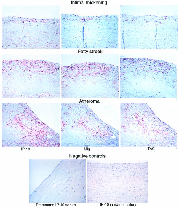
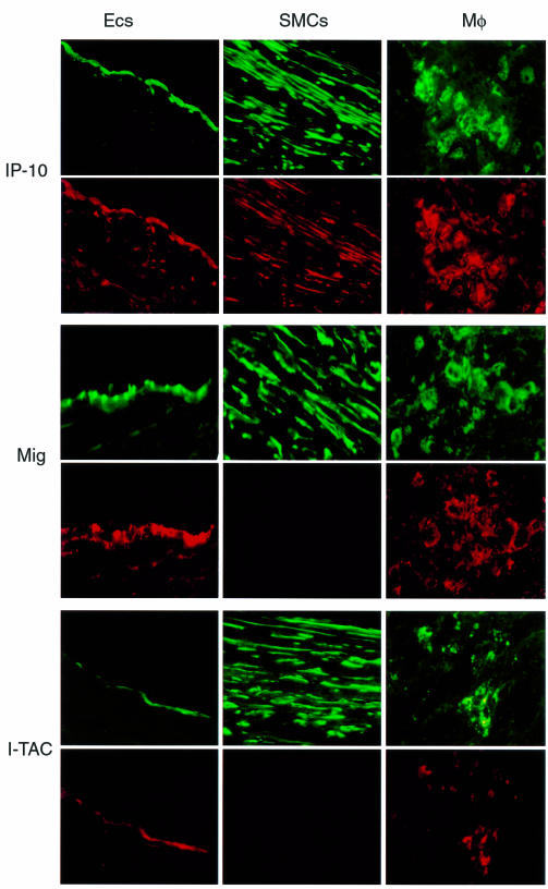
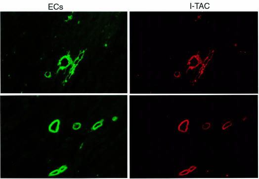
Immunohistochemical analysis of human atherosclerotic plaques showed expression of IP-10, Mig, and I-TAC within the lesion (Figure 1). Analysis of atherosclerotic lesions at different developmental stages from early intimal thickening (n = 5) through fatty streaks (n = 5) and fully developed atheroma lesions (n = 7) revealed the expression of the 3 IFN-γ–inducible chemokines. Intimal thickening showed sparse but reproducibly detectable expression of the chemokines. In contrast, fatty streaks and well-developed atheroma lesions consistently showed strong immunoreactivity for all 3 chemokines, most prominently at the luminal border and in the shoulder region of the plaque, the margin between the lesion and unaffected portion of the artery. No immunoreactivity was observed in normal vessels (n = 4) or in atherosclerotic lesions examined with control pre-immune serum as shown for IP-10 (Figure 1, lower panels), confirming the specificity of the antibodies used in these experiments. To characterize further the expression of these chemokines within atherosclerotic lesions, we performed double immunofluorescence staining (n = 3) using cell-specific antibodies. ECs, SMCs, and MØ highly expressed IP-10 within human carotid atherosclerotic plaques (Figure 2). In contrast, Mig was expressed mostly on ECs and MØ, and to a lesser extent on SMCs (Figure 2). The chemokine I-TAC was expressed only on ECs and MØ (Figure 2). Furthermore, ECs within neovessel formations in atherosclerotic plaques (n = 3) expressed I-TAC but little IP-10 or Mig (Figure 3).
Figure 1.
Expression of IP-10, Mig, and I-TAC in human atherosclerotic lesions in situ. Human carotid arteries sections were stained with specific antibodies to IP-10, Mig, and I-TAC. High-power view (×100) of atherosclerotic lesions from different stages (Intimal thickening, Fatty streak, and Atheroma) revealed the expression of the chemokines (red-brown reaction product). Adjacent sections of the same atherosclerotic carotid tissue stained for IP-10 was stained with the rabbit preimmune IP-10 serum (×100). Normal human artery exhibited no expression of IP-10 (×100). The lumen of the artery is at the upper left side of each photomicrograph. Analysis of 5–7 atheroma at each stage of lesion development from different donors, and normal tissue from 4 different donors showed similar results.
Figure 2.
Colocalization of IP-10, Mig, and I-TAC with ECs, SMCs, and MØ in human atherosclerotic lesions. High-power views (×400) of human carotid sections showed specific staining for IP-10, Mig, and I-TAC (Texas red staining) within the atherosclerotic lesions. Cell types were characterized by immunofluorescence staining with anti-CD31 mAb for ECs, anti–α-actin mAb for SMCs, and anti-CD68 mAb for MØ (FITC; green staining). The lumen of the artery is at the top of each photomicrograph. Analysis of atheroma from 3 different donors showed similar results.
Figure 3.
Expression of I-TAC by ECs of microvessels within human atherosclerotic lesions. High-power views (×400) of human carotid sections showed specific staining for I-TAC (Texas red staining). Cell types was characterized by immunofluorescence staining of CD31 for ECs within the atherosclerotic lesions (FITC; green staining). The lumen of the artery is at the top of each photomicrograph. Analysis of atheroma from 3 different donors showed similar results.
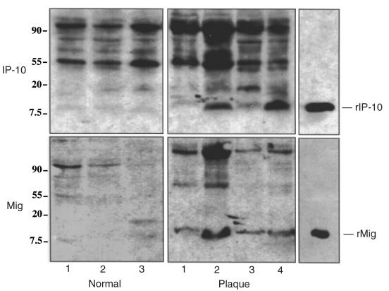
To support these immunohistochemical results, Western blot analyses were performed with the same chemokine antibodies used for immunohistochemistry on tissue extracts from human atherosclerotic (n = 6) and normal vessels (n = 4). The chemokines IP-10 and Mig were detected in 3 of 4 samples from atherosclerotic vessels and comigrated with the recombinant protein (Figure 4). Extracts of unaffected arteries contained neither IP-10 nor Mig.
Figure 4.
Western blot analysis of vascular tissue extracts. Samples (50 μg protein per lane) from normal human aortas (Normal) and human carotid atherosclerotic lesions (Plaque) were analyzed for IP-10 and Mig expression by Western blot. Analysis of 3 normal and 4 atherosclerotic tissues from different donors are shown. Human recombinant IP-10 (rIP-10) and Mig (rMig) were used as controls. The position of the molecular markers markers are indicated (kDa).
T lymphocytes within human atherosclerotic lesions express the chemokine receptor CXCR3.
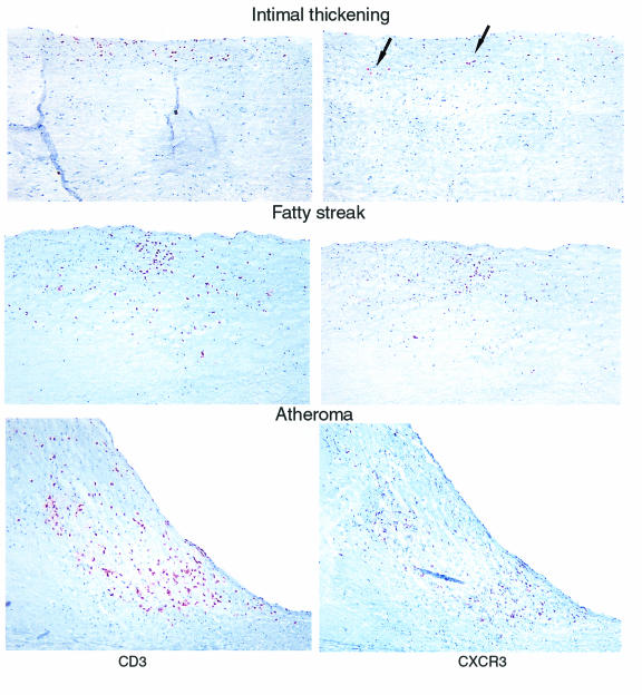
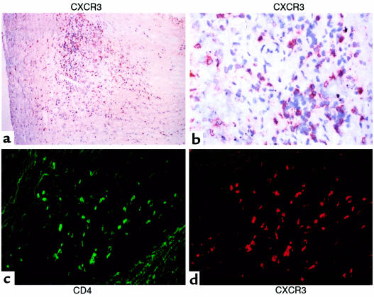
In view of the finding that atheroma-associated cells in situ expressed the 3 IFN-γ–inducible CXC chemokines — IP-10, Mig, and I-TAC — we investigated the expression of their receptor, CXCR3, in human atherosclerotic lesions. Serial sections of atherosclerotic lesions adjacent to the ones used for examining the expression of IP-10, Mig and I-TAC (Figure 1) were used to study the expression of CXCR3 (Figure 5). Immunohistochemical analysis revealed the expression of CXCR3+ cells within atherosclerotic lesions at all stages of lesion development examined (intimal thickening, fatty streak and atheroma; n = 5, 5, 7, respectively). Adjacent sections of the same atherosclerotic tissue stained for CD3 indicated that virtually all CD3+ cells expressed CXCR3 (Figure 5). A comparison with the pattern of expression seen for the chemokines (Figure 1), revealed the colocalization of CXCR3+ CD3+ T cells with the 3 CXCR3 ligands at all stages of atherogenesis. Double immunofluorescence staining (n = 4) with CD4 antibody indicated that virtually all CD4+ T cells expressed CXCR3+ (Figure 6). As demonstrated in previous studies (8), we also found that the vast majority of CD3+ cells were CD4+ lymphocytes (data not shown).
Figure 5.
Expression of the chemokine-receptor CXCR3 on T lymphocytes within human atherosclerotic lesions. Adjacent sections of human carotid arteries at different stages (Intimal thickening, Fatty streak, and Atheroma) were stained with anti-CD3 or anti-CXCR3 antibody (arrows). Photomicrographs (×100) reveal CD3 and CXCR3 expression (red-brown reaction product). The lumen of the artery is at the top of each photomicrograph. Analysis of 5–7 atheroma at each stage of lesion development from different donors showed similar results.
Figure 6.
Expression of CXCR3 on CD4+ T lymphocytes within human atherosclerotic lesions. Sections of human carotid arteries were stained with anti-CXCR3 antibody. (a) Low-power (×100) and (b) high-power (×400) views show CXCR3 expression (red-brown reaction product) in the shoulder region of the plaque. Colocalization of CD4+ lymphocytes (c) (FITC; green staining) with CXCR3 (d) (Texas red staining) was performed by immunofluorescence-double staining. The lumen of the artery is at the top of each photomicrograph. Analysis of atheroma from 4 different donors showed similar results
Atheroma-associated cells in vitro express the chemokines IP-10, Mig and I-TAC.
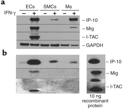
To characterize further IP-10, Mig and I-TAC expression by atheroma-associated cells, we performed in vitro experiments using human vascular ECs, SMCs and human monocyte-derived MØ. Quiescent cells lacked detectable IP-10, Mig, and I-TAC mRNA or protein expression (Figure 7). IFN-γ (1,000 U/mL for 18 hours) induced IP-10 mRNA accumulation in ECs and MØ and, to a lesser extent, in SMCs. Mig mRNA was detected in ECs and to a lesser extent in MØ and SMCs after IFN-γ stimulation. In contrast, I-TAC mRNA was highly expressed by ECs and was not detected in SMCs and MØ. IP-10, Mig and I-TAC mRNA induction by IFN-γ in ECs depended on the concentration and time of stimulation; mRNA for all 3 chemokines accumulated after stimulation with as little as 10 U/mL of IFN-γ and occurred as early as 2 hours after stimulation (data not shown). Viewing of Mig mRNA expression in SMCs required a longer exposure of the blots shown in Figure 7a. Western blot analysis for IP-10, Mig, and I-TAC extended Northern blot results to detection of all 3 proteins after IFN-γ stimulation in ECs (Figure 7b). The absence of I-TAC and Mig detection in SMCs and monocyte-derived MØ supernatants is likely due to lower concentrations compared with ECs supernatants. The control bands were loaded with 10 ng recombinant protein, enabling quantitative comparisons between levels of chemokine expression.
Figure 7.
Expression of IP-10, Mig, and I-TAC by human ECs, SMCs, and monocyte-derived MØ in vitro. (a) Northern blot and (b) Western analysis of unstimulated (–) or IFN-γ−stimulated (1,000 U/mL for 18 hours) (+) ECs, SMCs, and monocyte-derived MØ was performed using 20 μg total RNA per lane and 45 μL of unconcentrated cell supernatant per lane, respectively. GAPDH was used as a control for RNA loading. Recombinant chemokines (10 ng per lane) were used for controls in Western blots. For Northern Blots, the IP-10 and I-TAC blots were exposed for 7 hours, whereas the Mig blot was exposed for 70 hours. Similar results were obtained in independent experiments with cells from 4 different donors.
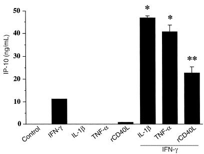
Stimulation of human ECs and SMCs with other cytokines implicated in atherosclerosis, such as IL-1β (10 ng/mL), TNF-α (50 ng/mL), and CD40L (5 μg/mL) each by itself, had no effect on IP-10 mRNA (data not shown) or protein expression (Figure 8). However, IL-1β, TNF-α, and CD40L synergized with IFN-γ in inducing the secretion of IP-10 protein in ECs as measured by an IP-10–specific sandwich ELISA (27) (compared with IFN-γ alone; 4.2-fold for IL-1β/IFN-γ; 3.7-fold for TNF-α/IFN-γ; and 2.0-fold for CD40L/IFN-γ).
Figure 8.
Secretion of IP-10 protein by human ECs in response to cytokine stimulation. Supernatants from human ECs unstimulated (–) or stimulated for 24 hours with IL-1β (10 ng/mL), TNF-α (50 ng/mL), IFN-γ (100 U/mL), or CD40L (5 μg/mL) were analyzed by ELISA for IP-10 levels. This figure is representative of 3 separate experiments using ECs from different donors. Error bars represent SD from triplicates (*P < 0.0001, **P < 0.005).
NO decreases IP-10, Mig, and I-TAC mRNA and protein expression by human vascular ECs.
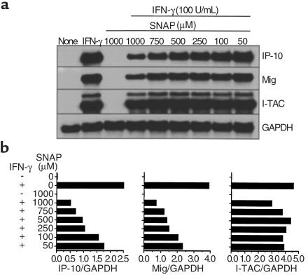
Incubation of human vascular ECs with the NO donor SNAP before IFN-γ stimulation caused a concentration-dependent decrease in IP-10, Mig, and I-TAC mRNA induction (Figure 8a). This effect was more pronounced for IP-10 and Mig than for I-TAC mRNA expression. The NO donor GSNO produced similar results (data not shown). Phosphoimager analysis showed that maximal effect (4.9-, 5.2-, and 1.5-fold decrease for IP-10, Mig, and I-TAC, respectively) was obtained with 1 mM SNAP (Figure 8b). The concentrations of NO donors described in these experiments were similar to those previously reported to act on vascular cells (47, 48), and the lowest concentration used (50 μM) still affected chemokine expression. Western blot analysis of supernatants from IFN-γ–stimulated ECs showed a similar decrease for IP-10 and Mig protein secretion after NO treatment (data not shown).
Discussion
Atherosclerotic lesions at all stages of development from fatty streaks to complicated plaques contain cells and molecules characteristic of immune-mediated processes (2). Macrophages and lymphocytes are the most numerous inflammatory cells found in atherosclerotic lesions. These cells elaborate growth factors and cytokines that mediate intimal hyperplasia and may therefore promote atherogenesis. Activated T lymphocytes accumulate early in atheroma formation and persist at sites of lesion growth and rupture, suggesting that they play an important role in atherogenesis (49–51). Moreover, the Th1-type cytokine IFN-γ is expressed in atherosclerotic lesions (10) and can promote atherosclerosis in vivo (11). In addition, experiments using mutant mice deficient in both apo E and RAG-1 (T- and B-cell deficient) suggest a functional role for lymphocytes in atherogenesis by revealing a 40% reduction in atherosclerotic lesions compared with immunocompetent apo E–deficient mice (52). Although the mechanism of monocyte recruitment within mouse atherosclerotic lesions involves the CC chemokine MCP-1 and its receptor CCR2 (36, 37), homing and accumulation of activated T lymphocytes in atherosclerotic lesions remain poorly understood. The present study investigated the expression of the 3 IFN-γ–inducible CXC chemokines, IP-10, Mig, and I-TAC, which specifically chemoattract activated T cells, through the receptor CXCR3.
Our data show expression of immunoreactive IP-10, Mig, and I-TAC within human atherosclerotic plaques but not within the normal vessel wall. We are unaware of previous reports of I-TAC expression related to a human disease or of the expression of the 3 IFN-γ–inducible CXC chemokines and their receptor CXCR3 simultaneously in the same diseased tissue. Using double immunofluorescence, we found that IP-10 was expressed by ECs, SMCs, and MØ, whereas Mig and I-TAC were predominantly expressed by ECs and MØ, consistent with an hypothesis that these 3 chemokines have nonredundant roles in T-cell trafficking in atherosclerotic lesions. In fact, I-TAC is a more efficacious chemoattractant than IP-10 or Mig for CXCR3+ T cells in vitro, and we have recently determined that among its ligands, only I-TAC can effectively downregulate CXCR3 expression on activated T cells (A. Sauty et al., unpublished observations). I-TAC may serve to capture and arrest activated T cells on the endothelium. The high expression of I-TAC, but not IP-10 or Mig, by ECs in neovessels within atherosclerotic plaques supports this hypothesis, as inflammatory cell recruitment into established atheroma may involve these neovessels. The ability of I-TAC to reduce cell surface CXCR3 expression may then facilitate T-cell diapedesis at this site. Moreover, the abundant expression of IP-10 by all 3 atheroma-associated cells, in particular SMCs, and the inability of IP-10 to limit CXCR3 expression, may facilitate T-cell retention within the lesion. In addition, the finding that all lesional T lymphocytes express CXCR3 agrees with the concept that these 3 IFN-γ–inducible chemokines play a role in the recruitment and retention of activated T lymphocytes within vascular wall lesions during the process of atherosclerosis.
Concordant with our in situ observations, we found strong expression of IP-10 mRNA and protein by IFN-γ–activated ECs, SMCs, and MØ in vitro. Lower levels of Mig mRNA were seen in activated ECs and MØ, but Mig protein was only detected in ECs supernatants. I-TAC mRNA and protein were detected only in IFN-γ–activated ECs. The discrepancy between I-TAC expression by macrophages in situ and by IFN-γ–activated peripheral blood monocyte–derived MØ in vitro likely reflects the activation of lesional macrophages by other stimuli in addition to IFN-γ. This study also examined the regulation of these chemokines in atheroma-associated cells by NO, another agent implicated in the pathogenesis of atherosclerosis. Normal vascular ECs secrete NO in response to shear stress, whereas ECs overlying atherosclerotic plaques produce less NO (41). In addition to its vascular relaxing effects, NO plays important immunoregulatory functions, such as inhibiting nuclear factor-κB (NF-κB) activation. We found that exogenous NO limits IP-10, Mig, and, to a lesser extent, I-TAC, mRNA accumulation induced by IFN-γ in ECs. Along these lines, exogenous NO inhibits (by 25%) IL-1–induced IL-8 secretion from ECs (53), and inhibition of basal NO production upregulates EC MCP-1 mRNA and protein expression (48). These results suggest that diminished secretion of NO by ECs leads to an increase in cytokine-induced chemokine expression by these cells and, therefore, to a greater stimulus for leukocyte recruitment into atherosclerotic lesions. In addition, we observed that the CD40 pathway, recently implicated in atherogenesis, potentiated the IFN-γ–induced secretion of IP-10 from ECs. CD40 ligation induces MIP-1α, MIP-1β, RANTES, and MCP-1 in macrophages (54) and, thus, may participate in atherogenesis through the release of multiple chemokines.
Chemokines may not only influence leukocyte recruitment within atherosclerotic lesions, but they may also regulate a number of vascular cell and leukocyte functions related to the acute and chronic manifestations of atherosclerosis. In particular, IP-10 mRNA was induced in the rat carotid artery after balloon angioplasty and was shown to be a mitogenic and chemotactic factor for vascular SMCs (55), suggesting that IP-10 might be involved in vascular remodeling during atherosclerosis. In addition, IP-10 inhibits neovascularization and wound healing in vivo (56), activities that may contribute to the necrosis associated with atherosclerotic lesions. Furthermore, IP-10 augments IFN-γ production from Th1 cells (57) and thus may help establish an autocrine loop that serves to drive the inflammatory response within the diseased vessel. Thus, in addition to recruiting activated T cells, these IFN-γ–inducible chemokines may play a role in the pathogenesis of atherosclerosis by regulating SMC and EC function.
In conclusion, we have demonstrated that atheroma-associated cells in situ differentially express the 3 IFN-γ–inducible CXC chemokines IP-10, Mig, and I-TAC, and that all CD4+ T lymphocytes within the same lesions express their receptor CXCR3. The coexpression of these chemokines and their receptor within atherosclerotic lesions suggests their involvement in the regulation of lymphocyte recruitment into atherosclerotic lesions, and that neutralization of this pathway in vivo may modulate immune cell migration within vascular wall during atherogenesis.
Figure 9.
NO regulation of IFN-γ–induced IP-10, Mig, and I-TAC from human vascular ECs. (a) Northern blot analysis of unstimulated (None) or IFN-γ–stimulated (100 U/mL for 24 hours) ECs pretreated with various concentration of the NO donor SNAP. GAPDH was used as a control for RNA loading. The IP-10, Mig, and I-TAC blots were exposed for 6 hours, 24 hours, and 20 hours, respectively. (b) Quantification of Northern blots expressed as the ratio of chemokine/GAPDH signal for each sample using a phosphoimager to normalize for differences in RNA loading between samples on a given blot. Because of differences in exposure times used for the quantification, comparisons of chemokine expression can only be made within a given blot. Similar results were obtained in independent experiments with cells from 4 different donors.
Acknowledgments
We thank E. Shvartz (Brigham and Women’s Hospital, Boston, Massachusetts, USA) for her skillful technical assistance, LeukoSite for providing mAb 1C6 to CXCR3, and J. Farber for providing the Mig cDNA probe. This work was supported in part by grants from the National Institutes of Health to P. Libby (HL-34636) and A.D. Luster (CA-69212), the Fonds National Suisse pour la Recherche Scientifique to F. Mach, and the Culpeper Medical Foundation to A.D. Luster.
Footnotes
François Mach and Alain Sauty contributed equally to this work.
References
- 1.Libby P. Molecular bases of the acute coronary syndromes. Circulation. 1995;91:2844–2850. doi: 10.1161/01.cir.91.11.2844. [DOI] [PubMed] [Google Scholar]
- 2.Ross R. Atherosclerosis: an inflammatory disease. N Engl J Med. 1999;340:115–126. doi: 10.1056/NEJM199901143400207. [DOI] [PubMed] [Google Scholar]
- 3.Hansson GK, et al. Localization of T lymphocytes and macrophages in fibrous and complicated human atherosclerotic plaques. Atherosclerosis. 1988;72:135–141. doi: 10.1016/0021-9150(88)90074-3. [DOI] [PubMed] [Google Scholar]
- 4.Hansson GK. Cell-mediated immunity in atherosclerosis. Curr Opin Lipidol. 1997;8:301–311. doi: 10.1097/00041433-199710000-00009. [DOI] [PubMed] [Google Scholar]
- 5.Stemme S, Hansson GK. Immune mechanisms in atherogenesis. Ann Med. 1994;26:141–146. doi: 10.3109/07853899409147881. [DOI] [PubMed] [Google Scholar]
- 6.Stemme S, Rymo L, Hansson GK. Polyclonal origin of T lymphocytes in human atherosclerotic plaques. Lab Invest. 1991;65:654–660. [PubMed] [Google Scholar]
- 7.Stemme S, et al. T lymphocytes from human atherosclerotic plaques recognize oxidized low density lipoprotein. Proc Natl Acad Sci USA. 1995;92:3893–3897. doi: 10.1073/pnas.92.9.3893. [DOI] [PMC free article] [PubMed] [Google Scholar]
- 8.Stemme S, Holm J, Hansson GK. T lymphocytes in human atherosclerotic plaques are memory cells expressing CD45RO and the integrin VLA-1. Arterioscler Thromb. 1992;12:206–211. doi: 10.1161/01.atv.12.2.206. [DOI] [PubMed] [Google Scholar]
- 9.Hansson GK, et al. T lymphocytes inhibit the vascular response to injury. Proc Natl Acad Sci USA. 1991;88:10530–10534. doi: 10.1073/pnas.88.23.10530. [DOI] [PMC free article] [PubMed] [Google Scholar]
- 10.Zhou X, Paulsson G, Stemme S, Hansson GK. Hypercholesterolemia is associated with a T helper (Th) 1/Th2 switch of the autoimmune response in atherosclerotic apo E-knockout mice. J Clin Invest. 1998;101:1717–1725. doi: 10.1172/JCI1216. [DOI] [PMC free article] [PubMed] [Google Scholar]
- 11.Gupta S, et al. IFN-gamma potentiates atherosclerosis in ApoE knock-out mice. J Clin Invest. 1997;99:2752–2761. doi: 10.1172/JCI119465. [DOI] [PMC free article] [PubMed] [Google Scholar]
- 12.Luster AD. Chemokines: chemotactic cytokines that mediate inflammation. N Engl J Med. 1998;338:436–445. doi: 10.1056/NEJM199802123380706. [DOI] [PubMed] [Google Scholar]
- 13.Clark-Lewis I, Dewald B, Geiser T, Moser B, Baggiolini M. Platelet factor 4 binds to interleukin 8 receptors and activates neutrophils when its N terminus is modified with Glu-Leu-Arg. Proc Natl Acad Sci USA. 1993;90:3574–3577. doi: 10.1073/pnas.90.8.3574. [DOI] [PMC free article] [PubMed] [Google Scholar]
- 14.Taub DD, et al. Recombinant human interferon-inducible protein 10 is a chemoattractant for human monocytes and T lymphocytes and promotes T cell adhesion to endothelial cells. J Exp Med. 1993;177:1809–1814. doi: 10.1084/jem.177.6.1809. [DOI] [PMC free article] [PubMed] [Google Scholar]
- 15.Loetscher M, et al. Chemokine receptor specific for IP10 and Mig: structure, function, and expression in activated T-lymphocytes. J Exp Med. 1996;184:963–969. doi: 10.1084/jem.184.3.963. [DOI] [PMC free article] [PubMed] [Google Scholar]
- 16.Liao F, et al. Human Mig chemokine: biochemical and functional characterization. J Exp Med. 1995;182:1301–1314. doi: 10.1084/jem.182.5.1301. [DOI] [PMC free article] [PubMed] [Google Scholar]
- 17.Farber JM. Mig and IP-10: CXC chemokines that target lymphocytes. J Leukoc Biol. 1997;61:246–257. [PubMed] [Google Scholar]
- 18.Cole KE, et al. Interferon-inducible T cell alpha chemoattractant (I-TAC): a novel non-ELR CXC chemokine with potent activity on activated T cells through selective high affinity binding to CXCR3. J Exp Med. 1998;187:2009–2021. doi: 10.1084/jem.187.12.2009. [DOI] [PMC free article] [PubMed] [Google Scholar]
- 19.Loetscher P, et al. CCR5 is characteristic of Th1 lymphocytes. Nature. 1998;391:344–345. doi: 10.1038/34814. [DOI] [PubMed] [Google Scholar]
- 20.Qin S, et al. The chemokine receptors CXCR3 and CCR5 mark subsets of T cells associated with certain inflammatory reactions. J Clin Invest. 1998;101:746–754. doi: 10.1172/JCI1422. [DOI] [PMC free article] [PubMed] [Google Scholar]
- 21.Sallusto F, Lenig D, Mackay CR, Lanzavecchia A. Flexible programs of chemokine receptor expression on human polarized T helper 1 and 2 lymphocytes. J Exp Med. 1998;187:875–883. doi: 10.1084/jem.187.6.875. [DOI] [PMC free article] [PubMed] [Google Scholar]
- 22.Bonecchi R, et al. Differential expression of chemokine receptors and chemotactic responsiveness of type 1 T helper cells (Th1s) and Th2s. J Exp Med. 1998;187:129–134. doi: 10.1084/jem.187.1.129. [DOI] [PMC free article] [PubMed] [Google Scholar]
- 23.Gottlieb AB, Luster AD, Posnett DN, Carter DM. Detection of a gamma interferon–induced protein IP-10 in psoriatic plaques. J Exp Med. 1988;168:941–948. doi: 10.1084/jem.168.3.941. [DOI] [PMC free article] [PubMed] [Google Scholar]
- 24.Kaplan G, Luster AD, Hancock G, Cohn ZA. The expression of a gamma interferon–induced protein (IP-10) in delayed immune responses in human skin. J Exp Med. 1987;166:1098–1108. doi: 10.1084/jem.166.4.1098. [DOI] [PMC free article] [PubMed] [Google Scholar]
- 25.Agostini C, et al. Involvement of the IP-10 chemokine in sarcoid granulomatous reactions. J Immunol. 1998;161:6413–6420. [PubMed] [Google Scholar]
- 26.Lahrtz F, et al. Chemotactic activity on mononuclear cells in the cerebrospinal fluid of patients with viral meningitis is mediated by interferon-gamma inducible protein-10 and monocyte chemotactic protein-1. Eur J Immunol. 1997;27:2484–2489. doi: 10.1002/eji.1830271004. [DOI] [PubMed] [Google Scholar]
- 27.Sauty A, et al. The T cell-specific CXC chemokines IP-10, Mig, and I-TAC are expressed by activated human bronchial epithelial cells. J Immunol. 1999;162:3549–3558. [PubMed] [Google Scholar]
- 28.Nelken NA, Coughlin SR, Gordon D, Wilcox JN. Monocyte chemoattractant protein-1 in human atheromatous plaques. J Clin Invest. 1991;88:1121–1127. doi: 10.1172/JCI115411. [DOI] [PMC free article] [PubMed] [Google Scholar]
- 29.Boisvert WA, Santiago R, Curtiss LK, Terkeltaub RA. A leukocyte homologue of the IL-8 receptor CXCR-2 mediates the accumulation of macrophages in atherosclerotic lesions of LDL receptor–deficient mice. J Clin Invest. 1998;101:353–363. doi: 10.1172/JCI1195. [DOI] [PMC free article] [PubMed] [Google Scholar]
- 30.Wang JM, et al. Expression of monocyte chemotactic protein and interleukin-8 by cytokine-activated human vascular smooth muscle cells. Arterioscler Thromb. 1991;11:1166–1174. doi: 10.1161/01.atv.11.5.1166. [DOI] [PubMed] [Google Scholar]
- 31.Berkhout TA, et al. Cloning, in vitro expression, and functional characterization of a novel human CC chemokine of the monocyte chemotactic protein (MCP) family (MCP-4) that binds and signals through the CC chemokine receptor 2B. J Biol Chem. 1997;272:16404–16413. doi: 10.1074/jbc.272.26.16404. [DOI] [PubMed] [Google Scholar]
- 32.Jordan NJ, et al. Chemokine production by human vascular smooth muscle cells: modulation by IL-13. Br J Pharmacol. 1997;122:749–757. doi: 10.1038/sj.bjp.0701433. [DOI] [PMC free article] [PubMed] [Google Scholar]
- 33.Klouche M, et al. Atherogenic properties of enzymatically degraded LDL: selective induction of MCP-1 and cytotoxic effects on human macrophages. Arterioscler Thromb Vasc Biol. 1998;18:1376–1385. doi: 10.1161/01.atv.18.9.1376. [DOI] [PubMed] [Google Scholar]
- 34.Wang N, et al. Interleukin 8 is induced by cholesterol loading of macrophages and expressed by macrophage foam cells in human atheroma. J Biol Chem. 1996;271:8837–8842. doi: 10.1074/jbc.271.15.8837. [DOI] [PubMed] [Google Scholar]
- 35.Rus HG, Vlaicu R, Niculescu F. Interleukin-6 and interleukin-8 protein and gene expression in human arterial atherosclerotic wall. Atherosclerosis. 1996;127:263–271. doi: 10.1016/s0021-9150(96)05968-0. [DOI] [PubMed] [Google Scholar]
- 36.Gu L, et al. Absence of monocyte chemoattractant protein-1 reduces atherosclerosis in low density lipoprotein receptor-deficient mice. Mol Cell. 1998;2:275–281. doi: 10.1016/s1097-2765(00)80139-2. [DOI] [PubMed] [Google Scholar]
- 37.Boring L, Gosling J, Cleary M, Charo IF. Decreased lesion formation in CCR2–/– mice reveals a role for chemokines in the initiation of atherosclerosis. Nature. 1998;394:894–897. doi: 10.1038/29788. [DOI] [PubMed] [Google Scholar]
- 38.Mach F, et al. Functional CD40 ligand is expressed on human vascular endothelial cells, smooth muscle cells, and macrophages: implications for CD40-CD40 ligand signaling in atherosclerosis. Proc Natl Acad Sci USA. 1997;94:1931–1936. doi: 10.1073/pnas.94.5.1931. [DOI] [PMC free article] [PubMed] [Google Scholar]
- 39.Mach F, Schonbeck U, Sukhova GK, Atkinson E, Libby P. Reduction of atherosclerosis in mice by inhibition of CD40 signalling. Nature. 1998;394:200–203. doi: 10.1038/28204. [DOI] [PubMed] [Google Scholar]
- 40.Wang BY, et al. Regression of atherosclerosis: role of nitric oxide and apoptosis. Circulation. 1999;99:1236–1241. doi: 10.1161/01.cir.99.9.1236. [DOI] [PubMed] [Google Scholar]
- 41.Kouretas PC, et al. Nonanticoagulant heparin prevents coronary endothelial dysfunction after brief ischemia-reperfusion injury in the dog. Circulation. 1999;99:1062–1068. doi: 10.1161/01.cir.99.8.1062. [DOI] [PubMed] [Google Scholar]
- 42.Luster AD, Greenberg SM, Leder P. The IP-10 chemokine binds to a specific cell surface heparan sulfate site shared with platelet factor 4 and inhibits endothelial cell proliferation. J Exp Med. 1995;182:219–231. doi: 10.1084/jem.182.1.219. [DOI] [PMC free article] [PubMed] [Google Scholar]
- 43.Mazzei GJ, et al. Recombinant soluble trimeric CD40 ligand is biologically active. J Biol Chem. 1995;270:7025–7028. doi: 10.1074/jbc.270.13.7025. [DOI] [PubMed] [Google Scholar]
- 44.Gimbrone MA, Jr, Cotran RS, Folkman J. Human vascular endothelial cells in culture. Growth and DNA synthesis. J Cell Biol. 1974;60:673–684. doi: 10.1083/jcb.60.3.673. [DOI] [PMC free article] [PubMed] [Google Scholar]
- 45.Libby P, O’Brien KV. Culture of quiescent arterial smooth muscle cells in a defined serum-free medium. J Cell Physiol. 1983;115:217–223. doi: 10.1002/jcp.1041150217. [DOI] [PubMed] [Google Scholar]
- 46.Galis ZS, Sukhova GK, Lark MW, Libby P. Increased expression of matrix metalloproteinases and matrix degrading activity in vulnerable regions of human atherosclerotic plaques. J Clin Invest. 1994;94:2493–2503. doi: 10.1172/JCI117619. [DOI] [PMC free article] [PubMed] [Google Scholar]
- 47.Peng HB, Rajavashisth TB, Libby P, Liao JK. Nitric oxide inhibits macrophage-colony stimulating factor gene transcription in vascular endothelial cells. J Biol Chem. 1995;270:17050–17055. doi: 10.1074/jbc.270.28.17050. [DOI] [PubMed] [Google Scholar]
- 48.Zeiher AM, Fisslthaler B, Schray-Utz B, Busse R. Nitric oxide modulates the expression of monocyte chemoattractant protein 1 in cultured human endothelial cells. Circ Res. 1995;76:980–986. doi: 10.1161/01.res.76.6.980. [DOI] [PubMed] [Google Scholar]
- 49.Emeson EE, Robertson AL., Jr T lymphocytes in aortic and coronary intimas. Their potential role in atherogenesis. Am J Pathol. 1988;130:369–376. [PMC free article] [PubMed] [Google Scholar]
- 50.Xu QB, Oberhuber G, Gruschwitz M, Wick G. Immunology of atherosclerosis: cellular composition and major histocompatibility complex class II antigen expression in aortic intima, fatty streaks, and atherosclerotic plaques in young and aged human specimens. Clin Immunol Immunopathol. 1990;56:344–359. doi: 10.1016/0090-1229(90)90155-j. [DOI] [PubMed] [Google Scholar]
- 51.Hansson GK, Holm J, Jonasson L. Detection of activated T lymphocytes in the human atherosclerotic plaque. Am J Pathol. 1989;135:169–175. [PMC free article] [PubMed] [Google Scholar]
- 52.Dansky HM, Charlton SA, Harper MM, Smith JD. T and B lymphocytes play a minor role in atherosclerotic plaque formation in the apolipoprotein E-deficient mouse. Proc Natl Acad Sci USA. 1997;94:4642–4646. doi: 10.1073/pnas.94.9.4642. [DOI] [PMC free article] [PubMed] [Google Scholar]
- 53.De Caterina R, et al. Nitric oxide decreases cytokine-induced endothelial activation. Nitric oxide selectively reduces endothelial expression of adhesion molecules and proinflammatory cytokines. J Clin Invest. 1995;96:60–68. doi: 10.1172/JCI118074. [DOI] [PMC free article] [PubMed] [Google Scholar]
- 54.Kornbluth RS, Kee K, Richman DD. CD40 ligand (CD154) stimulation of macrophages to produce HIV-1–suppressive β-chemokines. Proc Natl Acad Sci USA. 1998;95:5205–5210. doi: 10.1073/pnas.95.9.5205. [DOI] [PMC free article] [PubMed] [Google Scholar]
- 55.Wang X, Yue T-L, Ohlstein EH, Sung C-P, Feuerstein GZ. Interferon-inducible protein-10 involves vascular smooth muscle cell migration, proliferation, and inflammatory response. J Biol Chem. 1996;271:24286–24293. doi: 10.1074/jbc.271.39.24286. [DOI] [PubMed] [Google Scholar]
- 56.Luster AD, Cardiff RD, MacLean JA, Crowe K, Granstein RD. Delayed wound healing and disorganized neovascularization in transgenic mice expressing the IP-10 chemokine. Proc Assoc Am Physicians. 1998;110:183–196. [PubMed] [Google Scholar]
- 57.Gangur V, Simons FE, Hayglass KT. Human IP-10 selectively promotes dominance of polyclonally activated and environmental antigen-driven IFN-gamma over IL-4 responses. FASEB J. 1998;12:705–713. doi: 10.1096/fasebj.12.9.705. [DOI] [PubMed] [Google Scholar]
