Direct regulation of GAS6/AXL signaling by HIF promotes renal metastasis through SRC and MET (original) (raw)
Significance
Here we report a fundamental and previously unknown role for the receptor tyrosine kinase AXL as a direct hypoxia-inducible transcription factor target driving the aggressive phenotype in renal clear cell carcinoma through the regulation of the SRC proto-oncogene nonreceptor tyrosine kinase and the MET proto-oncogene receptor tyrosine kinase. Of therapeutic relevance, we demonstrate that inactivation of growth arrest-specific 6 (GAS6)/AXL signaling using a soluble AXL decoy receptor reversed the invasive and metastatic phenotype of clear cell renal cell carcinoma (ccRCC) cells. Furthermore, we define a pathway by which GAS6/AXL signaling utilizes lateral activation of MET through SRC to maximize cellular invasion. Our data provide an alternative model for SRC and MET activation by GAS6 in ccRCC and identify AXL as a therapeutic target driving the aggressive phenotype in renal clear cell carcinoma.
Keywords: targeted therapy, kidney cancer, VHL, hepatocellular carcinoma
Abstract
Dysregulation of the von Hippel–Lindau/hypoxia-inducible transcription factor (HIF) signaling pathway promotes clear cell renal cell carcinoma (ccRCC) progression and metastasis. The protein kinase GAS6/AXL signaling pathway has recently been implicated as an essential mediator of metastasis and receptor tyrosine kinase crosstalk in cancer. Here we establish a molecular link between HIF stabilization and induction of AXL receptor expression in metastatic ccRCC. We found that HIF-1 and HIF-2 directly activate the expression of AXL by binding to the hypoxia-response element in the AXL proximal promoter. Importantly, genetic and therapeutic inactivation of AXL signaling in metastatic ccRCC cells reversed the invasive and metastatic phenotype in vivo. Furthermore, we define a pathway by which GAS6/AXL signaling uses lateral activation of the met proto-oncogene (MET) through SRC proto-oncogene nonreceptor tyrosine kinase to maximize cellular invasion. Clinically, AXL expression in primary tumors of ccRCC patients correlates with aggressive tumor behavior and patient lethality. These findings provide an alternative model for SRC and MET activation by growth arrest-specific 6 in ccRCC and identify AXL as a therapeutic target driving the aggressive phenotype in renal clear cell carcinoma.
Kidney cancer is a leading cause of cancer-related deaths in the United States. Metastasis to distant organs including the lung, bone, liver, and brain is the primary cause of death in kidney cancer patients, as only 12% of patients with metastatic kidney cancer will survive past 5 y, in comparison with 92% of patients with a localized disease (1). Because kidney cancer is chemo- and radiation-resistant, targeted therapies are needed for the prevention and management of metastatic kidney cancer.
The von Hippel–Lindau (VHL)–hypoxia-inducible transcription factor (HIF) pathway is a critical regulator of clear cell renal cell carcinoma (ccRCC) tumor initiation and metastasis. VHL is a classic tumor suppressor controlling tumor initiation in ∼90% of ccRCC tumors (2, 3). VHL is the substrate recognition component of an E3 ubiquitin ligase complex containing the elongins B and C (4, 5), Cullin-2 (6), and Rbx1 (7) that targets the hydroxylated, oxygen-sensitive α-subunits of HIFs (HIF-1, -2, and -3) for ubiquitination and degradation by the 26S proteasome (8, 9). Thus, the primary function ascribed to VHL is the regulation of HIF protein stability. In VHL-deficient tumors, HIF transcriptional activity is constitutively active and contributes to both ccRCC tumor initiation and metastasis (8–11). Although many downstream HIF targets controlling ccRCC tumor initiation have been defined, key targets involved in ccRCC metastasis remain to be identified.
AXL, a member of the TAM family of receptor tyrosine kinases (RTKs), has recently been described as an essential mediator of cancer metastasis. Additionally, AXL has been reported to mediate RTK crosstalk and resistance to targeted kinase inhibitors in cancer (12–14). Although these findings implicate AXL as an emerging therapeutic target for advanced disease, the mechanisms by which AXL is overexpressed in tumors remains largely unknown. Furthermore, the functional role of AXL and therapeutic potential of AXL inhibitors in kidney cancer remains unknown.
In this report, we establish a molecular link between HIF stabilization and AXL expression in metastatic ccRCC. We demonstrate that AXL expression is directly activated by HIF-1 and HIF-2 in VHL-deficient and hypoxic cancer cells. These data provide a mechanism for AXL up-regulation in kidney cancer but also in cancers such as hepatocellular cancer, where activation of HIF through intratumoral hypoxia is a prominent feature. Importantly, AXL plays a significant role in ccRCC invasion and metastasis. Genetic inactivation of AXL in ccRCC cells significantly reduced tumor cell invasion and metastasis to the lung. Additionally, therapeutic blockade of AXL signaling using a soluble AXL (sAXL) decoy receptor blocked tumor invasion and metastatic progression in the lung. At the molecular level, we demonstrate that the growth arrest-specific 6 (GAS6)/AXL signaling is in a complex with SRC proto-oncogene nonreceptor tyrosine kinase and activates the met proto-oncogene (MET) receptor in an HGF-independent manner to optimize ccRCC migration and invasion. Clinically, AXL expression in primary ccRCC tumors correlates with an aggressive tumor phenotype and patient lethality. These data identify the GAS6/AXL signaling pathway as a therapeutic target to prevent and treat metastatic ccRCC.
Results
AXL Is Activated by HIF-1 and HIF-2 in VHL-Deficient and Hypoxic Cancer Cells.
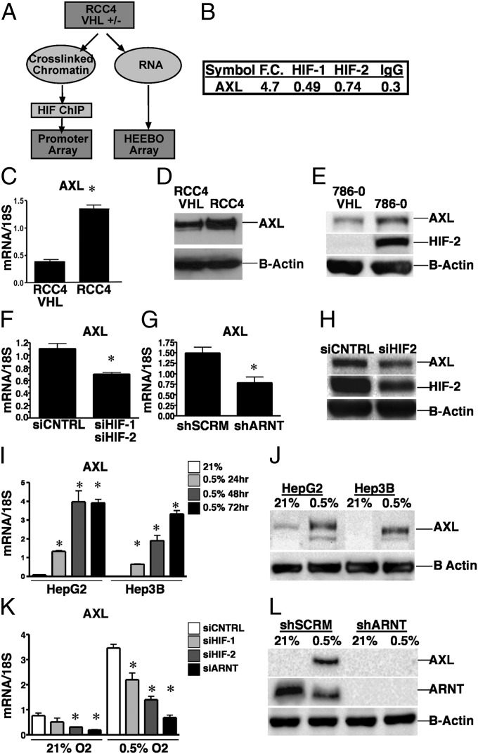
To identify novel molecular targets involved in ccRCC tumor progression and metastasis, we performed a directed screen by combining high-throughput chromatin immunoprecipitation (ChIP–chip) with gene expression analysis to identify functionally relevant HIF target genes (15) (Fig. 1_A_). The RCC4 ccRCC line was used as a model system based on its abundant normoxic expression of HIF-1 and HIF-2 due to genetic inactivation of VHL. Expression profiling was used to identify functional HIF target genes induced greater than 1.5-fold in RCC4 cells compared with VHL reconstituted RCC4 cells (RCC4–VHL) (16). In parallel, ChIP–chip analysis was used to identify genes bound by HIF-1 or HIF-2 within promoter regions of RCC4 cells. The screen identified several known HIF target genes, including vascular endothelial growth factor (VEGF), providing validity for our ChIP–chip analysis (15). We were particularly interested in identifying HIF targets that could be molecular targets for ccRCC therapy, including secreted factors, receptors, or kinases. Using these criteria, we found the RTK AXL was induced 4.7-fold in RCC4 cells in comparison with RCC4–VHL cells (Fig. 1_B_). By ChIP, we found that the AXL promoter was enriched for HIF-1 (0.49) and HIF-2 (0.74) binding in comparison with the IgG (0.3) control (Fig. 1_B_). Thus, we used an unbiased screen to identify the RTK AXL as a putative HIF target capable of therapeutic intervention.
Fig. 1.
AXL is regulated by HIF in VHL-deficient and hypoxic cancer cells. (A) Schematic representation of the ChIP–chip assay performed in RCC4 VHL wild-type (RCC4–VHL) and deficient (RCC4) cells. (B) Results from the ChIP–chip assay demonstrating AXL fold change with HIF-1 and HIF-2 enrichment in RCC4 compared with RCC4–VHL cells. (C) Real-time PCR analysis of AXL mRNA expression relative to 18S (n = 3 per group). (D and E) Western blot analysis of AXL protein levels in RCC4 (D) and 786-0 (E) cells. Beta actin (B-actin) was used as a protein loading control. (F and G) Real-time PCR analysis of AXL expression in RCC4 cells treated with siRNA against HIF-1 and HIF-2 or shRNA targeting scramble control (shSCRM) or ARNT (shARNT, n = 3 per group). (H) Western blot analysis of AXL, HIF-2, and B-actin in 786-0 cells treated with siRNA targeting HIF-2. (I) Real-time PCR analysis of AXL expression in HepG2 and Hep3B cells exposed to normoxia or hypoxia (n = 3 per group). (J) AXL protein levels in HepG2 and Hep3B cells exposed to hypoxia for 48 h. (K) Real-time PCR analysis of AXL expression in HepG2 cells treated with siRNA against HIF-1, HIF-2, or ARNT exposed to either normoxia (21% O2) or hypoxia (0.5% O2, n = 3 per group). Error bars represent ±SEM. *P ≤ 0.05. (L) Western blot analysis of AXL and ARNT in HepG2 cells with shSCRM or shARNT exposed to hypoxia for 48 h.
To validate AXL as a HIF-regulated gene in VHL-deficient cells, we confirmed the ChIP–chip data by quantitative real-time PCR analysis. AXL mRNA was significantly up-regulated (threefold) in RCC4 compared with RCC4–VHL cells, respectively (Fig. 1_C_). Similarly, AXL protein levels were increased in RCC4 and 786-0 VHL-deficient cells compared with their matched VHL reconstituted cells, verifying AXL as a VHL-regulated gene (Fig. 1 D and E). Repression of endogenous HIF signaling through siRNA-mediated inactivation of HIF-1 and HIF-2 or shRNA-mediated inactivation of ARNT, the common binding partner for HIF-1 and HIF-2, resulted in a significant repression of AXL expression (Fig. 1 F and G and Fig. S1 A–C) (17, 18). Similarly, knockdown of HIF-2 expression in 786-0 cells, which only express HIF-2, resulted in a decrease in AXL protein levels (Fig. 1_H_) (19).
In addition to VHL loss, hypoxia is another mechanism by which HIF signaling is activated in cancer cells. Therefore, we investigated whether HIF regulates AXL expression in hypoxic cancer cells. In contrast to kidney cancer, where activation of HIF primarily occurs through loss of VHL, activation of HIF through intratumoral hypoxia is a prominent feature of hepatocellular carcinoma (20). AXL expression was significantly induced by hypoxia in Hep3B and HepG2 cells (Fig. 1_I_). Furthermore, AXL protein was also induced by hypoxia in HepG2 and Hep3B cells (Fig. 1_J_). Genetic inactivation of HIF signaling using siRNAs targeting HIF-1, HIF-2, or ARNT demonstrated that similar to VEGF, deletion of HIF-1 or HIF-2 partially decreased the hypoxic induction of AXL, whereas inactivation of ARNT completely abolished the hypoxic induction of AXL (Fig. 1 K and L and Fig. S1_D_). Collectively, these findings demonstrate that HIF signaling regulates AXL expression in both VHL-deficient and hypoxic cancer cells.
The majority of AXL signaling occurs in a ligand-dependent manner mediated by GAS6. In cancer, GAS6/AXL signaling can be activated in an autocrine or paracrine manner with tumor cells as well as cells within the tumor microenvironment, including macrophages and endothelial cells producing biologically relevant sources of GAS6 (12). Therefore, we examined the expression of GAS6, AXL, and phosphorylated AXL in VHL-deficient and hypoxic cancer cells. In comparison with human embryonic kidney cells (293T) that do not express AXL, the majority (5/7) of ccRCC cell lines expressed high levels of the AXL receptor (Fig. S1 E and F). Phosphorylation of AXL in these cell lines was only present in those cell lines that also produced GAS6 (Fig. S1_F_). Furthermore, stimulation of GAS6-low ccRCC cells with exogenous GAS6 resulted in a robust stimulation of phospho-AXL (p-AXL) (Fig. S1_G_). These findings indicate that AXL kinase activity is modulated in a GAS6-dependent manner in ccRCC. Under hypoxia, we also observed that up-regulation of AXL was accompanied by an activation of p-AXL in HepG2 cells that express endogenous GAS6 (Fig. S1 H and I). Thus, up-regulation of AXL by VHL loss or hypoxia results in an increase in AXL protein that is activated by endogenous and exogenous sources of GAS6.
AXL Is a Direct HIF Target.
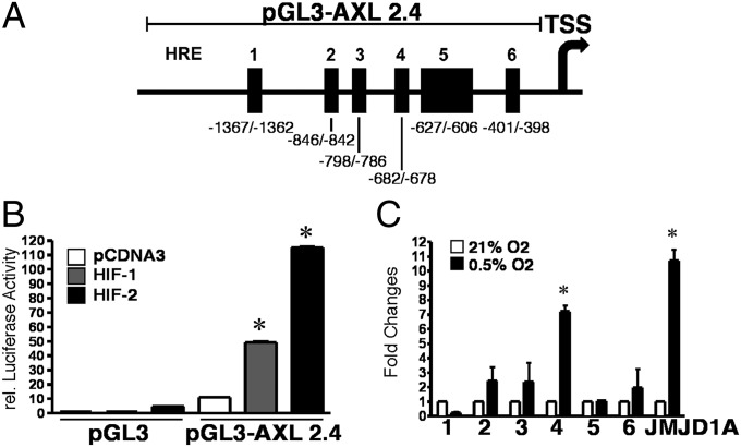
HIF can exert its transcriptional activity through both direct and indirect mechanisms (21–23). To determine if AXL is a direct HIF target, we searched the AXL promoter for consensus hypoxia-response elements (HREs) containing a conserved RCGTG sequence. Six consensus HRE sites were identified within a 2.4 kb fragment of the AXL promoter (Fig. 2_A_) (24). Luciferase assays using the 2.4 kb fragment of the AXL promoter demonstrated that expression of nondegradable HIF-1 and HIF-2 was sufficient to activate AXL promoter activity (Fig. 2_B_). To identify which HRE sites are bound by HIF in vivo, we performed ChIP assays in which RCC4–VHL cells were exposed to normoxia or hypoxia and DNA fragments bound by endogenous HIF-1 were immunoprecipitated. HIF-1 binding to HRE 4 (−682/−678) was enriched in cells treated with hypoxia (Fig. 2_C_). HIF-1 binding on the AXL promoter was comparable to binding within the JMJD1A HRE promoter, an established HIF target gene in ccRCC (Fig. 2_C_) (15). Similar results were observed in HepG2 cells where hypoxia induced binding of endogenous HIF-1 to the AXL HRE (Fig. S2_A_). Additionally, infection of an HA-tagged nondegradable HIF-2 into these cells also demonstrated a specific binding of HIF-2 at the HRE element in vivo (Fig. S2_B_) (25). These data demonstrate that HIF-1 and HIF-2 directly bind to and activate AXL expression.
Fig. 2.
AXL is a direct HIF-1 and HIF-2 target. (A) Schematic representation of the human AXL promoter sequence spanning 2.4 kb upstream of the transcriptional start site (TSS). Six potential HIF binding sites (HREs) are indicated with black boxes. (B) HIF-1 and HIF-2 are sufficient to activate AXL promoter activity. Luciferase reporter assay of the AXL promoter in HepG2 cells expressing the pCDNA3 control or nondegradable HIF-1 and HIF-2 proline mutants (n = 6). (C) HIF-1 directly binds to the HRE located within the AXL promoter. ChIP assay analysis of HIF-1 binding at HREs 1–6 in the AXL promoter in RCC4–VHL cells exposed to normoxia (21% O2) or hypoxia (0.5% O2). HIF-1 occupancy on the JMJD1A promoter HRE was used as the positive control. Enrichments were calculated as percentage of total input after subtraction of IgG signal and are presented as fold change in HIF-1 binding relative to normoxic control cells (21% O2). Data represent the averages from two independent experiments measured in triplicate ± SEM. *P ≤ 0.01.
Genetic and Therapeutic Inactivation of AXL Signaling in Metastatic ccRCC Cells Reversed the Invasive and Metastatic Phenotype in Vivo.
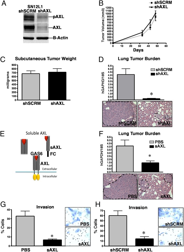
AXL has multiple protumorigenic properties, including regulating proliferation/survival, apoptosis, and invasion/metastasis (26). To investigate the functional role of AXL in ccRCC, we used the highly metastatic SN12L1 cell line selected for its increased metastatic colonization of the lung and its high expression of both AXL and GAS6 (Fig. S1_H_) (27). Genetic inhibition of AXL using shRNA targeting did not affect s.c. SN12L1 tumor growth, indicating that AXL signaling pathways are not essential for cell proliferation or survival of ccRCC cells (Fig. 3 A–C). In contrast to the primary tumor, metastatic colonization of the lung was significantly impaired in mice injected with AXL-deficient cells. Both histologic analysis and quantification of human GAPDH (hGAPDH) expression revealed decreased tumor burden in the lungs of mice injected with shAXL cells compared with mice injected with control cells (Fig. 3_D_). These findings demonstrate that AXL is a critical factor governing ccRCC invasion and metastatic colonization to the lung.
Fig. 3.
Genetic and therapeutic inhibition of AXL inhibits the metastatic phenotype of ccRCC cells. (A) Efficient inactivation of AXL in the highly metastatic SN12L1 ccRCC cell line. Western blot analysis of AXL and p-AXL expression in control (shSCRM) and AXL-deficient (shAXL) SN12L1 cells. B-actin was used as a protein loading control. (B) Genetic inactivation of AXL does not affect primary SN12L1 tumor growth. Average volume of s.c. SN12L1 tumors (n = 8 per group). Error bars represent SEM. (C) Total weight of s.c. SN12L1 tumors excised from mice at day 50 following injection (n = 8). (D) Genetic inactivation of AXL significantly reduces the metastatic potential of SN12L1 cells to the lung. Real-time PCR analysis (Upper) of hGAPDH expression in the lungs of mice 8 wk following i.v. injection of shSCRM or shAXL SN12L1 cells (n = 8). Hematoxylin and eosin staining (Lower) of lung taken from mice 8 wk following i.v. injection of shSCRM or shAXL SN12L1 cells. (E) Schematic representation of the sAXL decoy therapy. (F) Therapeutic inactivation of AXL significantly reduces the metastatic potential of SN12L1 cells to the lung. Real-time PCR analysis (Upper) of hGAPDH expression in the lungs of mice 8 wk following i.v. injection of SN12L1 cells (n = 8). Tumors were allowed to establish for 1 wk followed by vehicle or sAXL (5 mg/kg) treatment. Hematoxylin and eosin staining (Lower) of lung from mice treated with vehicle or sAXL 8 wk following i.v. injection of SN12L1 cells. Error bars represent ±SEM. *P ≤ 0.05. (G) sAXL inhibits ccRCC tumor cell invasion. Matrigel invasion assays of vehicle- or sAXL- (4 μg/mL) treated 786-0 cells in the presence of GAS6 (100 ng/mL). (H) Genetic inhibition of AXL inhibits ccRCC tumor cell invasion. Matrigel invasion assays of shSCRM or shAXL 786-0 cells in the presence of GAS6 (100 ng/mL). Results represent the normalized percentage of cells that invaded through matrigel transwells per field in three biologic replicates. All experiments were independently repeated three times.
The findings above identify a role for AXL in ccRCC metastasis, raising the intriguing possibility that therapeutic inhibition of AXL could be an effective strategy for the treatment of metastatic ccRCC. Several classes of AXL inhibitors have been developed and have shown efficacy in preclinical models of metastasis. Currently, two small-molecule AXL inhibitors (BGB324 BergenBio and S49076 Servier) are in phase I clinical trials for the treatment of advanced cancer (28). To selectively and specifically inhibit AXL activation directly, we developed a sAXL decoy receptor fused to human IgG1 (sAXL; Fig. 3_E_). sAXL is a potent and safe inhibitor of GAS6 signaling (29). To determine the efficacy of sAXL therapy in metastatic ccRCC, we treated mice with established SN12L1 metastatic lesions in the lung. Biweekly administration of sAXL (5 mg/kg) resulted in a significant reduction of metastatic tumor burden in the lungs of mice with established renal metastases compared with vehicle treatment (Fig. 3_F_). These data demonstrate that selective inhibition of GAS6/AXL signaling using a single-agent sAXL decoy receptor is an effective strategy to inhibit ccRCC metastatic tumor progression in the lung in a model where high levels of endogenous GAS6 are produced by the tumor epithelium.
Given the significant role of AXL on SN12L1 (VHL wild-type ccRCC) metastasis, we sought to investigate the role of AXL in VHL-deficient and hypoxia-mediated metastasis (30). Although the VHL-deficient ccRCC cell lines are poorly metastatic in vivo, 786-0 cells migrate and invade through ECM matrix proteins toward serum-containing media under serum-starved conditions. Inactivation of AXL signaling therapeutically with sAXL and genetically with shAXL significantly inhibited the ability of 786-0 cells to invade through matrigel in the presence of exogenous GAS6 (Fig. 3 G and H, 100 ng/mL). Furthermore, hypoxia-mediated invasion of Hep3B cells was also dependent on AXL expression (Fig. S3). These findings demonstrate that AXL is an important factor governing both VHL-deficient and hypoxic invasion. Importantly, these studies suggest that sAXL therapy is sufficient to inhibit the prometastatic properties of ccRCC cells that express high levels of endogenous GAS6 and ccRCC cells that express low levels of GAS6 but respond to exogenous sources of GAS6.
HGF-Independent Activation of MET by GAS6 Signaling Promotes ccRCC Invasion.
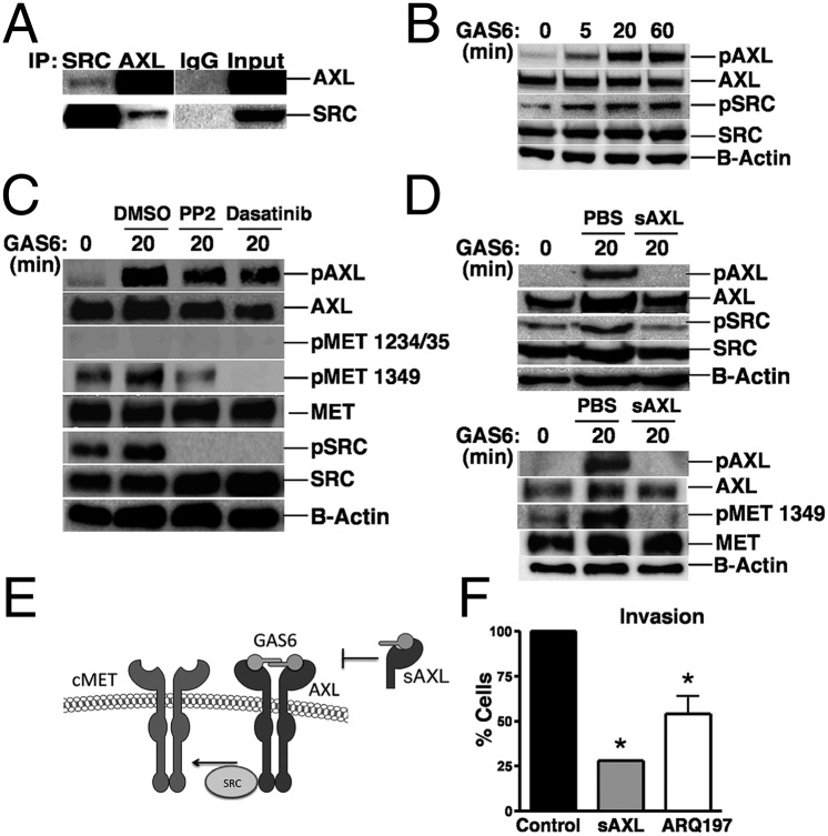
We next sought to investigate the molecular mechanisms by which GAS6/AXL signaling regulates ccRCC invasion and metastasis. The intracellular domain of AXL contains multiple tyrosine residues that serve as docking sites for signaling molecules including the non-RTK SRC family kinases (26). In particular, in vitro binding studies revealed that tyrosine 821 is a docking site for SRC and LCK (31). It is well established that SRC plays an important role in tumor growth, angiogenesis, and metastasis (32). Moreover, SRC is active in ccRCC and correlates with poor patient survival (30). However, the mechanisms for SRC activation in ccRCC remain unclear. We performed a series of experiments to determine if SRC activation is mediated through AXL in ccRCC. Immunoprecipitation studies in SN12L1 cells showed that AXL is complexed with SRC in ccRCC cells (Fig. 4_A_). Time course analysis of GAS6-treated cells revealed that similar to AXL phosphorylation, SRC phosphorylation occurs within 5 min and is sustained with maximal levels at 60 min (Fig. 4_B_). These findings indicate that SRC is a direct target of GAS6/AXL signaling in ccRCC cells.
Fig. 4.
GAS6/AXL signaling regulates ccRCC invasion and metastasis through the lateral activation of MET via SRC. (A) SRC coimmunoprecipitates with AXL in ccRCC cells. Lysates from SN12L1 cells were immunoprecipitated with anti-AXL, anti-SRC, and anti-rabbit (Lower) or goat (Upper) IgG antibodies and analyzed by Western blot. Lysates taken before immunoprecipitation (input) were used to determine total AXL and SRC levels. (B) GAS6 stimulates both p-AXL and phospho-SRC (p-SRC) activation. Western blot analysis of lysates from serum-starved SN12L1 cells treated with GAS6 (400 ng/mL). (C) GAS6 activates MET Y1349 in an SRC-dependent manner. Western blot analysis of serum-starved SN12L1 cells treated with either GAS6 (400 ng/mL) alone or in combination with SRC inhibitors PP2 (500 nM) or dasatinib (50 nM). (D) sAXL treatment prevents GAS6-mediated activation of pAXL, pSRC, and pMET 1349. Western blot analysis of starved SN12L1 cells treated with GAS6 (400 ng/mL) in the presence of vehicle or sAXL therapy (4 μg/mL). (E) Model for GAS6-dependent activation of MET in ccRCC cells. GAS6 signaling through AXL stimulates SRC phosphorylation and lateral activation of MET. sAXL is sufficient to inhibit GAS6-mediated activation of pAXL, pSRC, and pMET 1349. (F) Therapeutic inhibition of AXL and MET inhibits ccRCC tumor cell invasion. Matrigel invasion assays of SN12L1 cells treated with sAXL (4 μg/mL) or ARQ197 (500 nM). Results represent the normalized percentage of cells that invaded through matrigel transwells per field in three biologic replicates. Experiments were independently repeated three times. Error bars represent ±SEM. *P ≤ 0.05.
SRC is a key intermediary in regulating lateral RTK–RTK signaling. Once activated, SRC has the ability to phosphorylate the intracellular domain of neighboring RTKs to relieve autoinhibition of the kinase domain (33). We used the cBioPortal for Cancer Genomics database to analyze protein and phosphorylation changes of receptors in human ccRCC samples with high AXL expression (34, 35). Phosphorylation of the RTK MET is increased in ccRCC tumors expressing high levels of AXL, indicating that MET activity may be regulated by GAS6/AXL signaling. MET has previously been shown to be an intracellular target of SRC phosphorylation and is a key factor regulating epithelial–mesenchymal transition, invasion, and metastasis (36, 37). In ccRCC cells lacking HGF expression, we found that similar to inactivation of AXL, knockdown of MET expression resulted in a significant decrease in the expression of EMT-associated factors, including SLUG and SNAIL (Fig. S4 A and B). Additionally, genetic inhibition of MET inhibited ccRCC invasion in the absence of HGF, indicating a functional role for ligand-independent MET activation in ccRCC (Fig. S4_C_). Therefore, we hypothesized that lateral activation of MET may contribute to GAS/AXL-mediated EMT and metastasis in ccRCC. In support of this hypothesis, stimulation with GAS6 resulted in the activation of Y1349 in both SN12L1 and 786-0 cells, a key residue required for MET signaling (Fig. 4 C and D and Fig. S4_D_). This phosphorylation event occurred independent of the HGF-dependent phosphorylation at residues Y1234/Y1235 (Fig. 4_C_) (38). Because ccRCC cells express a number of SFKs, including SRC, CSK, FYN, LYN, and YES, we used small-molecule SFK inhibitors to determine if GAS6-mediated activation of MET occurs through SFK members (Fig. S4_E_). Preincubation with the kinase inhibitors PP2 (500 nM) and dasatinib (50 nM) that target SFKs abolished the GAS6-mediated increase in both SRC and MET Y1349 phosphorylation without affecting p-AXL or total MET levels (Fig. 4_C_). Similar results were observed using the more specific SFK inhibitor sarcatinib/AZD0530 (1 μM; Fig. S4_F_). Importantly, we used sAXL to demonstrate that therapeutic inhibition of GAS6 in ccRCC is sufficient to block GAS6-mediated activation of AXL, SRC, and MET (Fig. 4 D and E). These findings indicate that SFKs are required for GAS6 transphosphorylation of MET and targeting GAS6/AXL signaling may be a therapeutic strategy to inhibit AXL, SRC, and MET activity in metastatic ccRCC cells.
Given that AXL is upstream of MET, we compared the efficacy of AXL (sAXL, 4 μg/mL) and MET (ARQ197, 500 nM) inhibitors in GAS6-mediated invasion. Treatment with sAXL therapy reduced invasion by 70%, in comparison with a 40% reduction in cellular invasion using the MET inhibitor (Fig. 4_F_). These findings suggest that GAS6/AXL signaling uses lateral activation of MET to maximize cellular invasion through nonconical signaling mechanisms in kidney cancer cells.
AXL Is Associated with the Lethal Phenotype in ccRCC.
The results above identify the GAS6/AXL signaling pathway as a therapeutic target to block invasion and metastasis in ccRCC. Therefore, we analyzed AXL expression in human RCC tissue within The Cancer Genome Atlas (TCGA). Compared with normal kidney tissue, AXL expression is significantly increased in ccRCC tumor tissue (Fig. 5_A_). Furthermore, when analyzing AXL expression within molecular subgroups of tumor and normal samples, AXL is expressed at the highest levels in aggressive tumors (ccB) compared with tumors taken from patients with good prognosis (ccA), non–VHL-related tumors (cc3), or normal kidney (Fig. 5_B_). Moreover, RCC samples with strong AXL expression came from patients with reduced survival compared with patients whose samples had weak AXL expression (Fig. 5_C_, P = 0.004812). Most strikingly, 100% of patients with high AXL expression died within 80 mo following diagnosis, whereas 50% of patients with low AXL expression remained alive (Fig. 5_C_). These findings identify AXL as a prognostic marker for the lethal phenotype in ccRCC. Collectively, our findings identify AXL as a critical factor and therapeutic target driving the aggressive phenotype in renal clear cell carcinoma.
Fig. 5.
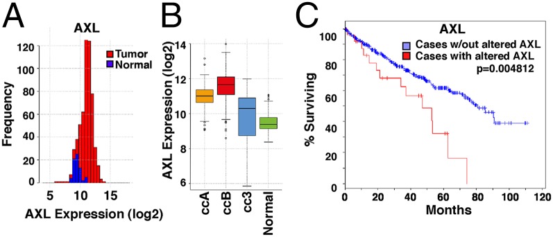
AXL is highly expressed in aggressive ccRCC tumors and is associated with poor outcome. (A) AXL expression in the ccRCC TCGA dataset. Cumulative histogram of the RNAseq rsem normalized gene expression (log2 base): tumors (red) and normal (blue). (B) Box plot of AXL gene expression with tumor and normal samples split into molecular subgroups. Tumors are classified into ccA (good prognosis), ccB (aggressive), and cc3 (non-VHL) and compared with normal kidney. (C) Kaplan–Meier plot of survival by AXL altered gene expression in the TCGA tumor samples.
Discussion
Here we demonstrate that the RTK AXL is a downstream target of HIF and plays a critical role in the metastatic phenotype of ccRCC. Importantly, we demonstrate that AXL is up-regulated by HIF signaling in both VHL-deficient and hypoxic tumor cells. Recently, a role for VHL in the regulation of AXL in ccRCC has been observed. Similar to our findings, Gustafsson et al. found that reconstitution with VHL resulted in down-regulation of AXL protein levels in VHL-deficient ccRCC cells (39). However, the mechanism by which VHL regulates AXL expression was not explored in these studies. Gustafsson et al. concluded that GAS6 activation of AXL resulted in a decrease in cell viability and migration associated with no overall effect on invasion (39). We observe a significant role for AXL in activating ccRCC invasion and metastasis. Differences in our conclusions are likely due to the quality and purity of GAS6. The GAS6 used in our studies was recombinant human GAS6 with >90% purity and <1.0 EU/1 μg of endotoxin. The purity of GAS6 used in Gustafsson et al. studies is unclear (39). Here we report that the transcription factors HIF-1 and HIF-2 directly bind to the AXL promoter and are necessary and sufficient to activate AXL in VHL-deficient and hypoxic cancer cells. AXL is overexpressed in a variety of tumor types, where its expression is correlated with tumor progression and metastasis (26). However, the mechanisms by which AXL expression is activated during tumor progression are poorly understood. Here we identify a functional HIF binding site within the AXL promoter, indicating that HIF signaling directly activates AXL expression (Fig. S5). As activation of HIF is a prominent feature of many solid tumors including kidney cancer, these findings provide a potential mechanism for AXL up-regulation in cancer either by genetic or microenvironmental activation of HIF.
We also define a pathway by which GAS6/AXL signaling regulates invasion and metastasis through the lateral activation of MET through SRC (Fig. S5). These findings provide an alternative model for SRC and MET activation by GAS6 in ccRCC. Previous reports have shown that SRC is elevated in RCC and correlates with poor patient survival (30). Although SRC plays a central role in mediating multiple protumorigenic signaling cascades, it is rarely mutated in cancers (32). In melanoma, HIF-1 and HIF-2 activate SRC through PDGFRα and FAK to mediate cellular invasion (40). Our data suggest that a mechanism for SRC activation in ccRCC occurs through HIF-mediated up-regulation of GAS6/AXL signaling where AXL directly binds to and activates SRC activity. Although previous studies have demonstrated that Y821 is a docking site for SRC and LCK, future studies are needed to delineate the role of Y821 in ccRCC invasion and metastasis (31). The ccRCC cell lines examined in our study expressed low to high levels of endogenous GAS6. Infiltrating immune cells including leukocytes also have the capacity to produce GAS6 and stimulate TAM receptor signaling on tumor cells (41). Thus, both autocrine and paracrine production of GAS6 may contribute to SRC activation in kidney cancer.
The RTK MET plays an important role in the pathogenesis of kidney cancer, where it regulates tumor growth, metastasis, and angiogenesis. Although mutations in the MET kinase domain drive constitutive activation of MET in papillary RCC, MET mutations in renal clear cell carcinoma have not been found (42). Nakaigawa et al. demonstrated that VHL loss in ccRCC induces constitutive phosphorylation of MET in the absence of HGF (43). However, the mechanisms driving HGF-independent activation of MET in ccRCC remain unknown. Here we demonstrate that GAS6 activates MET in a SRC-dependent manner. The activation of MET by GAS6 occurred in an HGF-independent manner, as the renal cell lines used in this study do not produce HGF and the upstream residue Y1235 remained unstimulated during GAS6 treatment. Our findings suggest lateral activation of MET by GAS6/AXL signaling in ccRCC. These findings highlight the importance of targeting GAS6/AXL signaling to inhibit oncogenic signaling pathways including SRC and MET.
AXL expression may be used as a valuable marker to predict the aggressive behavior and lethality of tumors in patients with ccRCC. AXL expression is associated with poor patient prognosis in ccRCC patients (44). Moreover, we demonstrate that therapeutic inhibition of GAS6/AXL signaling in metastatic ccRCC is sufficient to prevent metastatic tumor progression. Importantly, we demonstrate that sAXL therapy inhibits the prometastatic properties of ccRCC cells that express high levels of endogenous GAS6 (SN12L1) and ccRCC cells that express low levels of GAS6, but respond to exogenous sources of GAS6 (786-0). Previous studies have shown that both tumor- and stromal-derived GAS6 contributes to tumor growth and metastasis. Loges et al. used GAS6-deficient mice to demonstrate that bone marrow-derived GAS6 promotes the growth and metastasis of a variety of cancer cell lines deficient for endogenous GAS6 (41). In these models, the tumor microenvironment stimulated the up-regulation of GAS6 in leukocytes, which in turn mediate the growth and metastasis of GAS6-deficient tumor cells. Similar findings were observed in AML cells (45). Thus, our findings indicate that sAXL therapy may inhibit both autocrine and paracrine GAS6/AXL signaling in tumors. Anti-AXL therapy primarily functions as an antimetastatic agent in ccRCC, as sAXL therapy significantly reduced metastatic tumor burden but without affecting primary tumor growth. These findings indicate that anti-AXL therapy may be most effective when combined with current antiangiogenic agents in the treatment of advanced ccRCC. Collectively, our data provide the preclinical studies to support the use of AXL inhibitors for the treatment of metastatic kidney cancer.
Materials and Methods
Detailed analysis of cell lines and culture conditions; shRNA, siRNA, and cDNA constructs; recombinant protein production; adenovirus production; ChIP–chip assay; protein isolation and Western blot analysis; luciferase assay; matrigel invasion assay; ChIP assay; immunoprecipitation assay; real-time PCR; TCGA data analysis; s.c. tumor growth and lung metastasis assay; and statistical analysis is provided in SI Materials and Methods. Real-time PCR primer sequences can be found in Table S1. Statistical analysis was performed with ANOVA followed by two-tailed, unpaired Student t tests. P < 0.05 were considered statistically significant.
Supplementary Material
Supplementary File
Acknowledgments
We thank The Cancer Genome Atlas Research Network for making the renal clear cell carcinoma dataset available for analysis. This work was supported by National Institutes of Health (NIH) Grants CA-67166, CA-088480, and ARO65403 and the Sydney Frank Foundation (to A.J.G.).
Footnotes
The authors declare no conflict of interest.
This article is a PNAS Direct Submission.
References
- 1.Escudier B, Szczylik C, Porta C, Gore M. Treatment selection in metastatic renal cell carcinoma: Expert consensus. Nat Rev Clin Oncol. 2012;9(6):327–337. doi: 10.1038/nrclinonc.2012.59. [DOI] [PubMed] [Google Scholar]
- 2.Latif F, et al. Identification of the von Hippel-Lindau disease tumor suppressor gene. Science. 1993;260(5112):1317–1320. doi: 10.1126/science.8493574. [DOI] [PubMed] [Google Scholar]
- 3.Iliopoulos O, Kibel A, Gray S, Kaelin WG., Jr Tumour suppression by the human von Hippel-Lindau gene product. Nat Med. 1995;1(8):822–826. doi: 10.1038/nm0895-822. [DOI] [PubMed] [Google Scholar]
- 4.Kibel A, Iliopoulos O, DeCaprio JA, Kaelin WG., Jr Binding of the von Hippel-Lindau tumor suppressor protein to Elongin B and C. Science. 1995;269(5229):1444–1446. doi: 10.1126/science.7660130. [DOI] [PubMed] [Google Scholar]
- 5.Duan DR, et al. Inhibition of transcription elongation by the VHL tumor suppressor protein. Science. 1995;269(5229):1402–1406. doi: 10.1126/science.7660122. [DOI] [PubMed] [Google Scholar]
- 6.Pause A, et al. The von Hippel-Lindau tumor-suppressor gene product forms a stable complex with human CUL-2, a member of the Cdc53 family of proteins. Proc Natl Acad Sci USA. 1997;94(6):2156–2161. doi: 10.1073/pnas.94.6.2156. [DOI] [PMC free article] [PubMed] [Google Scholar]
- 7.Kamura T, et al. Rbx1, a component of the VHL tumor suppressor complex and SCF ubiquitin ligase. Science. 1999;284(5414):657–661. doi: 10.1126/science.284.5414.657. [DOI] [PubMed] [Google Scholar]
- 8.Jaakkola P, et al. Targeting of HIF-alpha to the von Hippel-Lindau ubiquitylation complex by O2-regulated prolyl hydroxylation. Science. 2001;292(5516):468–472. doi: 10.1126/science.1059796. [DOI] [PubMed] [Google Scholar]
- 9.Ivan M, et al. HIFalpha targeted for VHL-mediated destruction by proline hydroxylation: Implications for O2 sensing. Science. 2001;292(5516):464–468. doi: 10.1126/science.1059817. [DOI] [PubMed] [Google Scholar]
- 10.Staller P, et al. Chemokine receptor CXCR4 downregulated by von Hippel-Lindau tumour suppressor pVHL. Nature. 2003;425(6955):307–311. doi: 10.1038/nature01874. [DOI] [PubMed] [Google Scholar]
- 11.Vanharanta S, et al. Epigenetic expansion of VHL-HIF signal output drives multiorgan metastasis in renal cancer. Nat Med. 2013;19(1):50–56. doi: 10.1038/nm.3029. [DOI] [PMC free article] [PubMed] [Google Scholar]
- 12.Linger RM, Keating AK, Earp HS, Graham DK. Taking aim at Mer and Axl receptor tyrosine kinases as novel therapeutic targets in solid tumors. Expert Opin Ther Targets. 2010;14(10):1073–1090. doi: 10.1517/14728222.2010.515980. [DOI] [PMC free article] [PubMed] [Google Scholar]
- 13.Zhang Z, et al. Activation of the AXL kinase causes resistance to EGFR-targeted therapy in lung cancer. Nat Genet. 2012;44(8):852–860. doi: 10.1038/ng.2330. [DOI] [PMC free article] [PubMed] [Google Scholar]
- 14.Ruan GX, Kazlauskas A. Axl is essential for VEGF-A-dependent activation of PI3K/Akt. EMBO J. 2012;31(7):1692–1703. doi: 10.1038/emboj.2012.21. [DOI] [PMC free article] [PubMed] [Google Scholar]
- 15.Krieg AJ, et al. Regulation of the histone demethylase JMJD1A by hypoxia-inducible factor 1 alpha enhances hypoxic gene expression and tumor growth. Mol Cell Biol. 2010;30(1):344–353. doi: 10.1128/MCB.00444-09. [DOI] [PMC free article] [PubMed] [Google Scholar]
- 16.Maxwell PH, et al. The tumour suppressor protein VHL targets hypoxia-inducible factors for oxygen-dependent proteolysis. Nature. 1999;399(6733):271–275. doi: 10.1038/20459. [DOI] [PubMed] [Google Scholar]
- 17.Wang GL, Semenza GL. Purification and characterization of hypoxia-inducible factor 1. J Biol Chem. 1995;270(3):1230–1237. doi: 10.1074/jbc.270.3.1230. [DOI] [PubMed] [Google Scholar]
- 18.Jiang BH, Rue E, Wang GL, Roe R, Semenza GL. Dimerization, DNA binding, and transactivation properties of hypoxia-inducible factor 1. J Biol Chem. 1996;271(30):17771–17778. doi: 10.1074/jbc.271.30.17771. [DOI] [PubMed] [Google Scholar]
- 19.Jiang Y, et al. Gene expression profiling in a renal cell carcinoma cell line: Dissecting VHL and hypoxia-dependent pathways. Mol Cancer Res. 2003;1(6):453–462. [PubMed] [Google Scholar]
- 20.Dong ZZ, et al. Hypoxia-inducible factor-1alpha: Molecular-targeted therapy for hepatocellular carcinoma. Mini Rev Med Chem. 2013;13(9):1295–1304. doi: 10.2174/1389557511313090004. [DOI] [PubMed] [Google Scholar]
- 21.Wang GL, Semenza GL. Characterization of hypoxia-inducible factor 1 and regulation of DNA binding activity by hypoxia. J Biol Chem. 1993;268(29):21513–21518. [PubMed] [Google Scholar]
- 22.Keith B, Johnson RS, Simon MC. HIF1α and HIF2α: Sibling rivalry in hypoxic tumour growth and progression. Nat Rev Cancer. 2012;12(1):9–22. doi: 10.1038/nrc3183. [DOI] [PMC free article] [PubMed] [Google Scholar]
- 23.Semenza GL. Hypoxia-inducible factors in physiology and medicine. Cell. 2012;148(3):399–408. doi: 10.1016/j.cell.2012.01.021. [DOI] [PMC free article] [PubMed] [Google Scholar]
- 24.Mudduluru G, Allgayer H. The human receptor tyrosine kinase Axl gene—Promoter characterization and regulation of constitutive expression by Sp1, Sp3 and CpG methylation. Biosci Rep. 2008;28(3):161–176. doi: 10.1042/BSR20080046. [DOI] [PubMed] [Google Scholar]
- 25.Wei K, et al. A liver Hif-2α-Irs2 pathway sensitizes hepatic insulin signaling and is modulated by Vegf inhibition. Nat Med. 2013;19(10):1331–1337. doi: 10.1038/nm.3295. [DOI] [PMC free article] [PubMed] [Google Scholar]
- 26.Linger RM, Keating AK, Earp HS, Graham DK. TAM receptor tyrosine kinases: Biologic functions, signaling, and potential therapeutic targeting in human cancer. Adv Cancer Res. 2008;100:35–83. doi: 10.1016/S0065-230X(08)00002-X. [DOI] [PMC free article] [PubMed] [Google Scholar]
- 27.Saiki I, et al. Characterization of the invasive and metastatic phenotype in human renal cell carcinoma. Clin Exp Metastasis. 1991;9(6):551–566. doi: 10.1007/BF01768583. [DOI] [PubMed] [Google Scholar]
- 28.Sheridan C. First Axl inhibitor enters clinical trials. Nat Biotechnol. 2013;31(9):775–776. doi: 10.1038/nbt0913-775a. [DOI] [PubMed] [Google Scholar]
- 29.Rankin EB, et al. AXL is an essential factor and therapeutic target for metastatic ovarian cancer. Cancer Res. 2010;70(19):7570–7579. doi: 10.1158/0008-5472.CAN-10-1267. [DOI] [PMC free article] [PubMed] [Google Scholar]
- 30.Suwaki N, et al. A HIF-regulated VHL-PTP1B-Src signaling axis identifies a therapeutic target in renal cell carcinoma. Sci Transl Med. 2011;3(85):85ra47. doi: 10.1126/scitranslmed.3002004. [DOI] [PMC free article] [PubMed] [Google Scholar]
- 31.Braunger J, et al. Intracellular signaling of the Ufo/Axl receptor tyrosine kinase is mediated mainly by a multi-substrate docking-site. Oncogene. 1997;14(22):2619–2631. doi: 10.1038/sj.onc.1201123. [DOI] [PubMed] [Google Scholar]
- 32.Yeatman TJ. A renaissance for SRC. Nat Rev Cancer. 2004;4(6):470–480. doi: 10.1038/nrc1366. [DOI] [PubMed] [Google Scholar]
- 33.Dulak AM, Gubish CT, Stabile LP, Henry C, Siegfried JM. HGF-independent potentiation of EGFR action by c-Met. Oncogene. 2011;30(33):3625–3635. doi: 10.1038/onc.2011.84. [DOI] [PMC free article] [PubMed] [Google Scholar]
- 34.Gao J, et al. Integrative analysis of complex cancer genomics and clinical profiles using the cBioPortal. Sci Signal. 2013;6(269):pl1. doi: 10.1126/scisignal.2004088. [DOI] [PMC free article] [PubMed] [Google Scholar]
- 35.Cerami E, et al. The cBio cancer genomics portal: An open platform for exploring multidimensional cancer genomics data. Cancer Discov. 2012;2(5):401–404. doi: 10.1158/2159-8290.CD-12-0095. [DOI] [PMC free article] [PubMed] [Google Scholar]
- 36.Koochekpour S, et al. The von Hippel-Lindau tumor suppressor gene inhibits hepatocyte growth factor/scatter factor-induced invasion and branching morphogenesis in renal carcinoma cells. Mol Cell Biol. 1999;19(9):5902–5912. doi: 10.1128/mcb.19.9.5902. [DOI] [PMC free article] [PubMed] [Google Scholar]
- 37.Psyrri A, Arkadopoulos N, Vassilakopoulou M, Smyrniotis V, Dimitriadis G. Pathways and targets in hepatocellular carcinoma. Expert Rev Anticancer Ther. 2012;12(10):1347–1357. doi: 10.1586/era.12.113. [DOI] [PubMed] [Google Scholar]
- 38.Longati P, Bardelli A, Ponzetto C, Naldini L, Comoglio PM. Tyrosines1234-1235 are critical for activation of the tyrosine kinase encoded by the MET proto-oncogene (HGF receptor) Oncogene. 1994;9(1):49–57. [PubMed] [Google Scholar]
- 39.Gustafsson A, Boström AK, Ljungberg B, Axelson H, Dahlbäck B. Gas6 and the receptor tyrosine kinase Axl in clear cell renal cell carcinoma. PLoS ONE. 2009;4(10):e7575. doi: 10.1371/journal.pone.0007575. [DOI] [PMC free article] [PubMed] [Google Scholar]
- 40.Hanna SC, et al. HIF1α and HIF2α independently activate SRC to promote melanoma metastases. J Clin Invest. 2013;123(5):2078–2093. doi: 10.1172/JCI66715. [DOI] [PMC free article] [PubMed] [Google Scholar]
- 41.Loges S, et al. Malignant cells fuel tumor growth by educating infiltrating leukocytes to produce the mitogen Gas6. Blood. 2010;115(11):2264–2273. doi: 10.1182/blood-2009-06-228684. [DOI] [PubMed] [Google Scholar]
- 42.Harshman LC, Choueiri TK. Targeting the hepatocyte growth factor/c-Met signaling pathway in renal cell carcinoma. Cancer J. 2013;19(4):316–323. doi: 10.1097/PPO.0b013e31829e3c9a. [DOI] [PubMed] [Google Scholar]
- 43.Nakaigawa N, et al. Inactivation of von Hippel-Lindau gene induces constitutive phosphorylation of MET protein in clear cell renal carcinoma. Cancer Res. 2006;66(7):3699–3705. doi: 10.1158/0008-5472.CAN-05-0617. [DOI] [PubMed] [Google Scholar]
- 44.Gustafsson A, et al. Differential expression of Axl and Gas6 in renal cell carcinoma reflecting tumor advancement and survival. Clin Cancer Res. 2009;15(14):4742–4749. doi: 10.1158/1078-0432.CCR-08-2514. [DOI] [PubMed] [Google Scholar]
- 45.Ben-Batalla I, et al. Axl, a prognostic and therapeutic target in acute myeloid leukemia mediates paracrine crosstalk of leukemia cells with bone marrow stroma. Blood. 2013;122(14):2443–2452. doi: 10.1182/blood-2013-03-491431. [DOI] [PubMed] [Google Scholar]
Associated Data
This section collects any data citations, data availability statements, or supplementary materials included in this article.
Supplementary Materials
Supplementary File
