NO stimulation of ATP-sensitive potassium channels: Involvement of Ras/mitogen-activated protein kinase pathway and contribution to neuroprotection (original) (raw)
Abstract
ATP-sensitive potassium (KATP) channels regulate insulin release, vascular tone, and neuronal excitability. Whether these channels are modulated by NO, a membrane-permeant messenger in various physiological and pathological processes, is not known. The possibility of NO signaling via KATP channel modulation is of interest because both NO and KATP have been implicated in physiological functions such as vasodilation and neuroprotection. In this report, we demonstrate a mechanism that leads to KATP activation via NO/Ras/mitogen-activated protein kinase pathway. By monitoring KATP single-channel activities from human embryonic kidney 293 cell-attached patches expressing sulfonylurea receptor 2B and Kir6.2, we found KATP stimulation by NO donor Noc-18, a specific NO effect abolished by NO scavenger 2-phenyl-4,4,5,5-tetramethylimidazoline-1-oxyl-3-oxide (PTIO) but not guanylyl cyclase inhibitor 1_H_-[1,2,4]oxadiazolo[4,3-_a_]quinoxalin-1-one (ODQ). NO stimulation of KATP is indirect and requires Ras and mitogen-activated protein kinase kinase activities. Blockade of Ras activation by pharmacological means or by coexpressing either a dominant-negative or an S-nitrosylation-site mutant Ras protein significantly abrogates the effects of NO. Inhibition of mitogen-activated protein kinase kinase abolishes the NO activation of KATP but suppression of phosphatidylinositol 3-kinase does not. The NO precursor l-Arg also stimulates KATP via endogenous NO synthase and the Ras signaling pathway. In addition, in rat hippocampal neurons, the protective effect of ischemic preconditioning induced by oxygen-glucose deprivation requires KATP and NO synthase activity during preconditioning. Thus, neuroprotection caused by NO released during the short episode of sublethal ischemia may be mediated partly by KATP stimulation.
The ATP-sensitive potassium (KATP) channels respond to transmitters and changes in the internal metabolic state, thereby controlling insulin release and vascular tone and protecting neurons and muscles against hypoxic, hypoglycemic, or ischemic stress (1). Indeed, the role of KATP channels as critical sensors in protection against acute metabolic stress has been further supported by recent gene targeting studies (2).
Free radical NO is a biologically active messenger (3, 4). Physiological functions of NO include regulation of blood flow (5), cell-mediated immune response, neuronal signaling, synaptic plasticity (6, 7), and heart structure and function (8). Targets for NO include ion channels, soluble guanylyl cyclase (GC) (9), and Ras, a family of small monomeric GTPases (10), among others. Ras functions as molecular switches in the transduction of extracellular signals to the cytoplasm and nucleus (11, 12). Active Ras binds to and activates effector proteins, including members of the Raf family, phosphatidylinositol 3-kinase (PI3-kinase), and members of the Ral-specific guanine nucleotide exchange factor family (12, 13). Activation of Raf leads to activation of mitogen-activated protein kinase (MAPK) kinase (MEK) and extracellular signal-regulated kinase (ERK) (14). Neuronal ischemic preconditioning (IPC), modeled in vitro by oxygen-glucose deprivation (OGD) of cultured cortical neurons, requires the Ras/ERK cascade activated by NO (15). ERK is also activated in hippocampus after sublethal ischemia (16) and in myocardium after ischemia-reperfusion (17).
NO, Ras, and KATP channels appear to serve some common physiological functions, such as vasodilation and neuroprotection under ischemia. Whether KATP is a downstream effector of NO is not known. In this study, we first demonstrate that NO modulates Kir6.2/sulfonylurea receptor (SUR) 2B channels expressed in transfected human embryonic kidney (HEK) 293 cells. We then show that NO stimulates KATP channels via the Ras/MAPK pathway but not the PI3-kinase pathway; the activation of the GC pathway is not required. Finally, after investigating the role of KATP channels in OGD preconditioning of cultured rat hippocampal neurons, we report that the neuroprotection effect of late IPC in primary hippocampal cells requires the activities of NO synthase (NOS) and the KATP channel, which suggests a physiological role of the NO-KATP pathway in neuroprotection afforded by IPC.
Materials and Methods
Construction of cDNAs and Transient Transfection. Mutation of C118 on Ras to a serine residue was made by site-directed mutagenesis. cDNAs encoding SUR2B (rat) and Kir6.2 (mouse) in pcDNA3 (Invitrogen), as well as pCMV-Ras, pCMV-RasV12, pCMV-RasN17 (Clontech), and pCMV-RasC118S were prepared by using Qiagen (Valencia, CA) maxipreps. HEK293 cells were maintained and transiently transfected as described (18).
Preparations of Drugs. Noc-18, 2-phenyl-4,4,5,5-tetramethylimidazoline-1-oxyl-3-oxide (P TIO), 1_H_-[1,2,4]oxadiazolo[4,3-_a_]quinoxalin-1-one (ODQ), 8-Br-cGMP, farnesyltransferase (FPT) inhibitor III, PD98059, LY294002, l-Arg, and _N_G-monomethyl-l-arginine, monoacetate salt (l-NMMA) were obtained from Calbiochem, glibenclamide was obtained from Sigma, and the aliquots from frozen stocks prepared in appropriate solvents were diluted with bath recording solution to desired concentrations before use. Drugs were either applied through a gravity-driven bath-perfusion system while channel activities were monitored or added into the culture dish for preincubation.
Single-Channel Recordings and Data Analysis. The recording electrodes were prepared as described (18). The intracellular (bath) solution consisted of 110 mM KCl, 1.44 mM MgCl2, 30 mM KOH, 10 mM EGTA, and 10 mM Hepes, pH 7.2. The extracellular (intrapipette) solution consisted of 140 mM KCl, 1.2 mM MgCl2, 2.6 mM CaCl2, and 10 mM Hepes, pH 7.4. All salts were obtained from Sigma. Cell-attached single-channel recordings (19) were performed at room temperature 48-72 h after transfection. All patches were voltage-clamped at -60 mV intracellularly. Currents were recorded with Axopatch 200A amplifier (Axon Instruments, Foster City, CA) and were low-pass filtered (1 kHz) and digitized at 20 kHz. Single-channel events were detected by using fetchan 6.05 (pCLAMP; Axon Instruments) and analyzed with intrv5 (a gift from Barry S. Pallotta, University of North Carolina, Chapel Hill) as described (18).
OGD and Cell Viability Assays of Neurons. Primarily cultured hippocampal neurons were prepared from embryonic day 19 rat (20), and OGD experiments were performed at days 13-21 in vitro. Combined OGD was performed by changing media with deoxygenated glucose-free Earle's balanced salt solution, and cultures were kept in an anaerobic system during 10 (first OGD) and 90 (second OGD) min at 37°C with a 24-h interval. Cell viability assay was performed 24 h after the 90-min OGD. See Supporting Materials and Methods, which is published as supporting information on the PNAS web site, for details.
Statistics. Data were averaged and presented as mean ± SEM. Statistical comparisons were made by using Student's one-tailed, unpaired t tests (for absolute data) or one-sample t test (for paired normalized data). Significance was assumed when P < 0.05.
Results
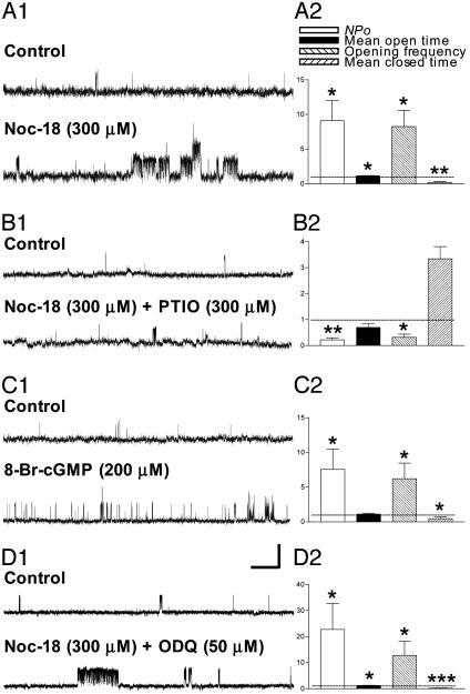
Stimulation of Kir6.2/SUR2B KATP Single-Channel Activity in HEK293 Cells by NO Donor Noc-18 Is Blocked by PTIO, a NO Scavenger. Single-channel activities were monitored continuously in cell-attached patches. Single-channel openings, most frequently at a conductance level around 63 pS (between -30 and -100 mV), appear as upward deflections (Fig. 1). Bath perfusion of the NO donor Noc-18 caused Kir6.2/SUR2B channel activities to increase over the course of several minutes. Because this stimulation could not be fully reversed by washout, we compared channel activities at the plateau of Noc-18 effect with controls obtained right before application of Noc-18. In contrast to the infrequent and brief channel openings in the control, the single-channel currents increased after Noc-18 treatment, resulting in simultaneous openings of three channels in this patch (Fig. 1 A1). Whereas the single-channel conductance level remained unaltered, Noc-18 treatment produced statistically significant changes in the single-channel properties: the open probability (NPo), opening frequency, and the mean open time increased whereas mean closed time decreased (five patches; Fig. 1 A2). The normalized fold changes (control as 1) are 9.11 ± 2.90 (P < 0.05), 8.26 ± 2.36 (P < 0.05), 1.14 ± 0.05 (P < 0.05), and 0.22 ± 0.11 (P < 0.001) for NPo, opening frequency, mean open time, and mean closed time, respectively (Fig. 1 A2). To determine whether the effect of Noc-18 was due to release of NO, we applied PTIO, a NO scavenger, together with Noc-18. Not only did coapplication of PTIO abolish the Kir6.2/SUR2B channel stimulation by Noc-18 (Fig. 1_B1_), but it also reduced NPo and opening frequency compared with controls obtained before the drug treatment (Fig. 1_B2_, four to six patches), probably because of the removal of endogenous NO by PTIO. These data indicate that NO released from NO donor Noc-18 significantly increases the single-channel activity of recombinant Kir6.2/SUR2B channels. In contrast, the NO effects on Kir6.2ΔC36 expressed in the absence of SUR (21) were much more variable, and only the opening frequency was significantly increased (4.89 ± 2.00, P < 0.05; nine patches; data not shown) by Noc-18, which implies a weaker effect of NO on the Kir6.2 subunit compared with Kir6.2/SUR2B. In addition, when Noc-18 was applied to the excised, inside-out membrane patches expressing Kir6.2/SUR2B channels, Noc-18 failed to stimulate channel activities (data not shown), indicating that the NO effects on Kir6.2/SUR2B channels are unlikely to result from direct channel modification.
Fig. 1.
Stimulation of Kir6.2/SUR2B channel function by NO does not require GC activity. (_A1_-D1) Single-channel current traces before (upper trace, control) and after (lower trace) bath perfusion of NO donor Noc-18 (300 μM) (A1), Noc-18 (300 μM) plus PTIO (300 μM) (B1), 8-Br-cGMP (200 μM) (C1), and Noc-18 (300 μM) plus ODQ (50 μM) (D1). Each pair of current traces was obtained from the same cell-attached patch from transfected HEK293 cells. Membrane potential was clamped at -60 mV. In this and all other figures, the scale bars represent 1 s and 4 pA. (A2-D2) Normalized fold changes of NPo, corrected mean open time, opening frequency, and mean closed time resulting from Noc-18 (five patches) (A2), Noc-18 plus PTIO (four to six patches) (B2), 8-Br-cGMP (seven patches) (C2), and Noc-18 plus ODQ (eight patches) (D2). Data were normalized as fractions of their corresponding controls obtained in individual patches (taken as 1, as indicated by dotted lines) and averaged, and are presented as mean ± SEM. *, P < 0.05; **, P < 0.001; ***, P < 0.0001.
KATP Channel Stimulation by Noc-18 Is Not Blocked by the GC Inhibitor. Some of the physiological actions of NO are thought to be mediated by cGMP because of the activation of GC (22, 23). Several studies have shown that KATP channel activity is either decreased (24) or increased (25) by the cGMP-dependent phosphorylation process. To test whether NO modulates Kir6.2/SUR2B channels via a cGMP-dependent pathway, we first applied 8-Br-cGMP, a membrane-permeable cGMP analog, and found that the single-channel activities of Kir6.2/SUR2B channels in cell-attached patches were stimulated (Figs. 1_C1_; seven patches in Fig. 1_C2_). We then examined whether KATP stimulation by Noc-18 was blocked by ODQ, a potent and selective inhibitor of GC. This GC inhibitor did not abolish NO stimulation (Fig. 1_D1_); NPo, opening frequency, and open time were all significantly increased (P < 0.05), whereas the mean closed time was decreased (P < 0.0001) (Fig. 1_D2_; eight patches). Thus, NO stimulation of Kir6.2/SUR2B channel activities appears to be mediated primarily via a pathway independent of GC activity; the GC pathway is not required.
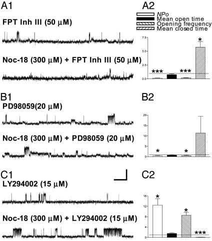
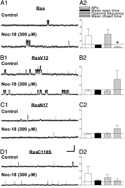
KATP Channel Stimulation by NO Is Diminished by Inhibition of Ras or MEK. To test whether NO stimulation of KATP channels was mediated by means of Ras, we examined the effects of FPT inhibitor III (FPT Inh III), a cell-permeable inhibitor of farnesyltransferase that is required for Ras processing (farnesylation) and plasma membrane association. Coapplication of Noc-18 and FPT Inh III after FPT Inh III pretreatment caused channel inhibition rather than stimulation (Fig. 2_A1_; five to seven patches in Fig. 2 A2). Another way to disrupt Ras function is to express RasN17, a dominant-negative form of Ras (26). Indeed, the Kir6.2/SUR2B single-channel activities in cells coexpressing RasN17 were not increased by Noc-18 (Fig. 3_C1_; four patches in Fig. 3_C2_). These data indicate that Ras activity is required for NO-mediated stimulation of Kir6.2/SUR2B channels.
Fig. 2.
Stimulation of Kir6.2/SUR2B channel function by NO donor Noc-18 requires Ras and MEK but not PI3-kinase activity. (A1-C1) Single-channel current traces before (control) and after perfusion of Noc-18 (300 μM) plus FPT inhibitor III (30 μM) (A1), Noc-18 (300 μM) plus PD98059 (20 μM) (B1), and Noc-18 (300 μM) plus LY294002 (15 μM) (C1). Cells were preincubated with the inhibitors for at least 15 min (A-C) before recordings. The openings of smaller conductance in Fig. 2_B1_ (and also Fig. 4_C1_) are endogenous, non-KATP channel activities that occasionally show in HEK cells. (A2-C2) Normalized fold changes of single-channel opening and closing properties resulting from Noc-18 plus FPT inhibitor III (five to seven patches) (A2), Noc-18 plus PD98059 (four patches) (B2), and Noc-18 plus LY294002 (four patches) (C2). Data were normalized as described in Fig. 1. *, P < 0.05; ***, P < 0.0001.
Fig. 3.
Enhancement of Kir6.2/SUR2B channel function by NO donor Noc-18 is abolished by coexpression of Ras mutants. (A1-D1) Single-channel current traces in individual cell-attached patches recorded before and after bath perfusion of Noc-18 (300 μM) when wild-type Ras (A1), RasV12 (B1), RasN17 (C1), or RasC118S (D1) was coexpressed. (A2-D2) Normalized fold changes of single-channel opening and closing properties resulting from Noc-18 treatment when wild-type Ras (three patches) (A2), RasV12 (five patches) (B2), RasN17 (four patches) (C2), or RasC118S (four patches) (D2) was coexpressed. Data were normalized and compared as described for Fig. 1. *, P < 0.05.
How might NO activate Ras to stimulate KATP? Ras can be modified directly by NO through S-nitrosylation at Cys-118 of Ras (27) by promoting the guanine nucleotide exchange of Ras (28). Coexpression of RasC118S with the channel prevented KATP channel stimulation by Noc-18 (Fig. 3_D1_; four patches in Fig. 3_D2_). How might Ras stimulate KATP? One of the signaling cascades downstream of Ras is the MAPK pathway (29). We tested the involvement of MAPK cascade by treating cells with PD98059, a selective and cell-permeable inhibitor of MEK. The kinase inhibitor caused KATP channels to be inhibited rather than stimulated by Noc-18 (Fig. 2_B1_; four patches in Fig. 2_B2_). Thus, like inhibition of Ras by FPT inhibitor III (Fig. 2 A2), inhibition of MEK by PD98059 unmasked an inhibitory effect of NO. Multiple MEK/MAPK pathways may exist in these cells; whereas NO stimulates the MEK/MAPK pathway that activates Kir6.2/SUR2B channels, other MEK/MAPK pathway(s) may mediate channel inhibition. Precedence for multiple MAPK pathways with shared and specific kinase components has been established in yeast (30).
NO Stimulation of SUR2B/Kir6.2 Channels Is Not Abolished by Blockade of PI3-Kinase Activity. Besides the MEK pathway, Ras activates PI3-kinase in antiapoptotic signaling (31). Treatment with LY294002, a specific PI3-kinase inhibitor, at room temperature for at least 15 min did not prevent channel stimulation by subsequent coapplication of Noc-18 and LY294002 (Fig. 2_C1_). The normalized fold changes are 12.49 ± 2.50 (P < 0.05), 8.59 ± 1.15 (P < 0.05), 1.54 ± 0.24, and 0.11 ± 0.01 (P < 0.0001) for NPo, opening frequency, mean open time, and mean closed time (Fig. 2_C2_; four patches), indicating that NO stimulation of Kir6.2/SUR2B channels does not require PI3-kinase.
Coexpression of Constitutively Active Ras Results in Elevated Activities of Kir6.2/SUR2B Channels and Occludes the NO Stimulation. Coexpression of RasV12, an activated form of Ras (32), with Kir6.2/SUR2B channels significantly raised the basal channel activity compared with cells expressing only the channels (Table 1, which is published as supporting information on the PNAS web site). Moreover, RasV12 coexpression prevented further stimulation of channel activities by NO donor (Fig. 3 B1 and B2; five patches). These findings reinforce the notion that NO stimulation of KATP is mediated by Ras. See Supporting Results, which is published as supporting information on the PNAS web site, for details.
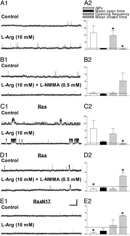
KATP Stimulation by l-Arg, Precursor of Endogenously Produced NO, Is Blocked by the NOS Inhibitor l-NMMA. NOS catalyzes the oxidation of l-arginine (l-Arg) to l-citrulline, leading to the generation of NO. Bath perfusion of l-Arg increased Kir6.2/SUR2B single-channel activity (Fig. 4_A1_). The opening frequency was significantly increased, whereas the mean closed time was reduced (Fig. 4_A2_)(P < 0.05 for both; two to five patches). To determine the specificity of such effects, we applied a competitive NOS inhibitor, l-NMMA, together with l-Arg. l-NMMA blocked the stimulatory effect of l-Arg on Kir6.2/SUR2B (Figs. 4_B1_; four patches in Fig. 4_B2_). Similar stimulatory effects of l-Arg were evident if the cells were transfected with cDNAs encoding Kir6.2, SUR2B, and wild-type Ras, although only the change of mean closed time reached statistical significance (Fig. 4_C1_; five to six patches in Fig. 4_C2_). No such trend of current increase was found when l-Arg was coapplied with l-NMMA to patches coexpressing Kir6.2, SUR2B, and Ras (Fig. 4_D1_). Instead, the NOS inhibitor reduced channel activity by decreasing NPo and lengthening the mean closed time (P < 0.05; three patches; Fig. 4_D2_). Consistent with our experiments with the NO scavenger PTIO (Fig. 1_B_), these data provide further evidence that NO generated by endogenous NOS stimulates KATP channels. Although HEK293 cells may not express a high amount of endogenous NOS, apparently it was sufficient for l-Arg to induce NO production and stimulate KATP in our experiments.
Fig. 4.
NO generated by endogenous NOS stimulates Kir6.2/SUR2B channels. (A1 and B1) Single-channel current traces in cell-attached patches recorded before and after bath perfusion of l-Arg (10 mM) (A1), or l-Arg (10 mM) plus l-NMMA (0.5 mM) (B1) without Ras coexpression. (C1-E1) Single-channel current traces recorded before and after l-Arg (C1), l-Arg plus l-NMMA (D1) when wild-type Ras was coexpressed, or before and after l-Arg (E1) when RasN17 was coexpressed. (A2 and B2) Normalized fold changes of single-channel opening and closing properties resulting from l-Arg (two to five patches) (A2), or l-Arg plus l-NMMA (four patches) (B2) without Ras coexpression. (C2-E2) Normalized fold changes of single-channel properties resulting from l-Arg (five to six patches) (C2), or l-Arg plus l-NMMA (three patches) (D2) when wild-type Ras was coexpressed, or before and after l-Arg (E2) when RasN17 was coexpressed (two to five patches). *, P < 0.05.
l-Arg, When Applied to Cells Coexpressing RasN17 and KATP Channels, Fails to Increase the Channel Activities. To test whether Ras mediates channel stimulation by NO produced by endogenous NOS, we examined the effect of dominant-negative Ras. When l-Arg was applied to the cells expressing not only Kir6.2 and SUR2B subunits but also RasN17, there was no increase of the channel function by l-Arg (Fig. 4_E1_; two to five patches in Fig. 4_E2_), indicating that KATP stimulation by endogenous NO relies on the Ras signaling pathway. Moreover, blockade of Ras activation by RasN17 revealed an inhibitory effect of NO, decreasing NPo and prolonging the mean closed time (P < 0.05), similar to the Noc-18-induced channel inhibition that was unmasked by inhibitor of Ras farnesylation (FPT inhibitor III) or by MEK inhibitor (PD98059) (Fig. 2 A and B). These findings indicate that Ras/MEK activation by NO overrides or overshadows a Ras-independent pathway for channel inhibition by NO and causes NO to increase channel openings severalfold.
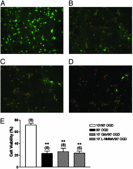
Blockade of KATP Activity Reduces Cell Viability in Late Neuronal IPC. Neurons may be protected from the lethal effects of prolonged ischemia by preceding brief periods of ischemia. The protective effects have been observed both early, within a few hours of IPC (early IPC), and late, days after IPC (late IPC) (33). This neuronal IPC can be modeled in vitro by OGD. In cortical neurons, OGD preconditioning induces Ras activation in an _N_-methyl-d-aspartate- and NO-dependent manner; moreover, ERK cascade downstream of Ras is required for late IPC (15). Given that many components of the signaling pathway for neuronal late IPC are also used during NO stimulation of KATP, we wondered whether the KATP channel plays a role in neuronal IPC. To test this, we treated primarily cultured hippocampal neurons from rats with glibenclamide (5 μM), a KATP channel inhibitor, during the short period of OGD and assayed the cell viability after a prolonged ischemia (see Materials and Methods). Exposure to a 10-min OGD given 24 h earlier (first OGD) greatly enhanced survival of hippocampal neurons after a 90-min OGD assault (second OGD) (normalized cell viability rate 71.80 ± 3.13%, n = 8; Fig. 5 A and E) compared with control without prior brief exposure to OGD (23.45 ± 3.84%, n = 8; Fig. 5 B and E) (P < 0.001). This protection requires KATP as well as NOS activity: the cell viability was significantly reduced by glibenclamide (26.33 ± 5.68%, n = 8; Fig. 5 C and E) (P < 0.001) or l-NMMA (23.75 ± 3.49%, n = 6; Fig. 5 D and E) (P < 0.001) applied during first OGD. Thus, NO stimulation of KATP in hippocampal neurons may promote neuronal survival in OGD preconditioning.
Fig. 5.
The protective effects of OGD preconditioning in hippocampal neurons is NO- and KATP-dependent. (A-D) Images of primary hippocampal neurons obtained after OGD treatments. Ten-minute OGD (preconditioning) given 24 hr before the lethal 90-min OGD (10′/90′ OGD) resulted in predominant cell survival (A), whereas 90-min OGD alone (90′ OGD) greatly reduced the proportion of viable cells (B). Blockade of KATP channel activity by glibenclamide (5 μM) (10′ Glib/90′ OGD) (C), or inhibition of NOS by l-NMMA (1 mM) (10′ l-NMMA/90′ OGD) (D) added at least 15 min before as well as during the 10-min OGD abolished the protective effect of OGD preconditioning and thus increased the number of dead cells. Cell viability was determined 24 hr after the last OGD, with viable cells stained green and dead cells stained red. (E) Normalized cell viability of neurons. Data obtained from six to eight experiments are presented as mean ± SEM. **, P < 0.001. Total neurons examined number 5,000-9,000 for each group.
Discussion
Both NO and KATP have been implicated in neuroprotection and blood flow regulation, raising the intriguing question of whether NO exerts its protective functions partly by modulating KATP channels. Our observations reveal that KATP is stimulated via the Ras signaling pathway in response to NO derived from either chemical NO donors or the natural NOS substrate l-Arg. Like Ras, the MEK in the Ras pathway is necessary for KATP channel activation by NO, but not PI3-kinase or GC. Hypoxia or ischemia may induce the production of NO. Our findings that the protective effect of IPC in hippocampal neurons was abolished by inhibition of KATP or NOS raise the possibility that NO stimulation of KATP channels via Ras/MAPK cascade could cause hyperpolarization, thereby increasing blood flow, reducing neuronal excitability, and protecting neurons against excitotoxicity, and implicate NO modulation of KATP in the neuroprotection afforded by late ischemic preconditioning. The mechanistic link between MAPK activation and KATP stimulation remains to be determined.
NO Enhances KATP Channel Activities. Application of the NO donor Noc-18, or SNAP (data not shown), or natural NOS substrate l-Arg increased Kir6.2/SUR2B channel activities. Whereas the NO effect on Ras or hemoglobin is reversible (28, 34), KATP stimulation by Noc-18 could not be easily reversed by washout, similar to the irreversible NO stimulation of the large-conductance Ca2+-activated potassium (BKCa) channels (35). NO may activate the BKCa channel either directly (36, 37) or indirectly by means of cGMP-dependent protein kinase (38). We have found no evidence for direct activation of KATP channels by NO; NO application to excised patches had no stimulatory effects on Kir6.2/SUR2B channels (data not shown). The slow time course of KATP channel stimulation would be consistent with the involvement of a multistep pathway downstream of NO.
Stimulation of KATP Channels Requires Ras and MEK/MAPK but Not PI3-Kinase or GC. Previously it had been shown that the KATP channel is regulated by a cGMP-dependent protein kinase-signaling pathway (24, 25, 39). In this study, although the Kir6.2/SUR2B channel is stimulated by a cGMP-dependent process, the action of NO to stimulate the channel does not require it. In line with this, Shinbo and Iijima (40) have suggested that NO-mediated potentiation of KATP activated by K+ channel openers is independent of the cGMP/cGMP-dependent protein kinase-signaling pathway in the heart. NO can modify proteins involved in the signaling mechanism by chemical reaction such as S-nitrosylation other than by means of activation of GC (41). Indeed, our data on RasC118S (Fig. 3_D_) suggest that the NO effects on the KATP channel require activation of Ras by means of S-nitrosylation. Downstream of Ras, the MAPK pathway rather than the PI3-kinase pathway is responsible for mediating NO modulation of KATP channels (Fig. 2 B and C). We also found that NO stimulation of KATP channel activity may require new protein synthesis (data not shown). Given the many evolutionarily conserved functions of MAPK, including the phosphorylation of transcription factors (42, 43), NO stimulation of KATP could conceivably result from altered transcription and translation or phosphorylation of either the channel proteins or other regulatory proteins, which remains to be determined. It is also worth noting that inhibition of the Ras/MEK signaling pathway unmasked an inhibitory effect of NO. This channel inhibition is not likely to be mediated by cGMP, because 8-Br-cGMP has a stimulatory effect on these channels. It is possible that NO activates KATP via the Ras pathway but inhibits KATP channels via a Ras-independent pathway, resulting in a net stimulation. Alternatively, Ras/MEK activation by NO may not only activate KATP but also suppress the ability of NO to inhibit KATP via an as yet unknown pathway. It would be of interest to explore conditions that differentially modulate the different NO effects on KATP channels.
NO and KATP Channels in the Brain: Role of KATP in Neuronal IPC. KATP channel activation may contribute to changes in neuronal excitability and protection of neurons from cell death caused by ischemia, hypoxia, or metabolic stress, by inducing membrane hyperpolarization (44). Indeed, pretreatment of rat neocortical brain slices with KATP openers is neuroprotective, counteracting hypoxia-induced cell injury (45). Studies of KATP channels in midbrain dopaminergic neurons in weaver mice further indicate that these channels may be protective against neurodegeneration (46).
The late preconditioning in the heart is mediated by increased synthesis of NO by inducible NOS (47). The mechanism whereby NO protects during late preconditioning remains speculative. In the brain, IPC protection against lethal ischemia is mediated largely by the activation of _N_-methyl-d-aspartate receptors through increases in intracellular Ca2+ (48). The activation of adenosine A1 receptors, PKC, or KATP channels may also be involved (49, 50). KATP channels may be involved in neuroprotection afforded by anoxic preconditioning in hippocampal slices (50). OGD preconditioning of cultured cortical neurons requires Ras and MAPK/ERK activation, which is induced in an _N_-methyl-d-aspartate receptor- and NO-dependent, but cGMP-independent, manner (15). In this study, we demonstrate that both KATP channel activation and NO generation are required for OGD preconditioning of rat hippocampal neurons (Fig. 5), implicating NO and KATP in ischemic preconditioning in vivo. One potential pathway of neuronal IPC may involve NO activation of KATP via the Ras/MAPK signaling cascade; follow-up studies are required to elucidate the detailed mechanism in neurons.
NO has been implicated in neuronal signaling and synaptic plasticity (6, 7). The Ras/MAPK pathway underlying NO-mediated enhancement of KATP channel function may play important roles in mediating or modulating activity-induced changes such as neuronal differentiation, synaptic strength, and neuronal survival, as well as neuroprotection in ischemic preconditioning. Mechanistic studies of NO stimulation of KATP may also facilitate treatments for ischemic or traumatic injury and neurodegenerative diseases.
Supplementary Material
Supporting Information
Acknowledgments
We thank Drs. Susumu Seino and Joseph Bryan for the kind gifts of cDNA clones of rat SUR2B and mouse Kir6.2. We are grateful to Dr. Barry Pallotta for the single-channel analysis program intrv5. Preparation of DNA samples was kindly assisted by Tong Chen and Sharon Fried. Y.N.J. and L.Y.J. are Howard Hughes Medical Institute investigators. This study was supported by National Institute of Mental Health Grant MH65334.
Abbreviations: ERK, extracellular signal-regulated kinase; FPT, farnesyltransferase; GC, guanylyl cyclase; HEK, human embryonic kidney cell; IPC, ischemic preconditioning; KATP, ATP-sensitive potassium channel; l-NMMA, _N_G-monomethyl-l-arginine, monoacetate salt; MAPK, mitogen-activated protein kinase; MEK, mitogen-activated protein kinase kinase; NOS, NO synthase; NPo, open probability; ODQ, 1_H_-[1,2,4]oxadiazolo[4,3-_a_]quinoxalin-1-one; OGD, oxygen-glucose deprivation; PI3-kinase, phosphatidylinositol 3-kinase; PTIO, 2-phenyl-4,4,5,5-tetramethylimidazoline-1-oxyl-3-oxide; SUR, sulfonylurea receptor.
References
- 1.Seino, S. & Miki, T. (2003) Prog. Biophys. Mol. Biol. 81**,** 133-176. [DOI] [PubMed] [Google Scholar]
- 2.Seino, S. & Miki, T. (2004) J. Physiol. 554**,** 295-300. [DOI] [PMC free article] [PubMed] [Google Scholar]
- 3.Stamler, J. S., Singel, D. J. & Loscalzo, J. (1992) Science 258**,** 1898-1902. [DOI] [PubMed] [Google Scholar]
- 4.Gross, S. S. & Wolin, M. S. (1995) Annu. Rev. Physiol. 57**,** 737-769. [DOI] [PubMed] [Google Scholar]
- 5.Palmer, R. M. J., Ferrige, A. G. & Moncada, S. (1987) Nature 327**,** 524-526. [DOI] [PubMed] [Google Scholar]
- 6.Fang, M., Jaffrey, S. R., Sawa, A., Ye, K., Luo, X. & Snyder, S. H. (2000) Neuron 28**,** 183-193. [DOI] [PubMed] [Google Scholar]
- 7.Schmidt, H. H. H. W. & Walter, U. (1994) Cell 78**,** 919-925. [DOI] [PubMed] [Google Scholar]
- 8.Barouch, L. A., Harrison, R. W., Skaf, M. W., Rosas, G. O., Cappola, T. P., Kobeissi, Z. A., Hobai, I. A., Lemmon, C. A., Burnett, A. L., O'Rourke, B., et al. (2002) Nature 416**,** 337-339. [DOI] [PubMed] [Google Scholar]
- 9.Ignarro, L. J. (1991) Blood Vessels 28**,** 67-73. [DOI] [PubMed] [Google Scholar]
- 10.Lander, H. M., Hajjar, D. P., Hempstead, B. L., Mirza, U. A., Chait, B. T., Campbell, S. & Quilliam, L. A. (1997) J. Biol. Chem. 272**,** 4323-4326. [DOI] [PubMed] [Google Scholar]
- 11.Satoh, T., Nakafuku, M. & Kaziro, Y. (1992) J. Biol. Chem. 267**,** 24149-24152. [PubMed] [Google Scholar]
- 12.Bos, J. L. (1998) EMBO J. 17**,** 6776-6782. [DOI] [PMC free article] [PubMed] [Google Scholar]
- 13.Rodriguez-Viciana, P., Warne, P. H., Vanhaesebroeck, B., Waterfield, M. D. & Downward, J. (1996) EMBO J. 15**,** 2442-2451. [PMC free article] [PubMed] [Google Scholar]
- 14.Burgering, B. M. T. & Bos, J. L. (1995) Trends Biochem. Sci. 20**,** 18-22. [DOI] [PubMed] [Google Scholar]
- 15.Gonzalez-Zulueta, M., Feldman, A. B., Klesse, L. J., Kalb, R. G., Dillman, J. F., Parada, L. F., Dawson, T. M. & Dawson, V. L. (2000) Proc. Natl. Acad. Sci. USA 97**,** 436-441. [DOI] [PMC free article] [PubMed] [Google Scholar]
- 16.Shamloo, M., Rytter, A. & Wieloch, T. (1999) Neuroscience 93**,** 81-88. [DOI] [PubMed] [Google Scholar]
- 17.Maulik, N., Watanabe, M., Zu, Y. L., Huang, C. K., Cordis, G. A., Schley, J. A. & Das, D. K. (1996) FEBS Lett. 396**,** 233-237. [DOI] [PubMed] [Google Scholar]
- 18.Lin, Y. F., Jan, Y. N. & Jan, L. Y. (2000) EMBO J. 19**,** 942-955. [DOI] [PMC free article] [PubMed] [Google Scholar]
- 19.Hamill, O. P., Marty, A., Neher, E., Sakmann, B. & Sigworth, F. J. (1981) Pflügers Arch. 391**,** 85-100. [DOI] [PubMed] [Google Scholar]
- 20.Ma, D., Zerangue, D., Raab-Graham, K., Fried, S. R., Jan, Y. N. & Jan, L. Y. (2002) Neuron 33**,** 715-729. [DOI] [PubMed] [Google Scholar]
- 21.Tucker, S. J., Gribble, F. M., Zhao, C., Trapp, S. & Ashcroft, F. M. (1997) Nature 387**,** 179-183. [DOI] [PubMed] [Google Scholar]
- 22.Kerwin, J. F., Jr., Lancaster, J. R. & Feldman, P. L. (1995) J. Med. Chem. 38**,** 4342-4362. [DOI] [PubMed] [Google Scholar]
- 23.Lohse, M. J., Forstermann, U. & Schmidt, H. H. H. W. (1998) Naunyn-Schmiedeberg's Arch. Pharmacol. 358**,** 111-112. [DOI] [PubMed] [Google Scholar]
- 24.Ropero, A. B., Fuentes, E., Rovira, J. M., Ripoll, C., Soria, B. & Nadal, A. (1999) J. Physiol. 521**,** 397-407. [DOI] [PMC free article] [PubMed] [Google Scholar]
- 25.Han, J., Kim, N., Kim, E., Ho, W. K. & Earm, Y. E. (2001) J. Biol. Chem. 276**,** 22140-22147. [DOI] [PubMed] [Google Scholar]
- 26.Feig, L. A. & Cooper, G. M. (1988) Mol. Cell. Biol. 8**,** 3235-3243. [DOI] [PMC free article] [PubMed] [Google Scholar]
- 27.Kim, P. K., Kwon, Y. G., Chung, H. T. & Kim, Y. M. (2002) Ann. N.Y. Acad. Sci. 962**,** 42-52. [DOI] [PubMed] [Google Scholar]
- 28.Lander, H. M., Ogiste, J. S., Pearce, S. F. A., Levi, R. & Novogrodsky, A. (1995) J. Biol. Chem. 270**,** 7017-7020. [DOI] [PubMed] [Google Scholar]
- 29.Mazzucchelli, C. & Brambilla, R. (2000) Cell. Mol. Life Sci. 57**,** 604-611. [DOI] [PMC free article] [PubMed] [Google Scholar]
- 30.Madhani, H. D. & Fink, G. R. (1998) Trends Genet. 14**,** 151-155. [DOI] [PubMed] [Google Scholar]
- 31.Dudek, H., Datta, S. R., Franke, T. F., Birnbaum, M. J., Yao, R., Cooper, G. M., Segal, R. A., Kaplan, D. R. & Greenberg, M. E. (1997) Science 275**,** 661-665. [DOI] [PubMed] [Google Scholar]
- 32.Joneson, T., White, M. A., Wigler, M. H. & Bar-Sagi, D. (1996) Science 271**,** 810-812. [DOI] [PubMed] [Google Scholar]
- 33.Takano, H., Tang, X.-L. & Bolli, R. (2000) Am. J. Physiol. 279**,** H2350-H2359. [DOI] [PubMed] [Google Scholar]
- 34.Stamler, J. S. (1994) Cell 78**,** 931-936. [DOI] [PubMed] [Google Scholar]
- 35.Ahern, G. P., Hsu, S.-F. & Jackson, M. B. (1999) J. Physiol. 520**,** 165-176. [DOI] [PMC free article] [PubMed] [Google Scholar]
- 36.Bolotina, V. M., Najibi, S., Paracino, J. J., Pagano, P. J. & Cohen, R. A. (1994) Nature 368**,** 850-853. [DOI] [PubMed] [Google Scholar]
- 37.Miyoshi, H. & Nakaya, Y. (1994) J. Mol. Cell. Cardiol. 26**,** 1487-1495. [DOI] [PubMed] [Google Scholar]
- 38.Robertson, B. E., Schubert, R., Hescheler, J. & Nelson, M. T. (1993) Am. J. Physiol. 265**,** C299-C303. [DOI] [PubMed] [Google Scholar]
- 39.Sachs, D., Cunha, F. Q. & Ferreira, S. H. (2004) Proc. Natl. Acad. Sci. USA 101**,** 3680-3685. [DOI] [PMC free article] [PubMed] [Google Scholar]
- 40.Shinbo, A. & Iijima, T. (1997) Br. J. Pharmacol. 120**,** 1568-1574. [DOI] [PMC free article] [PubMed] [Google Scholar]
- 41.Ahern, G. P., Klyachko, V. A. & Jackson, M. B. (2002) Trends Neurosci. 25**,** 510-517. [DOI] [PubMed] [Google Scholar]
- 42.Böhm, M., Schröder, H. C., Müller, I. M., Müller, W. E. & Gamulin, V. (2000) Biol. Cell. 92**,** 95-104. [DOI] [PubMed] [Google Scholar]
- 43.Baker, D. A., Mille-Baker, B., Wainwright, S. M., Ish-Horowicz, D. & Dibb, N. J. (2001) Nature 411**,** 330-334. [DOI] [PubMed] [Google Scholar]
- 44.Hogg, R. C. & Adams, D. J. (2001) J. Physiol. 534**,** 713-720. [DOI] [PMC free article] [PubMed] [Google Scholar]
- 45.Garcia de Arriba, S., Franke, H., Pissarek, M., Nieber, K. & Illes, P. (1999) Neurosci. Lett. 273**,** 13-16. [DOI] [PubMed] [Google Scholar]
- 46.Liss, B., Neu, A. & Roeper, J. (1999) J. Neurosci. 19**,** 8839-8848. [DOI] [PMC free article] [PubMed] [Google Scholar]
- 47.Bolli, R., Manchikalapudi, S., Tang, X. L., Takano, H., Qiu, Y., Guo, Y., Zhang, Q. & Jadoon, A. K. (1997) Circ. Res. 81**,** 1094-1107. [DOI] [PubMed] [Google Scholar]
- 48.Grabb, M. C. & Choi, D. W. (1999) J. Neurosci. 19**,** 1657-1662. [DOI] [PMC free article] [PubMed] [Google Scholar]
- 49.Heurteaux, C., Lauritzen, I., Widmann, C. & Lazdunski, M. (1995) Proc. Natl. Acad. Sci. USA 92**,** 4666-4670. [DOI] [PMC free article] [PubMed] [Google Scholar]
- 50.Pérez-Pinzón, M. A. & Born, J. G. (1999) Neuroscience 89**,** 453-459. [DOI] [PubMed] [Google Scholar]
Associated Data
This section collects any data citations, data availability statements, or supplementary materials included in this article.
Supplementary Materials
Supporting Information
