Vibrio fischeri lux Genes Play an Important Role in Colonization and Development of the Host Light Organ (original) (raw)
Abstract
The bioluminescent bacterium Vibrio fischeri and juveniles of the squid Euprymna scolopes specifically recognize and respond to one another during the formation of a persistent colonization within the host's nascent light-emitting organ. The resulting fully developed light organ contains brightly luminescing bacteria and has undergone a bacterium-induced program of tissue differentiation, one component of which is a swelling of the epithelial cells that line the symbiont-containing crypts. While the luminescence (lux) genes of symbiotic V. fischeri have been shown to be highly induced within the crypts, the role of these genes in the initiation and persistence of the symbiosis has not been rigorously examined. We have constructed and examined three mutants (luxA, luxI, and luxR), defective in either luciferase enzymatic or regulatory proteins. All three are unable to induce normal luminescence levels in the host and, 2 days after initiating the association, had a three- to fourfold defect in the extent of colonization. Surprisingly, these lux mutants also were unable to induce swelling in the crypt epithelial cells. Complementing, in trans, the defect in light emission restored both normal colonization capability and induction of swelling. We hypothesize that a diminished level of oxygen consumption by a luciferase-deficient symbiotic population is responsible for the reduced fitness of lux mutants in the light organ crypts. This study is the first to show that the capacity for bioluminescence is critical for normal cell-cell interactions between a bacterium and its animal host and presents the first examples of V. fischeri genes that affect normal host tissue development.
Bioluminescent bacteria are commonly found associated with marine animal tissues, as members of the enteric consortia, as opportunistic pathogens, or most notably as essentially pure cultures colonizing the light-emitting organs of certain squids and fishes (43). In at least some of these light organ associations, normal development of host tissues requires the presence of their species-specific luminous bacterial symbionts (28), which are obtained from the surrounding seawater by the newly hatched host (41, 52). The importance of these light organs to antipredatory defense and other behaviors of the hosts has been well documented (27). In contrast, two questions that focus on the initiation and development of light organ symbioses have remained unanswered: (i) why is it that only certain strains of bacteria are able to colonize and persist in these associations, and (ii) what are the bacterial signals that induce host tissue differentiation? We report here that the capacity to bioluminescence plays a critical part in the answers to both of these questions.
While relatively little is known about the symbiotic significance of light emission, much has been published about the biochemistry and regulation of bioluminescence in luminous bacteria. Bacterial luminescence is a product of the enzyme luciferase, which uses molecular oxygen from the surrounding environment to oxidize both an aliphatic aldehyde and a reduced flavin mononucleotide (reviewed in reference 30). The final products of this reaction are the corresponding aliphatic acid, oxidized flavin, and water, and an unstable intermediate in the reaction emits a photon of blue-green light.
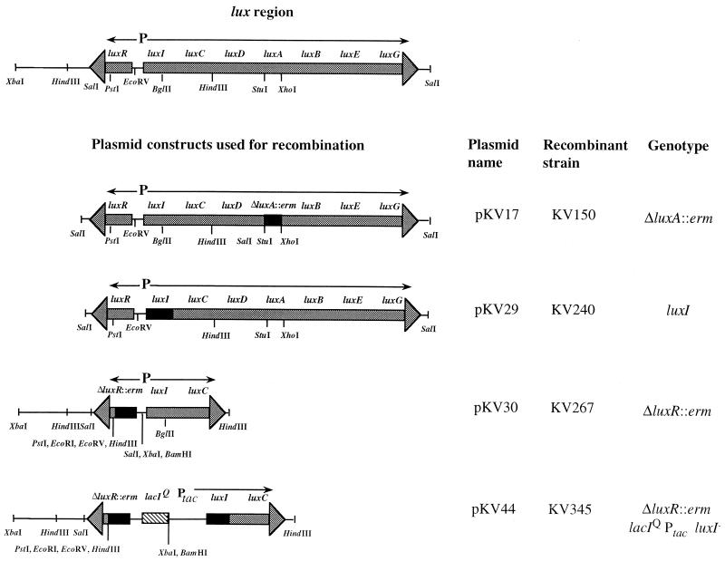
There are at least eight lux genes comprising a regulon that encodes the proteins essential for luminescence in the best-studied luminous bacterium, Vibrio fischeri (30). The lux operon (Fig. 1), encoding luciferase (luxAB) and proteins required to synthesize the aldehyde substrate (luxCDE), is controlled by a number of factors, most notably the quorum-sensing regulator LuxR (13) and the autoinducer molecule 3-oxohexanoyl l-homoserine lactone (3-oxo-C6-HSL), synthesized by LuxI (11, 20). V. fischeri cells also synthesize a second autoinducer molecule, octanoyl l-homoserine lactone (C8-HSL) that, under some conditions, may stimulate transcription of the lux genes (10, 14, 21).
FIG. 1.
Plasmids used for construction of lux mutants. The region of the chromosome containing the lux regulon is shown, with arrows demonstrating the direction of transcription of the two transcriptional units. Relevant restriction enzyme sites are indicated. Plasmid constructs that were used to make mutations in the chromosomal copy of the lux genes are also shown. Black boxes indicate the location of the mutation resulting from either a deletion and gene replacement with the erm gene or a frameshift mutation (see Materials and Methods); the box with diagonal stripes depicts the location of a lacI_q/P_tac cassette.
The process of quorum sensing, although first discovered in V. fischeri, is a widespread regulatory mechanism in gram-negative bacteria, particularly among a number of pathogens, which use various autoinducer molecules to modulate genes encoding virulence factors (5, 13, 37). To date, several organisms defective in their luxI homolog have been constructed, and they display a reduction in virulence (39, 47); the defect can be at least partially restored by exogenous addition of synthetic autoinducer. Such studies provide strong evidence that quorum sensing plays an important role in pathogenic bacterium-host interactions. While not a pathogen, V. fischeri produces a persistent, benign infection of specific light-emitting tissues of a number of species of squids and fishes (43). The best studied of these cooperative bacterium-host associations is that between V. fischeri and the Hawaiian sepiolid squid, Euprymna scolopes (reviewed in references 29 and 41). The bacteria reside as a monospecific culture in epithelial cell-lined crypts of the light organ, which is used in the host's nocturnal behavior (26).
Newly hatched juvenile squid are symbiont free and must acquire an inoculum of V. fischeri from the surrounding seawater (42, 52). As a result of colonization by V. fischeri, a number of morphological and biochemical changes are triggered in the nascent light organ, leading to the development of the functional adult structure (32, 33). Among these changes is a dramatic swelling of the epithelial cells that line the bacterium-containing crypts (8, 32). The swelling, as well as several other developmental events, does not occur in the absence of colonization by V. fischeri cells (32).
This study presents evidence that the bioluminescence of V. fischeri cells plays an important role during the development of a successful light organ association. Specifically, we have examined luxA, luxI, and luxR mutants of a symbiosis-competent strain of V. fischeri for their relative abilities to (i) colonize and persist in the squid light organ and (ii) induce normal host developmental morphogenesis. Our results indicate a direct role of bacterial luciferase and/or its bioluminescence activity in both the normal induction of host development and symbiont persistence in this cooperative bacterial association.
MATERIALS AND METHODS
Bacterial strains and media.
V. fischeri strain ESR1 (13), a rifampin-resistant derivative of wild-type strain ES114 (1), was used as the parent strain for all mutant constructions. Escherichia coli strain DH5α was used as the recipient for cloning experiments, and plasmids were passaged through a dam mutant E. coli strain (49) prior to introduction into V. fischeri.
V. fischeri strains were grown in either SWT (1), which contains 0.5% tryptone, 0.3% yeast extract, and 0.3% glycerol in 70% seawater, or LBS (9), which contains 1% (wt/vol) tryptone, 0.5% (wt/vol) yeast extract, 2% NaCl, and 0.3% (vol/vol) glycerol in 50 mM Tris-HCl (pH 7.5). E. coli strains were grown in LB (6) or Difco brain heart infusion medium. Conditioned medium (CM) was prepared as follows. V. harveyi strain B392, which removes inhibitory compounds from rich media without producing interfering autoinducers, was inoculated into 500 ml of luminous medium (34) (50 mM MgSO4, 10 mM CaCl2, 300 mM NaCl, 10 mM KCl, 333 μM K2HPO4, 18.7 mM NH4Cl, 0.5% yeast extract, 0.5% tryptone, and 0.3% glycerol in 50 mM Tris-HCl [pH 7.5]) and grown to an optical density at 600 nm (OD600) of approximately 0.8. Cells were removed from the spent medium by centrifugation and filtration through a 0.22-μm-pore-size membrane filter. Agar was added to a concentration of 1.5% to solidify media. Antibiotics and other supplements were added to media where appropriate to the following final concentrations: ampicillin, 100 μg/ml; erythromycin, 150 μg/ml for E. coli and 5 μg/ml for V. fischeri; chloramphenicol, 25 μg/ml for E. coli and 2.5 μg/ml for V. fischeri; isopropyl-β-d-thiogalactopyranoside (IPTG), 570 μg/ml; 3-oxo-C6-HSL, 200 to 600 ng/ml; and C8-HSL, 15 to 45 ng/ml.
Plasmid construction.
All cloning was performed using standard molecular biology techniques as follows. DNA fragments obtained by restriction digest were physically separated by agarose gel electrophoresis, and the desired fragments were extracted from gel slices using GeneClean (Bio101, Inc., Vista, Calif.). T4 DNA ligase was used to join two fragments together, the ligated fragments were transformed into DH5α cells made competent by CaCl2 treatment, and plasmid-carrying strains were isolated on selective media. Enzymes were obtained from either Promega (Madison, Wis.) or New England Biolabs (Beverly, Mass.). Plasmid pKV29 (Fig. 1) was constructed by insertion of the 8.8-kb _Sal_I fragment from pHV200I− (38) containing the lux genes from V. fischeri strain ES114 (17) into the _Sal_I site of pBC (Stratagene, La Jolla, Calif.). The luxI gene in that construct carries a 2-bp frameshift mutation near the end of the gene, rendering the protein product inactive (38). Because this strain regains luminescence activity with the addition of 3-oxo-C6-HSL, the product of LuxI (Table 1), the mutation does not have any significant polar effects on downstream lux genes.
TABLE 1.
Effects of autoinducer additions on the bioluminescence of V. fischeri ESR1 and its lux mutant derivatives
| Strain | Luminescence (quanta/s/cell at OD600 of 0.1–1.5) | Alteration of luminescence by the addition ofa: | |
|---|---|---|---|
| 3-oxo-C6-HSL | C8-HSL | ||
| ESR1 | 24 | 4,400 ± 1,400 | 2.4 ± 0.89 |
| KV150 (luxA) | <0.01 | <0.01 | <0.01 |
| KV240 (luxI) | 8.7 | 3,600 ± 1,500 | 1.7 ± 0.76 |
| KV267 (luxR) | 17 | 1.0 ± 0.11 | 0.74 ± 0.06 |
Plasmid pKV30 (Fig. 1) was constructed as follows. A 3-kb _Sal_I-_Hin_dIII DNA fragment carrying the luxR, luxI, and luxC genes was cloned into pUC19 (54) that had been digested with _Sal_I and _Hin_dIII. The _luxR_-encoding portion of the resulting plasmid, pVO13, was removed by a partial digestion with _Eco_RI and _Pst_I and replaced with the gene for erythromycin resistance (erm) contained on a 1.2-kb _Sma_I-_Pst_I fragment from pKV25 (50), resulting in pVO15. To facilitate the subsequent recombination of the luxR::erm mutation into the chromosome of V. fischeri, the region of the chromosome downstream of the luxR gene was cloned (K. Visick and E. G. Ruby, unpublished data). A _Pst_I-_Sal_I fragment carrying a portion of the luxR gene and approximately 2.5 kb of DNA downstream of luxR was cloned into pVO15 digested with _Pst_I and _Xba_I, resulting in pKV30 (Fig. 1).
Plasmid pKV44 (51) was obtained by first cloning a 1.6-kb _Xho_I fragment (carrying the _lacI_q gene and the tac promoter [P_tac_]) from WM2194 (7) into pKV30 digested with _Sal_I (Fig. 1). The plasmid construct was then digested with _Bgl_II, treated with T4 DNA polymerase to fill in the overhanging ends of the DNA, and self-ligated using T4 DNA ligase. In the resulting plasmid, pKV44, the luxR gene has a large deletion, the luxI gene is disrupted by a 2-bp frameshift mutation at the Bgl_II site, and transcription of luxCDABE is under the control of P_tac (Fig. 1). Construction of pKV19, which carries constitutively expressed copies of the wild-type luxA and luxB genes of V. harveyi, has been described previously (49).
Construction of V. fischeri mutants.
Construction of the V. fischeri lux mutants was begun by electroporation of the plasmid DNA carrying the lux mutation into strain ESR1, followed by antibiotic selection, as described previously for the luxA mutant (49). Strains that had undergone a single recombination event, i.e., had in their chromosome one wild-type and one mutant copy of either luxI or luxR, were identified by their increased levels of luminescence due to the carriage of duplicate copies of one of these regulatory genes. Strains that had undergone the second recombination event, i.e., had replaced the wild-type copy in the chromosome with the mutant luxI or luxR allele, were identified by a screen for decreased luminescence levels. The luxR mutant alleles retained erythromycin resistance, while the luxI mutant lost the chloramphenicol resistance encoded by the vector. Strain KV345 was similarly constructed using plasmid pKV44 (Fig. 1) as the donor DNA.
Southern analysis.
The putative mutations in strains KV240 (luxI), KV267 (luxR), and KV345 (luxR luxI P_tac_) were confirmed by Southern analysis (45) performed as previously described (49). Chromosomal DNA from either KV240 or its parent strain ESR1 was isolated and digested with _Sal_I, _Sal_I/_Bgl_II, and _Bgl_II. As expected, the _Sal_I fragment that carries the lux operon was the same in the two strains, but the _Sal_I/_Bgl_II and _Bgl_II fragments from KV240 were larger for the mutant strain, which carries a 2-bp frameshift at the _Bgl_II site. Chromosomal DNA from KV267 or ESR1 was digested with _Hin_dIII and _Sal_I. The lux probe-hybridizing fragment from the _Hin_dIII digest of KV267 DNA was approximately 1 kb shorter than that from the wild-type parent, due to the insertion of a _Hin_dIII site downstream of the erm cassette, and the _Sal_I lux fragment from KV267 was shorter by about 1 kb due to the insertion of a _Sal_I fragment upstream of the erm cassette. Similarly, the _Sal_I digests of chromosomal DNA from KV345 yielded fragments of lux DNA that were 1 kb larger than those obtained with the wild type, and the _Bam_HI fragment was approximately 1.8 kb larger than that of KV267, due to the insertion of the _lacI_q cassette.
Luminescence assays.
V. fischeri strains were grown in CM at 28°C in either the presence or absence of added autoinducer (either 3-oxo-C6-HSL or C8-HSL) (21). At approximately 30-min intervals, 1-ml aliquots were removed from the culture flask, and both bioluminescence and OD levels were quantified. The level of bioluminescence was measured with a Turner (Sunnyvale, Calif.) 20/20 luminometer.
Squid colonization assays.
Juveniles of E. scolopes were inoculated within 4 h of hatching as described previously (41) and exposed for at least 12 h either to single strains of V. fischeri or to pairs of competing strains. After this exposure, the animals were transferred to symbiont-free seawater and maintained for up to 3 days. At specified times after inoculation, the juveniles were homogenized, and dilutions of the homogenates were spread on SWT agar plates to determine the number of CFU in the homogenate. In mixed inoculation experiments, the medium contained erythromycin to distinguish between parent and lux mutant CFU. Luminescence measurements of juvenile squid colonized by either wild-type or lux mutant strains of V. fischeri were performed using either the Turner 20/20 luminometer or a scintillation counter modified to detect luminescence by single-photon counting.
Host developmental events.
Juveniles infected with wild-type or lux mutants were assayed for two of the benchmarks of squid development known to be triggered by the presence of the symbiotic bacteria: (i) apoptotic cell death and (ii) swelling of the epithelial cells lining the crypts of the light organ (32). Animals were exposed to V. fischeri cells and, at 14 h postinoculation, monitored for the normal pattern of apoptotic cell death. Briefly, the squid were first anesthetized in a 1:1 mixture of filter-sterilized seawater and 7.5% MgCl2 and then stained for 1 min in a solution containing 5 ng of acridine orange per ml of seawater. Following a ventral dissection, the exposed light organs were visualized by epifluorescence microscopy to determine whether the typical pattern of apoptotic cell death was evident.
For a determination of epithelial cell swelling, squid light organ tissues were prepared and analyzed with transmission electron microscopy (TEM) (31). Juvenile squid were placed in a 0.1 M sodium cacodylate–0.45 M NaCl buffer (pH 7.4) containing 2.5% glutaraldehyde and 2.5% paraformaldehyde and incubated for 12 h at 23°C. This fixation was followed with three 15-min washes with buffer alone, and the samples were then dehydrated in a graded ethanol series. Uranyl acetate (2%) was added to the 70% ethanol wash to increase contrast of the ultrathin sections. Next, the animals were infiltrated with 100% propylene oxide for 15 min and then placed in a 1:1 ratio of propylene oxide and unaccelerated Spurr embedding medium (46) for 30 min. After infiltration, specimens were left for 3 days in unaccelerated Spurr medium and then moved to accelerated Spurr medium for overnight incubation at 23°C. The animals were then embedded within freshly prepared accelerated Spurr medium at 62°C for 24 h. Ultrathin sections were stained first with a solution of 5% uranyl acetate for 10 min and then with a 0.3% solution of lead citrate for 5 min (31).
Volumetric measurements of crypt epithelial cells.
Stained ultrathin sections were visualized on a LEO 912 transmission electron microscope, and TEM images were captured with a Proscan-CCD frame transfer camera. The epithelial cells selected for measurement were taken from at least five different sections for each animal, all located deep in the interior of the crypt spaces. Previous studies have reported that the shape of crypt epithelial cells in juvenile squid is essentially columnar (8, 32); thus, we calculated whole-cell volumes from two-dimensional areas (height times width) by assuming that the width of the cell along the axis parallel to basal lamina is equal to its depth. The volume of the nucleus of each cell was similarly measured; unlike the case for total-cell volume (see below), no differences were observed in these values under any of the conditions reported in this study (data not shown). We therefore subtracted the nuclear volume from the whole-cell volume to obtain a calculated cytoplasmic volume.
Statistical analyses of cytoplasmic volumes.
Crypt cell cytoplasmic volumes were compared between groups of animals colonized by different bacterial strains. The volumes of each group were shown to be normally distributed, and a one-way analysis of variance was performed using a Tukey pairwise comparison at the 95% confidence level to determine whether significant differences in cell volumes occurred between the various groups.
RESULTS
Construction and luminescence phenotypes of V. fischeri lux mutants.
To investigate the role of luminescence in the light organ association of V. fischeri with E. scolopes, mutants defective either for luxA, encoding one of the subunits of luciferase, or for luxI or luxR, two regulatory genes controlling lux gene expression, were constructed in the symbiosis-competent strain ESR1. Some characteristics of the luxA mutant have been described previously (49). The other two mutations were made in plasmid-borne copies of the luxI and luxR genes (Fig. 1) and recombined into strain ESR1. The genetic and physiological nature of the resultant mutants was confirmed both by Southern analysis (see Materials and Methods) and by their luminescence phenotype. In laboratory culture (SWT or CM), the growth rates of the lux mutants were indistinguishable from that of their parent, strain ESR1. In contrast, when cultures were grown with an addition of 3-oxo-C6-HSL to achieve the full level of induction that occurs in the light organ (3), those strains that produced a very high level of luminescence (ESR1 and the luxI mutant) exhibited a slightly lower rate of growth than those whose light emission remained low or absent (the luxA or luxR mutants) (data not shown). These results are consistent with previous reports that the synthesis of luciferase by highly induced cells requires a significant energy commitment (30).
Luminescence levels of the parent and the three lux mutants were assayed by growing the cells in media that either did or did not contain additions of an autoinducer (either 3-oxo-C6-HSL or C8-HSL). V. fischeri strain ESR1, like its wild-type parent (1), produces low concentrations of 3-oxo-C6-HSL in culture and thus emits a relatively low level of luminescence at cell densities below about 108 per ml (i.e., OD600 < 1). Thus, as expected, the luminescence of ESR1 was stimulated several thousand-fold by the addition of 3-oxo-C6-HSL (Table 1); in contrast, cells of ESR1 that were supplemented with C8-HSL increased their luminescence level only about twofold. Because the luxA mutant produces no active luciferase, it remains nonluminescent in the presence or absence of either autoinducer (Table 1). In the absence of a functional luxI gene, V. fischeri cells produced a reduced, basal level of luminescence; however, upon addition of 3-oxo-C6-HSL, light emission of this strain was induced to essentially the same extent as it is in its parent strain (Table 1). Also like the parent strain, the luxI mutant showed only a slight induction of bioluminescence in response to the addition of C8-HSL. Interestingly, the luxR mutant produced a level of luminescence in culture that was very close to the uninduced level of the parent (Table 1). The addition of 3-oxo-C6-HSL to this mutant strain did not significantly alter bioluminescence levels, suggesting that the absence of LuxR prevented the cells from responding to the added autoinducer. Curiously, addition of C8-HSL to the culture caused a small decrease in the level of luminescence (Table 1); whether this effect is a significant one awaits further investigation.
Levels of colonization by the lux mutant strains.
The lux mutants were assayed for the ability to colonize newly hatched juvenile E. scolopes. Measurements of bioluminescence, a central product of the symbiotic association, typically provide a noninvasive but indirect measure of colonization (42). Juvenile squid exposed to strain ESR1 showed the typical induction of luminescence, which becomes apparent between 12 and 24 h postinfection. However, animals colonized by either the luxA (luciferase) or the luxR or luxI (regulatory) mutant failed to produce detectable levels of luminescence (Fig. 2). Although in the light organ the luxI and luxR mutants are likely to produce some small level of luminescence that is masked by the surrounding animal tissue, additional measurements with a sensitive photometer (data not shown) showed that this activity was undetectable, thus representing less than 0.1% of that produced by the parent strain. These data indicate that the luxI and luxR mutants are uninduced in the light organ and that it is the bacterium, not the host tissue, that is the primary source of any quorum-sensing inducers.
FIG. 2.
Relative luminescence over time of newly hatched E. scolopes juveniles exposed to either the parent strain ESR1 (○), the luxA mutant strain KV150 (■), the luxI mutant strain KV240 (⧫), the luxR mutant strain KV267 (◊), or the luxR luxI P_tac_ strain KV345 (□). A subset of the squid exposed to KV345 (■) were treated with IPTG (see Materials and Methods) to induce luminescence genes. ESR1-exposed animals treated with IPTG (●) served as a control for IPTG effects on the association.
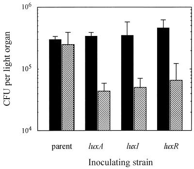
A direct measure of the extent of colonization that was achieved by each of the strains was obtained by determining the number of CFU present in juvenile light organs at various times after the association had been initiated. Approximately 24 h after initiation, all three of the mutants (luxA, luxI, and luxR) achieved colonization levels that were indistinguishable from that of the parent strain (Fig. 3). However, by about 48 h postinfection, the three lux mutant strains exhibited a three- to fourfold reduction in the level of colonization compared to that of the parent strain. This reduced level of colonization remained unchanged for at least another 24 h (data not shown). There is no evidence that the reduction in CFU results from the death of a significant number of the lux mutant bacteria in the light organ: an examination of both luxA mutant and parent cells released from the light organ crypts after 48 h showed that at least 90% of the cells of both groups were viable in the symbiosis (data not shown). Because the common characteristic of these mutants is that, unlike the parent strain, they do not produce induced levels of luminescence in the squid light organ (Fig. 2), we hypothesized that (i) luminescence is a requirement for the normal persistence of V. fischeri in the symbiosis and (ii) because of the role of the luxI and luxR genes in regulating luminescence induction, they are essential to the normal symbiotic competence of V. fischeri.
FIG. 3.
Symbiotic colonization levels achieved by lux mutant strains of V. fischeri and their parent, strain ESR1. The number of CFU present in the light organs of juvenile E. scolopes exposed to either lux mutant V. fischeri strains or the parent strain was determined at two times after inoculation, 24 h (black bars) or 48 h (striped bars). Each bar represents an average value obtained with at least four animals (standard error of the mean ranges are indicated). Similar results were obtained in three other independent trials.
Competitive colonization defects of the V. fischeri lux mutants.
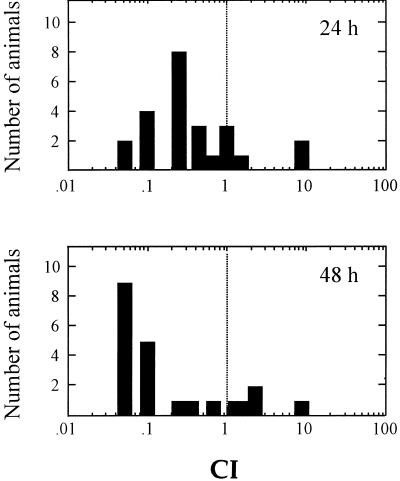
The ability of the luxA mutant strain to colonize the light organs of juvenile squid in coinoculations with the parent (lux+) strain was also investigated. When presented to the host animal in a 1:1 ratio with parent strain ESR1, the luxA mutant showed signs of being outcompeted by the parental strain as early as 24 h postinoculation (Fig. 4). This competitive advantage of ESR1 over the luxA mutant occurred in spite of the relatively slower growth of induced ESR1 cells discussed above. Not surprisingly, at 48 h the mutants continued to exhibit a significant competitive defect. A similar defect was observed in the luxR mutant when it was competed against the parent strain (data not shown). Because both of these mutants were constructed by the insertion of an erm cassette (Fig. 1), we were concerned that the carriage of the cassette was itself the basis for the defect; however, insertions of this cassette into other chromosomal loci had essentially no effect in competition experiments (data not shown). Taken together, these data provided support for the idea that luminescence plays a significant role in the ability of V. fischeri to colonize the light organ of E. scolopes juveniles and that this effect manifests itself as early as 24 h after colonization has been initiated.
FIG. 4.
Colonization of the E. scolopes light organ by a mixed inoculum of lux+ and luxA mutant V. fischeri cells. Forty-eight newly hatched E. scolopes juveniles were coinoculated with a 1:1 mixture of the luxA mutant (KV150) and its parent (ESR1). At two subsequent time points (24 and 48 h postinfection), the numbers of the two strains in the light organs of each of 24 animals were determined. The competitive index (CI) of the luxA mutants was calculated by dividing the number of KV150 mutant cells present in each organ by the number of ESR1 cells present. The number of animals with a given CI value is indicated by the bars. Symbiont populations of animals with a CI of <1 are dominated by lux+ cells.
Defects in the induction of host development by V. fischeri lux mutants.
We also tested whether carriage of the luxA, luxI, or luxR mutation resulted in a defect in the ability of the bacteria to trigger normal symbiont-induced changes in the host's program of light organ development. No differences were observed in the mutants' ability to induce the normal temporal and spatial pattern of apoptotic cell death in the ciliated surface of the light organ (data not shown). In contrast, defects were observed in the ability to trigger another morphological event that typically occurs in response to V. fischeri colonization, i.e., an increase in cytoplasmic volume, or cell swelling, within the epithelia that line the light organ crypt spaces. This swelling, or edema, results in the transformation of the initially columnar epithelial cells into more cuboidal ones. Cells of V. fischeri strain ESR1 (lux+) that infect juvenile squid induce the swelling within 48 h of inoculation, while the crypt epithelia of uninfected (aposymbiotic) juveniles remain unswollen (Fig. 5A and reference 32). In juvenile animals infected with either the luxA, luxI, or luxR mutant, the mean volume of the crypt cells in these animals was indistinguishable from that of uninfected animals (Fig. 5), demonstrating that all of the lux mutants are defective in triggering this specific host developmental event.
FIG. 5.
Effect of colonization by lux mutants on host epithelial cell morphology. (A) TEMs illustrating the ultrastructural morphology of crypt epithelial cells 48 h after inoculation with wild-type and mutant strains of V. fischeri (v, bacterial cells). (a) Epithelial cells of uninoculated aposymbiotic animals have a narrow, columnar shape (n, nucleus). (b) Epithelial cells of animals exposed to V. fischeri strain ESR1 have become swollen and cuboidal. (c) Epithelial cells exposed to the V. fischeri luxA mutant have retained a narrow, columnar shape. (d) Epithelial cells exposed to a serine auxotrophic mutant, which is defective in colonizing the light organ at normal levels, nevertheless become swollen and cuboidal. (Bar = 10 μm) (B) Cytoplasmic volumes of light organ epithelial cells in E. scolopes infected by either lux+ or lux mutant V. fischeri. Uninfected (aposymbiotic) E. scolopes, and those that had been exposed to lux mutants or their lux+ parent, were fixed for TEM (Materials and Methods) after 48 h of symbiotic infection. The average cytoplasmic volume of the epithelial cells flanking the symbiont population was determined for each condition of inoculation. Measurements from at least 10 epithelial cells were used to determine the average cytoplasmic volume (error bars indicate 95% confidence limits). Similar results were obtained in two other experiments.
One possible reason that the lux mutants were unable to induce host cell swelling was their reduced level of colonization. Thus, we determined the cell volumes of crypt epithelia from squid colonized by an amino acid auxotroph of V. fischeri. As described previously (16), we found that squid infected by this auxotrophic mutant contained 5 to 10% of the bacteria present in organs colonized by the parent strain, but unlike the lux mutants, the auxotroph produced easily detected levels of luminescence in the light organ. Colonization by the auxotroph induced a degree of epithelial cell swelling that was as great as that observed with the parent (Fig. 5A); thus, the defect in cell swelling exhibited by the lux mutants is more likely to be related to their failure to produce a normal level of luminescence than to their inability to achieve a normal extent of colonization.
Complementation of the luxA mutant colonization phenotype.
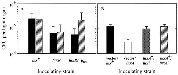
Because of the nature of the lux regulon and the mutations we had created in it, the possibility existed that the symbiotic defects described above were due to effects of the luxI or luxR mutations on some gene(s) other than those responsible for luminescence. To eliminate that possibility, we constructed a luxR luxI double mutant, strain KV345, in which transcription of the structural genes of the lux operon (luxCDABE) was placed under the control of P_tac_ in the chromosome (Fig. 1). In culture, the level of luminescence of KV345 was induced >100-fold by the addition of IPTG (51); when this strain was used to infect juvenile E. scolopes, IPTG addition to the surrounding seawater produced a >50-fold increase in bioluminescence emission from the squid (Fig. 2). The level of colonization established by KV345, in the presence and absence of IPTG, was assayed 48 h after the initiation of the association. While IPTG addition restored luminescence capability and wild-type levels of colonization to this strain, it did not affect the colonization levels of either the parent strain or the luxR mutant (Fig. 6A). These results suggest that the luxR and/or luxI gene products may not play a significant role in early symbiotic colonization, apart from their requirement for the normal induction of the luminescence genes (Fig. 2).
FIG. 6.
Complementation of the luminescence defect. (A) The average level of colonization of juvenile E. scolopes by the lux+ parent strain (ESR1), the luxR mutant (KV267), and the luxR luxI double mutant in which luxCDABE is under the control of P_tac_ (KV345) was determined after 46 h of infection. Throughout the course of the symbiotic infection, eight animals were exposed to IPTG (see Materials and Methods) (striped bars) and eight were not (black bars). (B) E. scolopes juveniles were exposed to one of four V. fischeri strains: a luxA mutant or its lux+ parent, carrying either a plasmid-borne copy of a complementing luxA+ gene or the parent vector alone. The number of CFU present in the light organs of 10 animals from each group was determined 48 h after inoculation. The bars represent the mean level of colonization (standard error of the mean ranges are indicated).
We were concerned that a gene downstream of the luxCDABE genes might be playing a role in the observed colonization phenotypes as a result of a polar effect caused by the luxA mutation. To look for evidence of polarity, we complemented this mutation in trans with a wild-type copy of the luxA gene carried on pKV19 (49). The presence of pKV19, but not the parent vector alone, restored both the luminescence phenotype of the complemented luxA mutant strain (data not shown) and normal levels of symbiotic colonization at 48 h (Fig. 6B). These data do not support the hypothesis of polar effects but instead provide evidence for the importance of luminescence itself in the ability of V. fischeri to successfully persist in the juvenile squid light organ.
Complementation of the epithelial cell swelling defect.
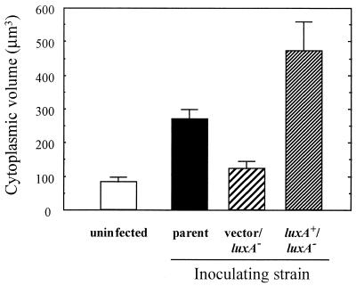
The formal possibility existed that the factors responsible for normal colonization and induction of host cell swelling are distinct. For example, the inability to induce host cell swelling may have resulted from a downstream polar effect of the lux mutations. Thus, we tested the ability of the luxA+ plasmid to also compensate for the deficiency of the luxA mutant in inducing host cell swelling. Measurements of the cytoplasmic volume of host light organ epithelial cells demonstrated that, in contrast to the luxA mutant strain, the complemented luxA strain was now proficient at inducing cell swelling to at least the same extent as its lux+ parent (Fig. 7). These data provide further evidence of a linkage between bacterial light emission and host cell swelling, and thus that the ability to bioluminesce is essential for the development of a normal symbiotic association between V. fischeri and E. scolopes.
FIG. 7.
Cytoplasmic volumes of light organ epithelial cells in E. scolopes juveniles infected by a complemented lux mutant of V. fischeri. Uninfected (aposymbiotic) E. scolopes juveniles or those that had been exposed to either the lux+ parent, a luxA mutant carrying a luxA+-complementing plasmid, or the vector alone were fixed for TEM after 48 h of symbiotic infection. The average cytoplasmic volume of the epithelial cells flanking the symbiont population was determined for each condition of inoculation. Measurements from at least 10 epithelial cells were used to determine the average cytoplasmic volume (error bars indicate 95% confidence limits).
DISCUSSION
Three important conclusions can be drawn from this study. First, when colonizing the E. scolopes light organ, luxA, luxI, or luxR mutants of V. fischeri express less than 0.1% of the luminescence of their lux+ parent strain. Second, as a result of this defect, these mutants cannot maintain a normal population size within the symbiotic association and are unable to successfully compete with a lux+ V. fischeri strain. Finally, and most strikingly, colonization by luxA, luxI, or luxR mutants fails to elicit cytoplasmic swelling in the epithelial cells that line the light organ crypts. This third finding constitutes the first report of V. fischeri genes that are required to induce a portion of the normal program of bacterium-triggered host differentiation in the light organ. While it has been long suspected that normal microbiota play an important role in the development of the tissues of their hosts (28), the genetic factors that underlie some of these relationships have only recently begun to be revealed (19). Similarly, we are just beginning to learn how these developmental events are related to the mechanisms by which such associations are maintained with fidelity.
Light organ symbioses are remarkably species specific: only V. fischeri and the closely related Vibrio logei are capable of initiating a light organ symbiosis with E. scolopes (12, 41). In addition to this taxon level of specificity, different V. fischeri isolates have various colonization efficiencies (24, 35), suggesting that selectivity is active at even more subtle levels. Our studies of the symbionts of E. scolopes have resulted in the intriguing observation that none of the thousands of strains of V. fischeri that have been isolated from light organs has been found to be a nonluminous variant (41). Thus, this specificity apparently extends to the selection of particular genotypes of V. fischeri, i.e., those with intact lux genes. A competitive disadvantage for V. fischeri strains that are lux mutants is unexpected because luminescence is not an essential trait of cells growing in laboratory culture, and in fact, light emission requires a considerable energy commitment by these bacteria (26). Thus, the absence of dark variants in symbiotic populations is surprising because the light organ is an environment in which different strains compete for dominance (24), and one would predict that by gaining a mutation in luminescence activity, a dark strain of V. fischeri might achieve a slight growth advantage.
For this reason the apparent selectivity for lux+ strains in nature suggested to us that maintaining the ability to luminesce might be of value not only to the host but to the bacterial symbionts as well. Indeed, the results presented here show that the symbiotic relationship in E. scolopes is profoundly affected by the inability of a colonizing symbiont to luminesce at normal levels. V. fischeri mutants defective for either structural (luxA) or regulatory (luxI or luxR) luminescence genes exhibited a three- to fourfold decrease in the extent of colonization within 48 h of initiating the symbiotic association. In fact, the defect could be detected as soon as 24 h under the competitive conditions of a mixed inoculum with the lux+ parent. While the basis for this more rapid appearance of the colonization defect is unknown, one possibility is that there may be a host challenge that is produced only as a response to the presence of luminescing cells. Colonization by the lux mutants alone would not elicit this additional challenge but a mixed infection would, exacerbating their defect.
These competition results further indicate that the presence in the symbiotic organ of wild-type, light-emitting cells does not complement the colonization defect of nonluminescing cells. Therefore, it is the luminescence activity of individual bacterial cells that is important, not any net effect of the symbiotic population as a whole. A similar competitive defect has been reported in experiments with a mutant defective for a periplasm-localized catalase enzyme (KatA), which is not rescued by the presence of wild-type V. fischeri cells (50). How this apparent cell-level selectivity is achieved remains to be determined.
A recent report has suggested that the products of luxI and luxR control the expression of several non-lux loci, including one required for V. fischeri to remain competitive in mixed symbiotic infections (4). Whether the _luxI/R_-dependent induction of these loci observed in culture occurs in the symbiosis, and/or is itself required for symbiotic competency, remains to be directly demonstrated. Our results indicate that the function of luxI and luxR during the establishment of a normal light organ colonization lies primarily with their role in the induction of bioluminescence. Examination of colonization by a luxI luxR double mutant that carried a tac promoter to control lux structural gene expression revealed that at least in juvenile squid, essentially wild-type levels of colonization could be achieved by this mutant as long as luminescence was artificially induced with IPTG. The direct role of the luxA gene was further substantiated by complementation studies that did not indicate a polar effect on any downstream gene critical for colonization. Taken together, these data suggest that in nature, V. fischeri cells that have lost their luminescence capability will be unlikely to sustain a symbiotic relationship, particularly in the face of coinfecting wild-type V. fischeri. Population biology theory would predict that the host would gain a benefit from imposing such a restriction (40); however, no evidence directly linking bioluminescence capacity to symbiotic success has been previously reported.
Perhaps the most novel and intriguing result presented in this report is the observation that the lux mutants exhibited defects in the ability to trigger a normal host tissue response. Upon colonization by wild-type V. fischeri, light organ epithelial cells in direct contact with bacterial symbionts initiate a program of differentiation, including a significant increase in cytoplasmic volume (23, 32). One consequence of crypt epithelial cell swelling is to effectively reduce the volume of the crypt spaces, thus forcing a greater percentage of the bacterial population to be in direct contact with the host epithelial surface. This change is bacterium induced; in the absence of V. fischeri, the epithelial cells do not swell, and if the colonization is cured, the swelling subsides (8). All three of the lux mutants that were examined were deficient in inducing epithelial edema. Complementation studies with the luxA mutant demonstrated that the presence of an intact copy of this gene coordinately restored both luminescence and normal epithelial development. V. fischeri cells that carry amino acid auxotrophy mutations making them unable to fully colonize the light organ (16), but that are lux+, were capable of inducing cell swelling (Fig. 5B). Thus, we believe that the lux mutations' effect on host development is due to a loss of luminescence function rather than to a reduced colonization level. We anticipate that there may be a number of bacterial activities that contribute to the induction of swelling and other host developmental events, and that the presence of luciferase and/or its luminescence activity is just one of them.
How does a lux mutation result in defects in both the effectiveness of colonization and the ability to induce host edema? Are these two symbiotic phenotypes linked and, if so, in what way? We hypothesize that the lux mutants express these symbiotic defects because they are less able to severely reduce the concentration of oxygen around them (44). Bacterial luciferase has an unusually high affinity for molecular oxygen, which is converted to water in the luminescence reaction. Specifically, its affinity constant (Km) for oxygen is in the nanomolar range, several orders of magnitude below that typical of cytochrome oxidase (44). Under the oxygen-limiting conditions in the symbiotic light organ (2), the high affinity of luciferase will give this enzyme an advantage over other oxidases for their required substrate. Thus, brightly luminescing bacteria can rapidly lower the ambient oxygen concentration (26), probably to a level below that resulting from the activities of nonluminescing bacteria. By creating a hypoxic condition around themselves, luminescing V. fischeri cells could potentially accomplish two goals: (i) reduce their exposure to host-generated reactive oxygen species (ROS) and (ii) gain access to host nutrients by a mechanism of hypoxia-induced epithelial exocytosis.
The presence of a host halide peroxidase in the light organ crypts (51), as well as the importance to V. fischeri of a periplasmic catalase (50), a bacterial defense against host-generated ROS, supports the hypothesis that the light organ environment is oxidatively stressful (48). Thus, it would not be surprising that symbionts might use the action of their luciferase to lower the ambient oxygen concentration, thereby reducing the potential for ROS generation. A mutant deficient in luciferase-catalyzed oxygen utilization might experience an increased steady-state oxygen concentration and become more exposed to damage by ROS (44).
The second phenotype of the lux mutants, failure to induce host epithelial cell edema, may also result because the typical hypoxic state is not being created in the light organ crypts by a normal level of luciferase activity. Hypoxia has been demonstrated to have an edemic effect on epithelial cells in other systems (18, 25) and to result in exocytosis of cytoplasmic material (36). Such exocytosed material may be a source of nutrients to support the growing population of luminous symbiotic bacteria colonizing the crypt surfaces. If such a mechanism does, in fact, supply host-derived nutrients to light organ symbionts, then V. fischeri lux mutants may face a growth disadvantage because they are unable to create the severe hypoxic conditions that lead to an attendant swelling of the crypt epithelium.
These predictions are being currently tested using a V. fischeri strain carrying an altered luciferase enzyme that consumes normal levels of oxygen but produces no luminescence. Such studies should further our understanding of the patterns of cell-cell signaling that occurs between V. fischeri and E. scolopes. In addition, they will encourage an examination of whether symbiont-induced hypoxia is a general mechanism used by other luminous, or highly respiratory, bacteria as they establish cooperative or pathogenic associations with their hosts.
ACKNOWLEDGMENTS
We thank V. Orlando for technical assistance and D. Millikan and E. Stabb for insightful comments.
This work was supported in part by National Institutes of Health grant RR-12294 to E.G.R. and M.M.-N. and by National Science Foundation grant IBN-9904601 to M.M.-N. and E.G.R. K.L.V. was supported by National Research Service Award F32GM17424-02 from NIH.
REFERENCES
- 1.Boettcher K J, Ruby E G. Depressed light emission by symbiotic Vibrio fischeri of the sepiolid squid Euprymna scolopes. J Bacteriol. 1990;172:3701–3706. doi: 10.1128/jb.172.7.3701-3706.1990. [DOI] [PMC free article] [PubMed] [Google Scholar]
- 2.Boettcher K J, Ruby E G. Detection and quantification of Vibrio fischeri autoinducer from symbiotic squid light organs. J Bacteriol. 1995;177:1053–1058. doi: 10.1128/jb.177.4.1053-1058.1995. [DOI] [PMC free article] [PubMed] [Google Scholar]
- 3.Boettcher K J, Ruby E G, McFall-Ngai M J. Bioluminescence in the symbiotic squid Euprymna scolopes is controlled by a daily biological rhythm. J Comp Physiol. 1996;179:65–73. [Google Scholar]
- 4.Callahan S M, Dunlap P V. LuxR- and acyl-homoserine-lactone-controlled non-lux genes define a quorum-sensing regulon in Vibrio fischeri. J Bacteriol. 2000;182:2811–2822. doi: 10.1128/jb.182.10.2811-2822.2000. [DOI] [PMC free article] [PubMed] [Google Scholar]
- 5.Chatterjee A, Cui Y, Liu Y, Dumenyo C K, Chatterjee A K. Inactivation of rmsA leads to overproduction of extracellular pectinases, cellulases, and proteases in Erwinia carotovora subsp. carotovora in the absence of the starvation/cell density-sensing signal, N-(3-oxohexanoyl)-l-homoserine lactone. Appl Environ Microbiol. 1995;61:1959–1967. doi: 10.1128/aem.61.5.1959-1967.1995. [DOI] [PMC free article] [PubMed] [Google Scholar]
- 6.Davis R W, Botstein D, Roth J R. Advanced bacterial genetics. Cold Spring Harbor, N.Y: Cold Spring Harbor Laboratory; 1980. [Google Scholar]
- 7.Diederich L, Roth A, Messer W. A versatile plasmid vector system for the regulated expression of genes in Escherichia coli. BioTechniques. 1994;16:916–923. [PubMed] [Google Scholar]
- 8.Doino J A. The role of light organ symbionts in signaling early morphological and biochemical events in the sepiolid squid Euprymna scolopes. Ph.D. thesis. Los Angeles: University of Southern California; 1998. [Google Scholar]
- 9.Dunlap P V. Regulation of luminescence by cyclic AMP in cya-like and crp-like mutants of Vibrio fischeri. J Bacteriol. 1989;171:1199–1202. doi: 10.1128/jb.171.2.1199-1202.1989. [DOI] [PMC free article] [PubMed] [Google Scholar]
- 10.Dunlap P V, Kuo A. Cell-density modulation of the Vibrio fischeri luminescence system in the absence of autoinducer and LuxR protein. J Bacteriol. 1992;174:2440–2448. doi: 10.1128/jb.174.8.2440-2448.1992. [DOI] [PMC free article] [PubMed] [Google Scholar]
- 11.Eberhard A, Burlingame A L, Eberhard C, Kenyon G L, Nealson K H, Oppenheimer N J. Structural identification of autoinducer of Photobacterium fischeri luciferase. Biochemistry. 1981;28:2444–2449. doi: 10.1021/bi00512a013. [DOI] [PubMed] [Google Scholar]
- 12.Fidopiastis P M, von Boletzky S, Ruby E G. A new niche for Vibrio logei, the predominant light organ symbiont of squids in the genus Sepiola. J Bacteriol. 1998;180:59–64. doi: 10.1128/jb.180.1.59-64.1998. [DOI] [PMC free article] [PubMed] [Google Scholar]
- 13.Fuqua C, Winans S C, Greenberg E P. Census and consensus in bacterial ecosystems: the LuxR-LuxI family of quorum-sensing transcriptional regulators. Annu Rev Microbiol. 1996;50:727–751. doi: 10.1146/annurev.micro.50.1.727. [DOI] [PubMed] [Google Scholar]
- 14.Gilson L, Kuo A, Dunlap P V. AinS and a new family of autoinducer synthesis proteins. J Bacteriol. 1995;177:6946–6951. doi: 10.1128/jb.177.23.6946-6951.1995. [DOI] [PMC free article] [PubMed] [Google Scholar]
- 15.Graf J, Dunlap P V, Ruby E G. Effect of transposon-induced motility mutations on colonization of the host light organ by Vibrio fischeri. J Bacteriol. 1994;176:6986–6991. doi: 10.1128/jb.176.22.6986-6991.1994. [DOI] [PMC free article] [PubMed] [Google Scholar]
- 16.Graf J, Ruby E G. Host-derived amino acids support the proliferation of symbiotic bacteria. Proc Natl Acad Sci USA. 1998;95:1818–1822. doi: 10.1073/pnas.95.4.1818. [DOI] [PMC free article] [PubMed] [Google Scholar]
- 17.Gray K M, Greenberg E P. Physical and functional maps of the luminescence gene cluster in an autoinducer-deficient Vibrio fischeri strain isolated from a squid light organ. J Bacteriol. 1992;174:4384–4390. doi: 10.1128/jb.174.13.4384-4390.1992. [DOI] [PMC free article] [PubMed] [Google Scholar]
- 18.Hierholzer C, Kelly E, Tsukada K, Loeffert E, Watkins S, Billiar T, Tweardy D. Hemorrhagic shock induces G-CSF expression in bronchial epithelium. Am J Physiol. 1997;273:L1058–L1064. doi: 10.1152/ajplung.1997.273.5.L1058. [DOI] [PubMed] [Google Scholar]
- 19.Hooper L V, Xu J, Falk P G, Midtvedt T, Gordon J I. A molecular sensor that allows a gut commensal to control its nutrient foundation in a competitive ecosystem. Proc Natl Acad Sci USA. 1999;96:9833–9838. doi: 10.1073/pnas.96.17.9833. [DOI] [PMC free article] [PubMed] [Google Scholar]
- 20.Kaplan H B, Greenberg E P. Diffusion of autoinducer is involved in regulation of the Vibrio fischeri luminescence system. J Bacteriol. 1985;163:1210–1214. doi: 10.1128/jb.163.3.1210-1214.1985. [DOI] [PMC free article] [PubMed] [Google Scholar]
- 21.Kuo A, Blough N V, Dunlap P V. Multiple N-acyl-l-homoserine lactone autoinducers of luminescence in the marine symbiotic bacterium Vibrio fischeri. J Bacteriol. 1994;176:7558–7565. doi: 10.1128/jb.176.24.7558-7565.1994. [DOI] [PMC free article] [PubMed] [Google Scholar]
- 22.Kuo A, Callahan S M, Dunlap P V. Modulation of luminescence operon expression by N-octanoyl-l-homoserine lactone in ainS mutants of Vibrio fischeri. J Bacteriol. 1996;178:971–976. doi: 10.1128/jb.178.4.971-976.1996. [DOI] [PMC free article] [PubMed] [Google Scholar]
- 23.Lamarcq L H, McFall-Ngai M J. Induction of a gradual, reversible morphogenesis of its host's epithelial brush border by Vibrio fischeri. Infect Immun. 1998;66:777–785. doi: 10.1128/iai.66.2.777-785.1998. [DOI] [PMC free article] [PubMed] [Google Scholar]
- 24.Lee K-H, Ruby E G. Competition between Vibrio fischeri strains during initiation and maintenance of a light organ symbiosis. J Bacteriol. 1994;176:1985–1991. doi: 10.1128/jb.176.7.1985-1991.1994. [DOI] [PMC free article] [PubMed] [Google Scholar]
- 25.Mairbaurl H, Wodopia R, Eckes S, Schulz S, Bartsch P. Impairment of cation transport in A549 cells and rat alveolar epithelial cells by hypoxia. Am J Physiol. 1997;273:L797–L806. doi: 10.1152/ajplung.1997.273.4.L797. [DOI] [PubMed] [Google Scholar]
- 26.Makemson J C. Luciferase-dependent oxygen consumption by bioluminescent vibrios. J Bacteriol. 1986;165:461–466. doi: 10.1128/jb.165.2.461-466.1986. [DOI] [PMC free article] [PubMed] [Google Scholar]
- 27.McFall-Ngai M J. Crypsis in the pelagic environment. Am Zool. 1990;30:175–188. [Google Scholar]
- 28.McFall-Ngai M J. The development of cooperative associations between animals and bacteria: establishing détente between domains. Am Zool. 1998;38:3–18. [Google Scholar]
- 29.McFall-Ngai M J. Consequences of evolving with bacterial symbionts: lessons from the squid-vibrio associations. Annu Rev Ecol Syst. 1999;30:235–256. [Google Scholar]
- 30.Meighen E A, Dunlap P V. Physiological, biochemical and genetic control of bacterial bioluminescence. Adv Microb Physiol. 1993;34:1–67. doi: 10.1016/s0065-2911(08)60027-2. [DOI] [PubMed] [Google Scholar]
- 31.Montgomery M K, McFall-Ngai M. Embryonic development of the light organ of the sepiolid squid Euprymna scolopes Berry. Biol Bull. 1993;184:296–308. doi: 10.2307/1542448. [DOI] [PubMed] [Google Scholar]
- 32.Montgomery M K, McFall-Ngai M. Bacterial symbionts induce host organ morphogenesis during early postembryonic development of the squid Euprymna scolopes. Development. 1994;120:1719–1729. doi: 10.1242/dev.120.7.1719. [DOI] [PubMed] [Google Scholar]
- 33.Montgomery M K, McFall-Ngai M J. The inductive role of bacterial symbionts in the morphogenesis of a squid light organ. Am Zool. 1995;35:372–380. [Google Scholar]
- 34.Nealson K H. Isolation, identification, and manipulation of luminous bacteria. Methods Enzymol. 1978;57:153–166. [Google Scholar]
- 35.Nishiguchi M K, Ruby E G, McFall-Ngai M J. Competitive dominance among strains of luminous bacteria provides an unusual form of evidence for parallel evolution in sepiolid squid-vibrio symbioses. Appl Environ Microbiol. 1998;64:3209–3213. doi: 10.1128/aem.64.9.3209-3213.1998. [DOI] [PMC free article] [PubMed] [Google Scholar]
- 36.Okada Y, Setoyama H, Matsumoto S, Imaoka A, Nanno M, Kawaguchi M, Umesaki Y. Effects of fecal microorganisms and their chloroform-resistant variants derived from mice, rats, and humans on immunological and physiological characteristics of the intestines of ex-germfree mice. Infect Immun. 1994;62:5442–5446. doi: 10.1128/iai.62.12.5442-5446.1994. [DOI] [PMC free article] [PubMed] [Google Scholar]
- 37.Passador L, Cook J M, Gambello M J, Rust L, Iglewski B H. Expression of the Pseudomonas aeruginosa virulence genes requires cell-to-cell communication. Science. 1993;260:1127–1130. doi: 10.1126/science.8493556. [DOI] [PubMed] [Google Scholar]
- 38.Pearson J P, Gray K M, Passador L, Tucker K D, Eberhard A, Iglewski B H, Greenberg E P. Structure of the autoinducer required for expression of Pseudomonas aeruginosa virulence genes. Proc Natl Acad Sci USA. 1994;91:197–201. doi: 10.1073/pnas.91.1.197. [DOI] [PMC free article] [PubMed] [Google Scholar]
- 39.Pirhonen M, Flego D, Heikinheimo R, Palva E T. A small diffusible signal molecule is responsible for the global control of virulence and exoenzyme production in the plant pathogen Erwinia carotovora. EMBO J. 1993;12:2467–2476. doi: 10.1002/j.1460-2075.1993.tb05901.x. [DOI] [PMC free article] [PubMed] [Google Scholar]
- 40.Read A F. The evolution of virulence. Trends Microbiol. 1994;2:73–76. doi: 10.1016/0966-842x(94)90537-1. [DOI] [PubMed] [Google Scholar]
- 41.Ruby E G. Lessons from a cooperative, bacterial-animal association: the Vibrio fischeri-Euprymna scolopes light organ symbiosis. Annu Rev Microbiol. 1996;50:591–624. doi: 10.1146/annurev.micro.50.1.591. [DOI] [PubMed] [Google Scholar]
- 42.Ruby E G, Asato L M. Growth and flagellation of Vibrio fischeri during initiation of the sepiolid squid light organ symbiosis. Arch Microbiol. 1993;159:160–167. doi: 10.1007/BF00250277. [DOI] [PubMed] [Google Scholar]
- 43.Ruby E G, Lee K-H. The Vibrio fischeri Euprymna scolopes light organ association: current ecological paradigms. Appl Environ Microbiol. 1998;64:805–812. doi: 10.1128/aem.64.3.805-812.1998. [DOI] [PMC free article] [PubMed] [Google Scholar]
- 44.Ruby E G, McFall-Ngai M J. Oxygen-utilizing reactions and symbiotic colonization of the squid light organ by Vibrio fischeri. Trends Microbiol. 1999;7:414–419. doi: 10.1016/s0966-842x(99)01588-7. [DOI] [PubMed] [Google Scholar]
- 45.Southern E M. Detection of specific sequences among DNA fragments separated by gel electrophoresis. J Mol Biol. 1975;98:503–517. doi: 10.1016/s0022-2836(75)80083-0. [DOI] [PubMed] [Google Scholar]
- 46.Spurr A. A low-viscosity epoxy resin embedding medium for electron microscopy. J Ultrastruct Res. 1969;26:31–43. doi: 10.1016/s0022-5320(69)90033-1. [DOI] [PubMed] [Google Scholar]
- 47.Tang H B, DiMango E, Bryan R, Gambello M, Iglewski B H, Goldberg J B, Prince A. Contribution of specific Pseudomonas aeruginosa virulence factors to pathogenesis of pneumonia in a neonatal mouse model of infection. Infect Immun. 1996;64:37–43. doi: 10.1128/iai.64.1.37-43.1996. [DOI] [PMC free article] [PubMed] [Google Scholar]
- 48.Visick K L, McFall-Ngai M J. An exclusive contract: specificity in the Vibrio fischeri-Euprymna scolopes partnership. J Bacteriol. 2000;182:1779–1787. doi: 10.1128/jb.182.7.1779-1787.2000. [DOI] [PMC free article] [PubMed] [Google Scholar]
- 49.Visick K L, Ruby E G. Construction and symbiotic competence of a luxA-deletion mutant of Vibrio fischeri. Gene. 1996;175:89–94. doi: 10.1016/0378-1119(96)00129-1. [DOI] [PubMed] [Google Scholar]
- 50.Visick K L, Ruby E G. The periplasmic, group III catalase of Vibrio fischeri is required for normal symbiotic competence and is induced both by oxidative stress and by approach to stationary phase. J Bacteriol. 1998;180:2087–2092. doi: 10.1128/jb.180.8.2087-2092.1998. [DOI] [PMC free article] [PubMed] [Google Scholar]
- 51.Visick K L, Ruby E G. Temporal control of lux gene expression in the symbiosis between Vibrio fischeri and its squid host. In: Le Gal Y, Halvorsen H O, editors. New developments in marine biotechnology. New York, N.Y: Plenum Press; 1998. pp. 277–279. [Google Scholar]
- 52.Wei S L, Young R E. Development of symbiotic bacterial bioluminescence in a nearshore cephalopod, Euprymna scolopes. Mar Biol. 1989;103:541–546. [Google Scholar]
- 53.Weis V M, Small A L, McFall-Ngai M J. A peroxidase related to the mammalian antimicrobial protein myeloperoxidase in the Euprymna-Vibrio mutualism. Proc Natl Acad Sci USA. 1996;93:13683–13688. doi: 10.1073/pnas.93.24.13683. [DOI] [PMC free article] [PubMed] [Google Scholar]
- 54.Yanisch-Perron C, Vieira J, Messing J. Improved M13 phage cloning vectors and host strains: nucleotide sequences of the M13mp18 and pUC19 cloning vectors. Gene. 1985;33:103–119. doi: 10.1016/0378-1119(85)90120-9. [DOI] [PubMed] [Google Scholar]
