Genetic Determinants of Altered Virulence of Taiwanese Foot-and-Mouth Disease Virus (original) (raw)
Abstract
In 1997, a devastating outbreak of foot-and-mouth disease (FMD) in Taiwan was caused by a serotype O virus (referred to here as OTai) with atypical virulence. It produced high morbidity and mortality in swine but did not affect cattle. We have defined the genetic basis of the species specificity of OTai by evaluating the properties of genetically engineered chimeric viruses created from OTai and a bovine-virulent FMD virus. These studies have shown that an altered nonstructural protein, 3A, is a primary determinant of restricted growth on bovine cells in vitro and significantly contributes to bovine attenuation of OTai in vivo.
Foot-and-mouth disease (FMD) is an extremely contagious viral disease of cloven-hoofed animals, most notably cattle, pigs, and sheep. FMD is characterized by fever, vesicular lesions, and erosions of the epithelium of the mouth, tongue, nares, muzzle, feet, and teats (22). Despite recent success in controlling the disease in Europe and portions of South America, FMD remains one of the most important infectious diseases of farm animals due to the impact an outbreak can have on trade in animals and animal products (13). Foot-and-mouth disease virus (FMDV), a positive-stranded RNA virus, is the type member of the Aphthovirus genus of the family Picornaviridae, a family which includes many important pathogens of humans and domestic animals. There are seven recognized serotypes of FMDV, but new subtypes with altered antigenic properties frequently emerge due to the well-characterized genetic instability of the virus (5).
Recent FMD outbreak in Taiwan.
In 1997, a devastating and unusual outbreak of FMD, characterized by disease in swine but not in cattle, occurred in Taiwan. The outbreak, which was caused by a serotype O virus (OTai) with an atypical porcinophilic phenotype (6), rapidly developed into a massive epizootic, resulting in cessation of export of all pork products from the country. This outbreak devastated the Taiwanese swine industry (approximately 4 million swine were destroyed) and had a severe impact on the national economy due to costs of control and trade restrictions (estimated at over 6 billion U.S. dollars). During the course of this outbreak, no cattle were reported to have been affected, and virus isolated from infected swine was unable to infect bovine thyroid cells in vitro or to cause typical disease in bovines following intradermal inoculation in the tongue (6).
Genomic regions responsible for host range specificity in vitro.
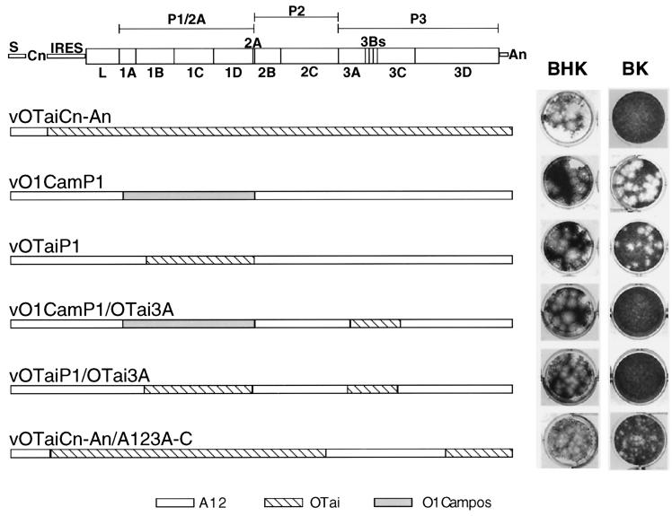
To define the genetic components of OTai responsible for its porcinophilic phenotype, we have generated and evaluated recombinant viruses with sequences of OTai substituted for sequences of an FMDV strain of high virulence in bovines (25). The engineered chimeric viruses produced for these studies are derived from in vitro-generated RNA transcripts of genome-length cDNAs by using standard techniques (17, 23). Specifically, all chimeric viruses used in these studies were generated by high-efficiency transfection of BHK cells (17), and experiments were performed with low-passage (less than five) stocks of virus produced by high-multiplicity infection of BHK cell cultures. Figure 1 shows schematic diagrams of the genomes of selected chimeric viruses along with photographs of plaques formed by these viruses in monolayer cultures of BHK or bovine kidney (BK) cells. All viruses that formed plaques on BHK cells also formed plaques on both IB-RS-2 cells (a porcine kidney cell line [6]) and porcine kidney cells (data not shown).
FIG. 1.
Structures of genetically engineered virus genomes and plaque assays of viruses containing these genomes on BHK and BK cells. Linear representations of the 8.4-kb chimeric genomes show the source of their sequences as indicated in the key at the bottom of the figure. Genetic elements: S, small fragment of the genome; Cn, poly(C) tract; IRES, internal ribosome entry site; L, leader proteinase-encoding region; P1/2A, capsid precursor coding region; P2 and P3, nonstructural protein precursor-encoding regions; An, poly(A) tract. Viral RNA recovered from porcine lesions was reverse transcribed, and specific portions of the resulting cDNA were amplified by PCR amplification with a high-fidelity polymerase (27). The segment of the genome between the poly(C) tract and the 3′ poly(A) tract was amplified and molecularly cloned in E. coli in three separate portions. The portion extending from the poly(C) tract to the beginning of the coding sequence for 1B was amplified with an oligonucleotide containing an _Avr_II site found at the border of the poly(C) tract of the serotype A12 genome followed by 28 nucleotides of the adjacent sequence (23) and an antisense oligonucleotide corresponding to codons 13 to 22 of protein 1B of OTai, containing mutations in codons 13 to 15, that produce an _Ssp_I site without altering the coding capacity of the sequence. The portion extending from 1B to 2A was amplified by using an oligonucleotide corresponding to codons 13 to 21 of protein 1B of O1 Campos (containing the same mutations needed to produce an _Ssp_I site) and an antisense oligonucleotide corresponding to codons 12 to 18 of 2A (containing mutations to produce an _Xma_I site in codons 17 and 18, without altering their coding capacity). The final portion of the genome was amplified by using an oligonucleotide corresponding to codons 17 and 18 of 2A (with the _Xma_I site described above) and the first six codons of OTai 2B and an oligo(T) oligonucleotide containing 15 T's and a _Not_I site (23). These fragments were assembled into serotype A12 or serotype A12/OTai chimeric cDNAs and used to generate viruses (see text) (17, 23). Chimeras with exchanged 3A sequences were created by utilizing existing _Nco_I sites at codons 215 to 217 in 2C and 110 to 112 of 3D shared by OTai and A12 or by introducing _Eco_RI and _Eco_RV sites (12) into OTai sequences at codons 24 and 25 of 3A and 98 and 99 of 3C, respectively, in a manner that preserved the coding capacity and permitted in-frame fusion to existing sites in A12 cDNA. Monolayer cultures of BHK and BK cells were infected side-by-side with 10-fold dilutions of virus stocks prepared from each chimeric genome and stained 2 days after infection to reveal plaques (23). The paired BHK and BK monolayers selected for display on the right side of this figure corresponded to those in which the virus dilutions produced 10 to 50 plaques on BHK cells.
The virus whose structure is shown first in Fig. 1, vOTaiCn-An, is derived from a full-length cDNA containing the entire OTai protein-encoding sequence. This chimera contains the A12 S fragment and poly(C) tract found in all of the viruses used in this study (23), with the remainder of the genome [poly(C) tract to poly(A) tail] derived by PCR amplification of cDNA reverse transcribed from OTai virus RNA extracted from lesions of swine infected with a field isolate of OTai (see legend to Fig. 1). As expected, vOTaiCn-An displays the growth restriction of field isolates of OTai on bovine cells (6). Immunostaining of BK cells infected with vOTaiCn-An showed that this virus could establish a limited infection in bovine cells, as indicated by few FMDV antigen-positive cells and no spread of infection to surrounding cells (data not shown). The virus whose structure is shown second in Fig. 1, vO1CamP1, is a chimeric virus previously generated (referred to as vCRM8 in references 1, 20, and 25) by inserting the capsid-encoding region (proteins 1A, 1B, 1C, and 1D) of serotype O1 Campos (from a 1958 bovine isolate from Brazil) in the genetic background of a high-passage serotype A12 virus (from a 1932 bovine isolate from the United Kingdom) (23, 25). vO1CamP1 displays a high level of virulence in cattle (25) and swine (1) and efficiently forms plaques in BHK and BK cell monolayers (Fig. 1). The virus whose structure is shown after vO1CamP1 in Fig. 1, vOTaiP1, encodes the surface-exposed capsid proteins (1B, 1C, and 1D) of OTai in the background of the serotype A12 virus. Interestingly, this virus, whose capsid differs significantly from the South American O1 capsid (Table 1), displays plaquing ability similar to that of vO1CamP1 on both BHK and BK cells (Fig. 1). These findings indicate that the basis of growth restriction in BK cells in vitro was not due to alterations in the capsid, although our previous work has shown a profound influence of FMDV capsid sequences on receptor specificity and tropism in cell culture and virulence in animals (20, 25). Furthermore, results obtained by evaluation of other chimeras failed to show a role for either the OTai Leader proteinase coding region (known to contribute to virus virulence in bovines [2, 18]) or the OTai translation initiation site (the internal ribosome entry site region, well known for its role in determining the virulence of the related picornavirus that causes poliomyelitis [reviewed in reference 28]) in restricting replication in bovine cell cultures (results not shown).
TABLE 1.
Summary of differences in predicted amino acid sequences between the capsid-encoding regions of O1 Campos and OTai
| Capsid protein | No. of altered amino acids/total amino acids (%)a | No. of surface-exposed amino acids/no. of altered amino acids (%)b |
|---|---|---|
| 1B | 7/218 (3.2) | 3/7 (42.9) |
| 1C | 7/220 (3.2) | 4/7 (57.1) |
| 1D | 32/211 (12.2) | 24/32 (75) |
To further map the basis of the altered host range of OTai, a series of viruses were generated to dissect the nonstructural protein-encoding regions (P2 and P3). Sequence data obtained during the construction of these chimeras revealed a significant difference among the 3A coding regions of the OTai, A12, and O1 Kaufbeuren (O1K) viruses (Table 2). The comparisons in Table 2 show that the dramatic differences in the 3A coding regions of the two type O genomes stand in sharp contrast to the modest differences detected between coding regions for 2B, 2C, 3B, 3C, and 3D between these viruses, which are similar to the differences seen between the P2 and P3 coding regions of O1K and the A12 virus.
TABLE 2.
Comparison of differences between nonstructural protein-encoding segments of the FMDV genomes of OTai, O1K, and A12
| Virus | % Differences at the amino acid level relative to O1Ka | |||||
|---|---|---|---|---|---|---|
| 2B | 2C | 3A | 3B3 | 3C | 3D | |
| OTaib | 4.5 | 3.1 | 28.6 | 2.8 | 2.8 | 1.4 |
| A12c | 3.9 | 1.9 | 4.6 | 4.2 | 3.8 | 2.6 |
To test the importance of the 3A region in the bovine growth restriction of OTai, three additional chimeras, shown at the bottom of Fig. 1, were constructed. These three viruses contained either the entire A12 3A coding region or the mutated portion of 3A of OTai (see below), along with some additional regions. In all three chimeras shown at the bottom of Fig. 1, the exchanged regions extend beyond the 3A coding region, and in two of the cases, the first 24 codons of the OTai 3A coding region were not included in the chimeras (the amino acids encoded by A12 and OTai are identical in this 24-codon region) (see Fig. 2). However, in the extensions beyond the borders of 3A, the substituted codons correspond to a small number of well-spaced single amino acid substitutions that are largely conservative in nature (in the _Nco_I fragment exchanged to form virus vOTaiCn-An/A123A-C [Fig. 1], a total of 3 substitutions in 2C, 5 substitutions in 3BBB, 12 changes in 3C, and 5 changes in 3D were incorporated along with 40 changes clustered in the latter half of 3A; in the _Eco_RI-_Eco_RV fragment exchanged to construct vO1CamP1/Tai3A or vOTaiP1/OTai3A [see Fig. 1], 5 changes in 3BBB and 1 change in 3C were incorporated in addition to the 40 changes clustered in the latter half of 3A). Thus, the only significant changes between the last three viruses in Fig. 1 and their corresponding parents are in the 3A coding region. A comparison of the abilities of these three viruses to form plaques on BK cells revealed that substitution of the OTai 3A coding region into vO1CamP1 or vOTaiP1 produced viruses that were unable to form plaques on BK cells, whereas the substitution of the A12 3A into O1TaiCn-An produced a virus capable of forming plaques on these cells (Fig. 1). The plaques formed on BK cells by this latter virus were smaller than those produced by vOTaiP1 and vO1CamP1, suggesting that other regions of the genome contribute to OTai growth restriction on BK cells. Taken together, the data in Fig. 1 demonstrate that the OTai 3A coding region is the primary determinant of the growth restriction of OTai on BK cells.
FIG. 2.
Alignment of the deduced 3A amino acid sequences (shown in single-letter code) of OTai, O1 Campos (9), A12 (24), and an egg-adapted O1 Campos (O1C-O/E) (9) reveal significant differences among their C-terminal halves (a period indicates identity to O1 Campos; a dash indicates a deletion). The hydrophobic domain located at residues 61 to 76 (underlined) is thought to function in membrane binding (29).
As pointed out in Table 2, sequence analyses of the genome of OTai revealed a high degree of identity to O1 and A12, except for the 3A coding region. The divergence in 3A sequences among these viruses reflects two dramatic differences in the OTai 3A coding region. The first is a 10-amino-acid deletion corresponding to codons 93 through 102 of the European and South American 3A coding regions, and the second is a large number of mutation located between codons 128 and 147 of the European and South American 3A coding regions. Interestingly, the deletion in the OTai 3A is similar to a deletion found in egg-passaged derivatives of O1 Campos (Fig. 2) and C3 Resende (9). Both of these egg-passaged viruses, which were developed and used as live-attenuated FMD vaccines in bovines in South America (21, 26), exhibited a greatly reduced ability to form plaques on BK cells, and the C3 derivative was reported to have maintained its virulence in swine (26). Evaluation of selected other 3A sequences (C3 Argentina, 1985 (7); SAT-2, Kenya, 1957 [J. W. I. Newman, A. M. Q. King, and S. Ortlep, personal communication]; and sequences obtained from PCR-amplified cDNAs of several other serotypes or subtypes of FMDV [Asia1, Lebanon, 1983; O11, Indonesia, 1962]) showed that none of these isolates contained the deleted or mutated segments identified in OTai.
Genomic regions responsible for host range specificity in vivo.
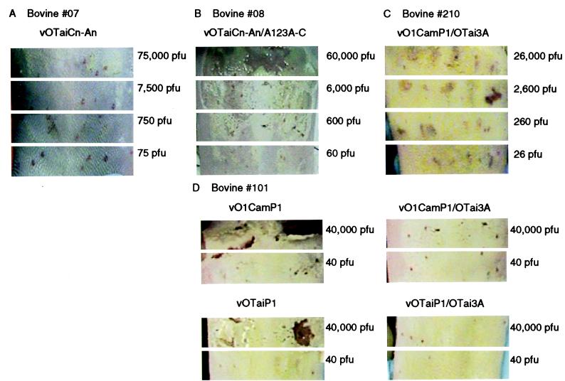
In vivo studies were conducted to establish the role of OTai 3A as a determinant of bovine virulence. vO1CamP1, which encodes the typical, full-length 3A of serotype A12, has been shown in previous studies to very efficiently cause vesicle formation on bovine tongues following intradermal inoculation, with a 50% infectious dose of 5 to 50 PFU (25). Furthermore, the animals inoculated with vO1CamP1 developed a systemic infection (25). A similar intradermal inoculation study performed with vOTaiCn-An in bovine 07 showed a significantly different outcome (Fig. 3A). In this case, lesions were detected only at doses of 750,000 and 75,000 PFU, and the quality of the lesions was significantly different than that observed with vO1CamP1. Specifically, the lesions caused by the high doses of vOTaiCn-An did not progress into sores denuded of epidermis, which were characteristic of the vesicles formed with all inoculations of over 50 PFU of vO1CamP1 (or other cattle-virulent FMDV). Moreover, animal 07 did not display lameness or vesicles on any feet during the 3-week observation period following inoculation. Thus, our results with the genetically engineered form of OTai are consistent with those obtained with the field isolate (6).
FIG. 3.
Vesicle-forming ability of chimeric viruses in bovine tongues. Bovines were sedated by intramuscular injection of xylazine at 22 mg/100 kg (body weight). The tongue was extended from the oral cavity, washed with warm water, and given five 0.05-ml intradermal inoculations of each virus dilution tested with a 22-gauge needle. Segments of each panel correspond to photographs taken at 48 h postinoculation of sections of the dorsal surface of the tongue given five inoculations of the indicated number of PFU. (A to C) Bovine 07, 08, and 210 were inoculated with dilutions of the indicated viruses; (D) bovine 101 was given two different dilutions of the four indicated viruses.
Inoculation of bovine 08 with vO1TaiCn-An/A123A-C demonstrated that the addition of the full-length, wild-type form of 3A to this OTai derivative produced a bovine-virulent virus (Fig. 3B). Forty-eight hours after tongue inoculation with this virus, vesicles were detected at all doses greater than 6 PFU, showing that this virus was highly efficient in establishing infectious foci in the epidermis of the tongue (Fig. 3B). Interestingly, bovine 08 did not display other signs of systemic disease, including pedal lesions or lameness, over the 3-week observation period.
Addition of the OTai 3A coding sequence to vO1CamP1 was capable of attenuating the infection caused by this virus. Bovine 210 was inoculated with graded dilutions of vO1CamP1/OTai3A and observed for 3 weeks. At 48 h, lesions were detected at the 260-PFU dose, but these lesions consisted of white swollen vesicles rather than the severe erosions detected with vO1CamP1 and vO1TaiCn-An/A123A-C (Fig. 3C). However, the following day, erosions appeared (although only at doses greater than 260 PFU), and this animal showed a systemic disease, characterized by spread of the infection to the feet. Infection of a second bovine with dilutions of vO1CamP1/OTai3A produced similar signs of disease (results not shown).
To confirm that our observations were not significantly influenced by individual responses of animals to inoculation, a single bovine was inoculated with two doses each of four different viruses. Bovine 101 was inoculated with vO1CamP1, vO1CamP1/OTai3A, vOTaiP1, and vOTaiP1/OTai3A (Fig. 3D) at doses of 40,000 or 40 PFU. Both vO1CamP1 and vOTaiP1 were able to cause severe lesions at high and low doses (Fig. 3D). vO1CamP1/OTai3A was able to cause swelling at the high dose, but no lesions were detected at the low dose. vOTaiP1/OTai3A was unable to cause signs of infection at either dose (Fig. 3D). Taken together, these in vivo data demonstrate that the mutated 3A coding region of OTai plays a major role in the attenuation of this virus in bovines.
Although FMDV can be distinguished from other members of the Picornaviridae by a much longer 3A protein (153 amino acids versus 87 for poliovirus), all picornavirus 3A proteins contain a 15- to 20-amino-acid hydrophobic domain approximately 60 to 80 residues from their N termini that is thought to function in binding the viral RNA replication machinery to cellular membranes (29). Furthermore, the interaction of 3A with cellular membranes has been shown to cause a cytopathic effect, prevent surface expression of proteins, and to inhibit protein secretion from 3A-expressing cells (3, 4). Thus, changes in the interaction of 3A with the host cell could affect the outcome of infection by directly altering RNA replication or by altering the ability of the cell or the host to mount a response to infection at the level of cytokine secretion or display of viral antigens in the context of the major histocompatibility complex. In the case of hepatitis A virus, changes in 3A have been documented during adaptation to cell culture (10, 11, 15, 19), and for poliovirus there is molecular genetic evidence that changes in 3A alter host range in vitro (14).
The activities of 3A in virus-infected cells, and their implication in alteration of host range of other picornaviruses, support a role for OTai 3A in the altered virulence of OTai. Moreover, the changes we have detected in OTai, including a deletion similar to those previously reported for egg-adapted FMDVs (9), suggest that under some circumstances altered 3A genes could have a selective advantage. Determining if these changes contribute to the high level of virulence of OTai in swine, and discovering the time of appearance of these changes in field isolates of FMDV, will contribute to our understanding of how a bovine-attenuated FMDV emerged to cause this recent devastating outbreak in swine.
Acknowledgments
A portion of this work was supported by a grant from the National Pork Producers Council (no. 1998/48).
We thank K. Beard for assisting in sequence data collection and analyses and D. Gregg, Foreign Animal Disease Diagnostic Laboratory, APHIS, USDA, PIADC, for supplying vesicular fluid obtained from a swine inoculated with OTai.
REFERENCES
- 1.Almeida M R, Rieder E, Chinsangaram J, Ward G, Beard C, Grubman M J, Mason P W. Construction and evaluation of an attenuated vaccine for foot-and-mouth disease: difficulty adapting the leader proteinase-deleted strategy to the serotype O1 virus. Virus Res. 1998;55:49–60. doi: 10.1016/s0168-1702(98)00031-8. [DOI] [PubMed] [Google Scholar]
- 2.Brown C C, Piccone M E, Mason P W, McKenna T S, Grubman M J. Pathogenesis of wild-type and leaderless foot-and-mouth disease virus in cattle. J Virol. 1996;70:5638–5641. doi: 10.1128/jvi.70.8.5638-5641.1996. [DOI] [PMC free article] [PubMed] [Google Scholar]
- 3.Doedens J R, Kirkegaard K. Inhibition of cellular protein secretion by poliovirus proteins 2B and 3A. EMBO J. 1995;14:894–907. doi: 10.1002/j.1460-2075.1995.tb07071.x. [DOI] [PMC free article] [PubMed] [Google Scholar]
- 4.Doedens J R, Giddings T H, Jr, Kirkegaard K. Inhibition of endoplasmic reticulum-to-Golgi traffic by poliovirus protein 3A: genetic and ultrastructural analysis. J Virol. 1997;71:9054–9064. doi: 10.1128/jvi.71.12.9054-9064.1997. [DOI] [PMC free article] [PubMed] [Google Scholar]
- 5.Domingo E, Mateu M G, Escarmis C, Martinez-Salas E, Andreu D, Giralt E, Verdaguer N, Fita I. Molecular evolution of aphthoviruses. Virus Genes. 1996;11:197–207. doi: 10.1007/BF01728659. [DOI] [PubMed] [Google Scholar]
- 6.Dunn C S, Donaldson A I. Natural adaptation to pigs of a Taiwanese isolate of foot-and-mouth disease virus. Vet Rec. 1997;141:174–175. doi: 10.1136/vr.141.7.174. [DOI] [PubMed] [Google Scholar]
- 7.Escarmís C, Carrillo E E C, Ferrer M, Arriaza J F G, Lopez N, Tami C, Verdaguer N, Domingo E, Franze-Fernandez M T. Rapid selection in modified BHK-21 cells of a foot-and-mouth disease virus variant showing alterations in cell tropism. J Virol. 1998;72:10171–10179. doi: 10.1128/jvi.72.12.10171-10179.1998. [DOI] [PMC free article] [PubMed] [Google Scholar]
- 8.Forss S, Strebel K, Beck E, Schaller H. Nucleotide sequence and genome organization of foot-and-mouth disease virus. Nucleic Acids Res. 1984;12:6587–6601. doi: 10.1093/nar/12.16.6587. [DOI] [PMC free article] [PubMed] [Google Scholar]
- 9.Giraudo A T, Beck E, Strebel K, Augé de Mello P, La Torre J L, Scodeller E A, Bergmann I E. Identification of a nucleotide deletion in parts of polypeptide 3A in two independent attenuated aphthovirus strains. Virology. 1990;177:780–783. doi: 10.1016/0042-6822(90)90549-7. [DOI] [PubMed] [Google Scholar]
- 10.Graff J, Normann A, Feinstone S M, Flehmig B. Nucleotide sequence of wild-type hepatitis A virus GBM in comparison with two cell culture-adapted variants. J Virol. 1994;68:548–554. doi: 10.1128/jvi.68.1.548-554.1994. [DOI] [PMC free article] [PubMed] [Google Scholar]
- 11.Graff J, Kasang C, Normann A, Pfisterer-Hunt M, Feinstone S M, Flehmig B. Mutational events in consecutive passages of hepatitis A virus strain GBM during cell culture adaptation. Virology. 1994;204:60–68. doi: 10.1006/viro.1994.1510. [DOI] [PubMed] [Google Scholar]
- 12.Higuchi R, Krummel B, Saiki R K. A general method of in vitro preparation and specific mutation of DNA fragments: study of protein and DNA interactions. Nucleic Acids Res. 1988;16:7351–7367. doi: 10.1093/nar/16.15.7351. [DOI] [PMC free article] [PubMed] [Google Scholar]
- 13.Kitching R P. Foot-and-mouth disease: current world situation. Vaccine. 1999;17:1772–1774. doi: 10.1016/s0264-410x(98)00442-3. [DOI] [PubMed] [Google Scholar]
- 14.Lama J, Sanz M A, Carrasco L. Genetic analysis of poliovirus protein 3A: characterization of a non-cytopathic mutant virus defective in killing Vero cells. J Gen Virol. 1998;79:1911–1921. doi: 10.1099/0022-1317-79-8-1911. [DOI] [PubMed] [Google Scholar]
- 15.Lemon S M, Murphy P C, Shields P A, Ping L H, Feinstone S M, Cromeans T, Jansen R W. Antigenic and genetic variation in hepatitis A virus variants arising during persistent infection: evidence for genetic recombination. J Virol. 1991;65:2056–2065. doi: 10.1128/jvi.65.4.2056-2065.1991. [DOI] [PMC free article] [PubMed] [Google Scholar]
- 16.Logan D, Abu-Ghazaleh R, Blakemore W, Curry S, Jackson T, King A, Lea S, Lewis R, Newman J, Parry N, Rowlands D, Stuart D, Fry E. Structure of the major immunogenic site on foot-and-mouth disease virus. Nature. 1993;362:566–568. doi: 10.1038/362566a0. [DOI] [PubMed] [Google Scholar]
- 17.Mason P W, Rieder E, Baxt B. RGD sequence of foot-and-mouth disease virus is essential for infecting cells via the natural receptor but can be bypassed by an antibody-dependent enhancement pathway. Proc Natl Acad Sci USA. 1994;91:1932–1936. doi: 10.1073/pnas.91.5.1932. [DOI] [PMC free article] [PubMed] [Google Scholar]
- 18.Mason P W, Piccone M E, McKenna T S, Chinsangaram J, Grubman M J. Evaluation of a live-attenuated foot-and-mouth disease virus as a vaccine candidate. Virology. 1997;227:96–102. doi: 10.1006/viro.1996.8309. [DOI] [PubMed] [Google Scholar]
- 19.Morace G, Pisani G, Beneduce F, Divizia M, Panà A. Mutations in the 3A genomic region of two cytopathic strains of hepatitis A virus isolated in Italy. Virus Res. 1993;28:187–194. doi: 10.1016/0168-1702(93)90135-a. [DOI] [PubMed] [Google Scholar]
- 20.Neff S, Carvalho D S, Rieder E, Mason P W, Blystone S D, Brown E J, Baxt B. Foot-and-mouth disease virus virulent for cattle utilizes the integrin alpha(v)beta3 as its receptor. J Virol. 1998;72:3587–3594. doi: 10.1128/jvi.72.5.3587-3594.1998. [DOI] [PMC free article] [PubMed] [Google Scholar]
- 21.Parisi J M, Costa Giomi P, Grigera P, Augé de Mello P, Bergmann I E, La Torre J L, Schodeller E A. Biochemical characterization of an aphthovirus type O1 strain Campos attenuated for cattle by serial passages in chicken embryos. Virology. 1985;147:61–71. doi: 10.1016/0042-6822(85)90227-2. [DOI] [PubMed] [Google Scholar]
- 22.Pereira H G. Foot-and-mouth disease. In: Gibbs E P J, editor. Virus diseases of food animals. New York, N.Y: Academic Press, Inc.; 1981. pp. 333–363. [Google Scholar]
- 23.Rieder E, Bunch T, Brown F, Mason P W. Genetically engineered foot-and-mouth disease viruses with poly(C) tracts of two nucleotides are virulent in mice. J Virol. 1993;67:5139–5145. doi: 10.1128/jvi.67.9.5139-5145.1993. [DOI] [PMC free article] [PubMed] [Google Scholar]
- 24.Robertson B H, Grubman M J, Weddell G N, Moore D M, Welsh J D, Fischer T, Dowbenko D J, Yansura D G, Small B, Kleid D G. Nucleotide and amino acid sequence coding for polypeptides of foot-and-mouth disease virus type A12. J Virol. 1985;54:651–660. doi: 10.1128/jvi.54.3.651-660.1985. [DOI] [PMC free article] [PubMed] [Google Scholar]
- 25.Sá-Carvalho D, Rieder E, Baxt B, Rodarte R, Tanuri A, Mason P W. Tissue culture adaptation of foot-and-mouth disease virus selects viruses that bind to heparin and are attenuated in cattle. J Virol. 1997;71:5115–5123. doi: 10.1128/jvi.71.7.5115-5123.1997. [DOI] [PMC free article] [PubMed] [Google Scholar]
- 26.Sagedahl A, Giraudo A T, Augé de Mello P, Bergmann I E, La Torre J L, Schodeller E A. Biochemical characterization of an aphthovirus type C3 strain Resende attenuated for cattle by serial passages in chicken embryos. Virology. 1987;157:366–374. doi: 10.1016/0042-6822(87)90279-0. [DOI] [PubMed] [Google Scholar]
- 27.Tellier R, Bukh J, Emerson S U, Purcell R H. Amplification of full-length hepatitis A virus genome by long reverse transcription-PCR and transcription of infectious RNA directly from the amplicon. Proc Natl Acad Sci USA. 1996;93:4370–4371. doi: 10.1073/pnas.93.9.4370. [DOI] [PMC free article] [PubMed] [Google Scholar]
- 28.Wimmer E, Hellen C U T, Cao X M. Genetics of poliovirus. Annu Rev Genet. 1993;27:353–436. doi: 10.1146/annurev.ge.27.120193.002033. [DOI] [PubMed] [Google Scholar]
- 29.Xiang W, Cuconati A, Hope D, Kirkegaard K, Wimmer E. Complete protein linkage map of poliovirus P3 proteins: interaction of polymerase 3Dpol with VPg and with genetic variants of 3AB. J Virol. 1998;72:6732–6741. doi: 10.1128/jvi.72.8.6732-6741.1998. [DOI] [PMC free article] [PubMed] [Google Scholar]


