A new heart for a new head in vertebrate cardiopharyngeal evolution (original) (raw)
. Author manuscript; available in PMC: 2016 Apr 29.
Published in final edited form as: Nature. 2015 Apr 23;520(7548):466–473. doi: 10.1038/nature14435
Abstract
It has been more than 30 years since the publication of the new head hypothesis, which proposed that the vertebrate head is an evolutionary novelty resulting from the emergence of neural crest and cranial placodes. Neural crest generates the skull and associated connective tissues, whereas placodes produce sensory organs. However, neither crest nor placodes produce head muscles, which are a crucial component of the complex vertebrate head. We discuss emerging evidence for a surprising link between the evolution of head muscles and chambered hearts — both systems arise from a common pool of mesoderm progenitor cells within the cardiopharyngeal field of vertebrate embryos. We consider the origin of this field in non-vertebrate chordates and its evolution in vertebrates.
In their influential 1983 paper, Gans and Northcutt1 proposed that early vertebrates evolved from invertebrates principally through innovations in the head. These include the muscularization of the ventrolateral mesoderm, or hypomere, to form branchiomeric muscles and the emergence of two novel ectodermal structures: the neurogenic placodes and the neural crest. Neural crest cells produce most of the cartilage, bone, dentine and other connective tissues of the vertebrate head, whereas the placodes give rise to the sensory neurons that are essential for the formation of vertebrates’ complex sensory systems2–4. The new head hypothesis proposed that these evolutionary innovations were associated with a shift from passive filter-feeding to active predation. Increased sensory capabilities and a muscularized pharynx arguably permitted more efficient prey detection and capture, as well as higher rates of respiratory gas exchange, which accompany the predatory lifestyle. This major behavioural and ecological transition also coincided with the emergence of a chambered heart, which presumably allowed for the increased growth and metabolism that was demanded by active predation. However, the new head hypothesis was primarily concerned with derivatives of neural crest and placodes, which are better represented in the fossil record than soft tissues such as muscles5,6. In this Review, we provide an up-to-date multidisciplinary discussion of the origin and evolution of vertebrate head muscles, taking into account surprising new evidence for shared developmental origins of several head muscles and the heart, and the ancient (pre-vertebrate) origin of this association.
The emerging concept of the cardiopharyngeal field
The cardiopharyngeal field (CPF) is a developmental domain that gives rise to the heart and branchiomeric muscles (Box 1 and Figs 1, 2). The amniote heart is made up of cardiomyocytes derived from two adjacent progenitor cell populations in the early embryo7. Early differentiating cardiac progenitor cells of the first heart field (FHF) give rise to the linear heart tube and later form the left ventricle and parts of the atria8,9. Subsequently, second-heart-field (SHF) progenitors, located in pharyngeal mesoderm, produce cardiac muscle tissue (myocardium) of the outflow tract, right ventricle and parts of the atria10–12 (Fig. 2). The SHF can be divided into anterior and posterior progenitor cell populations that contribute to the arterial and venous poles of the heart, respectively8. Cells from pharyngeal mesoderm can form either cardiac or skeletal muscles, depending on signals from adjacent pharyngeal endoderm, surface ectoderm and neural crest cells9,13–16. The latter have important roles in regulating the development of the CPF — they are required for the deployment of SHF-derived cells to the heart's arterial pole, and neural-crest-derived mesenchyme patterns branchiomeric muscle formation and gives rise to associated fascia and tendons17–19.
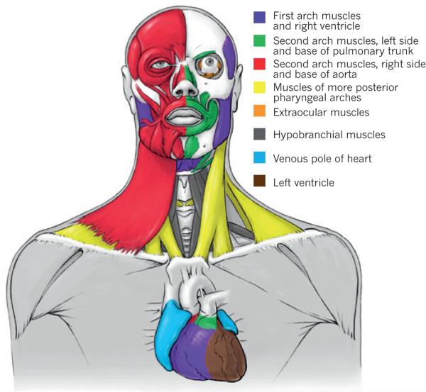
Figure 1. The striking heterogeneity of the human head and heart musculature.
The head includes at least six different muscle groups, all arising from the cardiopharyngeal field and being branchiomeric, except the hypobranchial and perhaps the extraocular muscles. On the left side of the body (right part of figure) the facial expression muscles have been removed to show the masticatory muscles. The six groups are: first/mandibular arch muscles, including cells clonally related to the right ventricle; left second/hyoid arch muscles related to myocardium at the base of the pulmonary trunk; right second/hyoid arch muscles, related to myocardium at the base of the aorta; muscles of the most posterior pharyngeal arches, including muscles of the pharynx and larynx and the cucullaris-derived neck muscles trapezius and sternocleidomastoideus; extraocular muscles, which are often not considered to be branchiomeric, but according to classic embryological studies and recent retrospective clonal analyses in mice contain cells related to those of the branchiomeric mandibular muscles; and hypobranchial muscles, including tongue and infrahyoid muscles that derive from somites and migrate into the head and neck36,38,70.
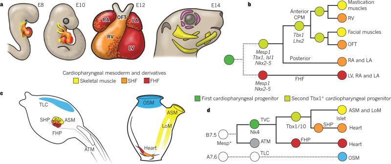
Figure 2. An evolutionarily conserved cardiopharyngeal ontogenetic motif.
a, Mouse embryos at embryonic days (E)8 and 10, the four-chambered mouse heart at E12, and the mouse head at E14. First heart field (FHF)-derived regions of heart (left ventricle (LV) and atria) are in red; second heart field (SHF)-derived regions of heart (right ventricle (RV), left atrium (LA), right atrium (RA) and outflow tract (OFT)) are in orange; branchiomeric skeletal muscles are in yellow; extraocular muscles are in purple. b, Lineage tree depicting the origins of cardiac compartments and branchiomeric muscles in mice. All cells derive from common pan-cardiopharyngeal progenitors (dark green) that produce the FHF, precursors of the left ventricle and atria, and the second Tbx1+ cardiopharyngeal progenitors (light green). Broken lines indicate that the early common FHF and SHF progenitor remains to be identified in mice. In anterior cardiopharyngeal mesoderm (CPM), progenitor cells activate Lhx2, self-renew and produce the SHF-derived RV and OFT, and first and second arch branchiomeric muscles (including muscles of mastication and facial expression). c, Cardiopharyngeal precursors in Ciona intestinalis hatching larva (left) and their derivatives in the metamorphosed juvenile (right). The first heart precursors (FHP) (red) and second heart precursors (SHP) (orange) contribute to the heart (red and orange mix), whereas atrial siphon muscle precursors (ASM, yellow) form atrial siphon and longitudinal muscles (LoM, yellow). Oral siphon muscles (OSM, blue) derive from a heterogenous larval population of trunk lateral cells (TLC, blue). ATM, anterior tail muscles. CPM is bilaterally symmetrical around the midline (dotted line). d, Lineage tree depicting clonal relationships and gene activities deployed in C. intestinalis cardiopharyngeal precursors. All cells derive from Mesp+ B7.5 blastomeres, which produce ATM (grey, see also left panel of c) and trunk ventral cells (TVC, dark green). The latter pan-cardiopharyngeal progenitors express Nk4 and divide asymmetrically to produce the FHP (red) and second TVCs, the Tbx1/10+ second cardiopharyngeal progenitors (second TVC, light green disk). The latter divide again asymmetrically to produce SHP (orange) and the precursors of ASM and LoM, which upregulate Islet. The OSM arise from A7.6-derived trunk lateral cells (TLC, light blue).
A suite of regulatory factors integrates the intercellular signals that coordinate the formation of cardiac and branchiomeric muscles from a common pool of mesodermal progenitor cells. Within the CPF there is considerable overlap in the expression of genes that encode cardiogenic regulatory factors (for example, Isl1 (also known as Islet1) and Nkx2-5) and those that specify head muscles (for example, Tbx1, Tcf21 (also known as capsulin), Msc (also known as MyoR) and Pitx2)13,15,20.
Importantly, many of the intercellular signalling pathways and transcription factors that control branchiomeric myogenesis upstream of the MyoD family of myogenic determination factors differ fundamentally from those operating in the trunk21,22. Here we focus on Isl1, Nkx2-5 and Tbx1. The LIM-homeodomain protein Isl1 is required in a broad subset of cardiovascular progenitor cells in mouse embryos23 and it is expressed in pharyngeal mesoderm, including the pharyngeal arches and SHF. Isl1+ progenitor cells substantially contribute to the heart and branchiomeric muscles, but not to hypobranchial (for example, tongue) or extraocular (eye) muscles13,24. Expression and functional studies indicate that Isl1 delays differentiation of bran-chiomeric muscles13,24; Isl1 thus marks a subset of CPF cells and plays an important part in the development of distinct cardiovascular and skeletal muscle progenitors24. The cardiac transcription factor Nkx2-5 regulates proliferation in the SHF and acts with Isl1 to modulate SHF progenitor-specific gene expression25–27. Tbx1 is required within the CPF for both heart and head muscle development, and is the major candidate gene for the congenital condition DiGeorge syndrome (or 22q11.2 deletion syndrome), which is characterized by a spectrum of cardiovascular defects and craniofacial anomalies. Like Isl1, Tbx1 has a crucial and conserved role in extending the heart's arterial pole by promoting proliferation and delaying differentiation of SHF cells28–31. Tbx1 is also required for activation of branchiomeric myogenesis and may directly regulate the myogenic determination gene MyoD32–34. Tbx1 acts upstream of the LIM-homeodomain protein Lhx2 within an intricate regulatory network that specifies cardiopharyngeal progenitors. Genetic ablation of these factors, alone or in combination, results in cardiac and head muscle defects; including DiGeorge syndrome phenotypes35. Thus, evolutionarily conserved regulatory factors maintain a pool of cardiopharyngeal progenitor cells for SHF-specific cardiogenesis and branchiomeric myogenesis.
Confirmation that multipotent progenitor cells give rise to branchiomeric skeletal muscles and SHF-derived regions of the heart comes from retrospective clonal analyses in mice, a method for analysing cell lineage in intact embryos36. These experiments demonstrated the existence of a series of common cardiopharyngeal progenitors along the anteroposterior axis that contribute to heart-tube growth and branchiomeric muscle morphogenesis. Interestingly, comparative anatomists suggested decades ago that branchiomeric muscles are related to muscles derived from the ‘visceral’ mesoderm (for example, of the heart and anterior gut)37,38, a view supported by the recent genetic and developmental studies reviewed here. Moreover, mouse clonal analyses revealed relationships between specific regions of the heart and subsets of branchiomeric muscles that go beyond the predictions of early comparative anatomists. SHF-derived regions of the heart, for example, are developmentally more closely related to branchiomeric muscles than to FHF-derived regions of the heart7,36. In support of such a grouping, the cardiac lineages contributing to the FHF and SHF have been shown to diverge before expression of Mesp1 during early gastrulation39,40. Taken together, recent findings provide a new paradigm for exploring the collinear emergence of cardiac chambers and branchiomeric muscles that underlies the early evolution and diverse origins of the vertebrate head9,21,22,41,42.
Origins and diversity of cardiopharyngeal structures
The heads of mammals, including humans, contain more than 60 muscles43, which control eye movements and allow food uptake, respiration, and facial and vocal communication44–46. Strikingly, the human head includes at least six different groups of muscles with distinct developmental origins and evolutionary histories35,37,44 (Fig. 1). Full recognition and detailed knowledge of this heterogeneity has enormous basic science and clinical implications because long accepted anatomy concepts, mainly based on adult function and physiology (for example, skeletal compared with cardiac muscles) do not correspond to the true developmental and evolutionary origins of body structures. Even the conventional classification of head muscle groups based on topographical relations masks the true heterogeneity of muscle origins and progenitor fates (for example, molecular profiling of early determinative signalling molecules and transcription factors reveals almost as much heterogeneity within each group — such as, branchial, extraocular and tongue — as between them43).
Comparative anatomical studies identified homologues of many amniote branchiomeric muscles in gnathostome (jawed) fish such as sharks, suggesting that they have ancient origins47,48 (Fig. 3). Cyclostomes (hagfish and lampreys49–52) lack some of these muscles (for example, the cucullaris group), but like some chondrichthyans (Selachii and Holocephali) they possess an additional, seventh group of head muscles: epibranchial muscles, which are derived from anterior somites53. Thus, extraocular, branchiomeric, and both hypobranchial and epibranchial somite-derived muscles were integral parts of the heterogenous head musculature of early vertebrates54–57 (Fig. 3). Moreover, lamprey embryos express homologues of Isl1, Nkx2-5 and Tbx1 in seemingly overlapping anterior and ventral mesodermal domains58–61, comparable with the patterns of their homo-logues in the amniote CPF. Interestingly, the emergence of heterogeneous head-muscle groups at the base of vertebrates coincided with the emergence of chambered hearts62,63 (Fig. 3). This intriguing correlation suggests that the two innovations are linked by their common developmental origin in the CPF.
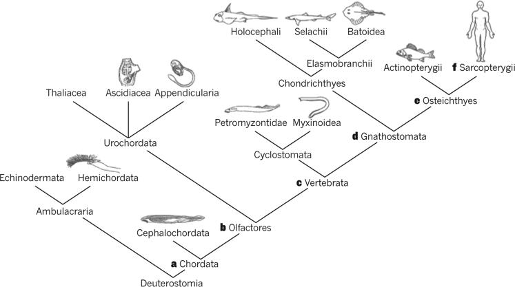
Figure 3. Some of the synapomorphies of the Chordata and its subgroups, according to our own data and review of the literature.
a, Somites and branchiomeric muscles. b, Placodes, neural-crest-like cells and cardiopharyngeal field (CPF) (although within invertebrates, conclusive evidence for these features was only reported in urochordates, some of these features may have been already present in the last common ancestor of extant chordates) giving rise to first- and second-heart-field-derived parts of the heart and to branchiomeric muscles (possibly not all of them, that is, inclusion of oral/velar muscles into CPF might have occurred during vertebrate evolution). c, Skull, cardiac chambers, and differentiation of epibranchial and hypobranchial somitic muscles. d, Jaws and differentiation between hypaxial and epaxial somitic musculature; paired appendages and fin muscles; origin of the branchiomeric muscle cucullaris. e, Loss of epibranchial muscles; cucullaris divided into levatores arcuum branchialium (going to pharyngeal arches) and protractor pectoralis (going to pectoral girdle), an exaptation that later allowed the emergence of the tetrapod neck. f, Within sarcopterygians, the protractor pectoralis gave rise to the amniote neck muscles trapezius and sternocleidomastoideus.
Studies indicate that specific branchiomeric muscles were crucial for evolutionary innovations among vertebrates, such as the emergence of the tetrapod neck. The amniote neck muscles trapezius and sternocleidomastoideus (Fig. 1) derive from the cucullaris, a muscle that probably appeared in early gnathostomes and was found in fossil placoderms5,6,48,64,65. Among extant gnathostomes, some of the anatomical and developmental characteristics of the cucullaris are shared with branchiomeric and somite-derived limb, epibranchial and hypobranchial muscles57,66,67. Most available data, however, indicate that the cucullaris is a branchiomeric muscle derived from the posterior-most pharyngeal arches, as suggested by Edgeworth22,68–71. Like other bran-chiomeric muscles, in most gnathostomes the cucullaris is attached to neural-crest-derived tendinous and skeletal elements38,64,65,70,72. Further-more, Tbx1 is active in core branchiomeric muscles (for example, the first and second arch muscles) and in the cucullaris-derived trapezius, whereas Pax3 is required in the somites for limb, diaphragm, tongue, infrahyoid and trunk-muscle formation, but not for trapezius formation22,73. These findings may also support Gegenbaur's hypothesis that the pectoral appendage, to which the cucullaris and its derivatives usually attach, probably originated as an integral part of the head74,75. Thus, the evolutionary history of the cucullaris-related muscles illustrates the roles that branchiomeric muscles had in fostering anatomical and functional innovations during vertebrate evolution. Future studies are needed to investigate whether the emergence of the cucullaris at the base of gnathostomes coincided with cardiovascular innovations and, if so, whether this muscle also shares a common origin with a specific heart region (Fig. 1).
A urochordate cardiopharyngeal ontogenetic motif
Recent phylogenetic studies place the urochordates — not the cephalochordates (for example, amphioxus) — as the sister group of the vertebrates76,77. On the basis of these results, urochordates provide important insights for our understanding of the origin of vertebrates’ evolutionary innovations, particularly from molecular and developmental perspectives. For instance, the new head hypothesis proposed that the emergence of branchiomeric muscles occurred during the transitions that led to the origin of vertebrates, and was associated with a shift from ‘passive’ filtration to more active feeding modes1,4,78,79 and the emergence of crest- and placode-derived sensory organs. However, recent studies have identified neural-crest-like cells, placodes and a CPF in tadpole-like larvae of the ascidian Ciona intestinalis, a model urochordate (Figs 2, 4). The pan-placodal regulatory gene Six1/2 is expressed in a crescent of cells straddling the anterior-most region of the developing neural tube in C. intestinalis embryos, comparable with the sites of origin of cranial placodes in the fate maps of vertebrates80–82. Ectodermal thickenings derived from this domain express placodal regulatory genes, including Six3/6, Pitx and Eya. For example, the atrial siphon placode shares extensive similarities with the vertebrate otic placode3,80,81 (Fig. 4), whereas the stomodeum (the oral siphon primordium) expresses regulatory genes implicated in the specification of the vertebrate olfactory and adenohypophyseal placodes, including Six, Eya and the anterior placode markers Pitx83–85 and Dlx. These new findings argue for homologies between urochordate siphon primordia and vertebrate placodes and suggest that; although certain placodes (profundal, maxillomandibular, epibranchial and lens) evolved by diversification within the vertebrate lineage3, others (adenohypophyseal, olfactory and otic) appeared before the separation of vertebrates and urochordates (Figs 3, 4).
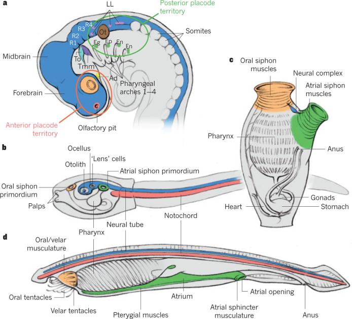
Figure 4. Homology hypotheses of placodes and branchiomeric muscles within chordates.
a, Location of ectodermal placodes in the vertebrate head according to Graham and Shimeld's3 hypothesis (anterior to the left): olfactory placode or pit (red) at the tip of the forebrain; lens placodes (orange) form posteriorly as part of eye; adenohypophyseal placode (Ad, yellow) lies ventrally to forebrain; trigeminal placodes form alongside the anterior hindbrain at the levels of rhombomeres 1 and 2 (R1 and R2), the anterior one being the ophthalmic placode (To, light blue) and the posterior one the maxillomandibular placode (Tmm, purple); otic placode (Ot, brown) forms opposite the central domain of hindbrain; lateral line placodes (LL, pink) form anteriorly and posteriorly to otic placode; epibranchial placodes (green) — geniculate (Eg), petrosal (Ep) and nodose (En) — form as part of pharyngeal series. Forebrain, midbrain and R1–4, and neural tube are shown in dark blue. b, Urochordate tadpole-like larva (anterior to the left). The notochord is in red and two siphon primordia are in green and orange, with putative relationships to the anterior and posterior placode territories shown in a. c, Adult urochordate showing siphon primordia after metamorphosis. d, Adult cephalochordate showing the urochordate–cephalochordate muscle homology hypotheses proposed in the present Review. Figures based on images from refs 3, 22, 105.
Ascidians and other urochordates possess a surprisingly sophisticated beating heart (Figs 2, 4), which shares several features with vertebrate hearts, including localized pacemakers that drive a regular, rhythmic beat. The ascidian heart is derived from two Mesp+ cells in early embryos. These produce four trunk ventral cells, which express homologues of Nkx2-5, Gata4, 5 and 6 and Hand, and migrate towards the pharyngeal endoderm86–92. They subsequently divide asymmetrically to produce medial heart precursors and secondary trunk ventral cells that divide again to produce second heart precursors and atrial siphon muscle precursors, which migrate towards the atrial siphon placode93–95 (Figs 2, 4). Thus, trunk ventral cells are multipotent cardiopharyngeal progenitors that produce bona fide heart and pharyngeal muscles, following a clonal pattern evocative of that seen in mice (Fig. 2). Gene-expression profiling data are also consistent with the idea that the trunk ventral cells are homologous to the vertebrate cardiopharyngeal progenitors: trunk ventral cells express Nk4, the homologue of Nkx2-5, and secondary trunk ventral cells also express Tbx1/10, which is active in vertebrate pharyngeal mesoderm. Furthermore, the regulatory network governing interactions among the cardiopharyngeal specification genes seems to be highly conserved in ascidians and vertebrates. For example, cross-repressive interactions between Tbx1/10 and Nk4/Nkx2-5 delineate atrial siphon muscles and heart, respectively95. Isl is also expressed in the CPF, although there are differences from the precise expression profile seen in vertebrates, where Isl1 is thought to delay muscle differentiation24. It is nonetheless striking that all of the identified molecular determinants of the vertebrate SHF are expressed in ascidian trunk ventral cells.
There are additional parallels between the CPFs of ascidians and vertebrates in the regulatory circuitry underlying the differentiation of specialized muscles (Fig. 2). COE/Ebf functions downstream of Tbx1/10 and upstream of both Mrf/MyoD and Notch signalling to promote either early muscle differentiation or maintain undifferentiated precursors that produce most later atrial siphon and longitudinal muscles93,96 (Fig. 2). Atrial siphon muscle precursors also associate with the Dlx+ atrial siphon placodes to form a ring of cells underlying the rosette-shaped placode in C. intestinalis swimming larvae80,81,93,97. These events parallel the migration of vertebrate branchiomeric muscle precursors into pharyngeal arches, their association with Dlx+ cranial neural crest cells, and the maintenance and growth of a pool of undifferentiated progenitor cells24,98. It is noteworthy that the ascidian FHF and SHF are each initially composed of four cells that independently arise from one of four multipotent cardiopharyngeal progenitors following a sequence of conserved regulatory interactions onto a stereotyped clonal pattern, producing FHF precursors and more closely related SHF and pharyngeal muscle precursors95. We refer to this clonal sequence of cell divisions, gene expression and cell-fate choices as a cardiopharyngeal ontogenetic motif95 (Fig. 2).
Chordate origins of branchiomeric muscles
Studies using cephalochordates further probed the early chordate origins of branchiomeric-like pharyngeal muscles (Figs 3, 4). In the cephalochor-date amphioxus, the larval mouth and unpaired primary gills develop five groups of orobranchial muscles99,100. This musculature is anatomically reminiscent of the vertebrate branchiomeric muscles, and disappears through apoptosis during metamorphosis to give way to adult oral, velar and pterygial muscles99 (Fig. 4), which are even more similar to vertebrate adult branchiomeric muscles. The oral and velar muscles, in particular, share anatomical similarities with the oral and velar muscles of lampreys and hagfish (Fig. 4), although the pterygial muscles have a branchiomeric-like innervation pattern99. Gans79 recognized this latter point and noted that this could mean that the branchiomeric muscles evolved before the last common ancestor (LCA) of vertebrates, as suggested by earlier authors22, but contrary to the original new head hypothesis1. Vestigial muscles appear transiently with secondary gill formation in amphioxus, providing additional evidence that bilateral muscular gills and a segmental pattern of branchiomeric muscles were already present in the LCA of extant chordates22.
Molecular studies suggest that the amphioxus homologues of Tbx1, Nkx2-5 and Isl1 are expressed in overlapping mesodermal domains in the pharyngeal region101–103. This domain includes cells that also express the vertebrate cardiac markers Hand and Tbx20 (refs 59, 104) and is thought to produce the branchial artery, a possible — but controversial — homo-logue of the heart with diffuse contractility105. These observations raise the possibility that the LCA of extant chordates had a CPF. However, contrary to urochordates and vertebrates, cephalochordates have a rather diffuse heart-like vasculature and their branchial muscles seem to develop independently of Ebf and Mrf homologues94,106,107. Amphioxus Mrf homo-logues seem to be expressed exclusively in somites, overlapping with the Pax3/7 homologue106,108, but also with the Tbx1 homologue102, suggesting the presence of distinct Tbx1+, Pax3/7+, Mrf+ somitic and Tbx1+, Pax3/7−, Mrf− pharyngeal mesodermal domains in ancient chordates.
Branchiomeric-like muscles, such as the cephalochordate oral, velar and pterygial muscles (Fig. 4), thus probably predate the origin of a CPF as defined in urochordates and vertebrates (Fig. 3). Comparative anatomical studies suggest that the pterygial and orovelar muscles of adult amphioxus probably correspond to the atrial and oral siphon muscles of urochordates, respectively (Fig. 4). Remarkably, the ascidian oral siphon muscles (Fig. 4), which control mouth movements in post-metamorphic animals, do not derive from cardiopharyngeal progenitors93,109,110 (Fig. 2). This is in contrast with the anterior oral muscles controlling mouth movements and in particular jaw opening (first (mandibular) arch muscles) in gnathostomes, which are CPF derivatives (Fig. 2). Comparative studies of basal chordates, including that of the fossil Haikouella, suggested that their pharyngeal arch series started with the second (hyoid) arch and that only during early vertebrate evolution did parts of the anterior mesoderm become incorporated into the pharyngeal series by forming a new, _Hox_-independent first arch111,112. Therefore, it is possible that the incorporation of the more anterior (first) arch in this series during vertebrate evolution was accompanied by integration of the associated oral and velar muscles into the CPF. This evolutionary scenario implies that the amphioxus orovelar muscles and urochordate oral siphon muscles may be homologous to the cyclostome orovelar muscles and gnathostome mandibular muscles, which could potentially explain why these muscles are derived from the CPF only in vertebrates.
Bilaterian roots of the cardiopharyngeal network
We have argued that the presence of a CPF, with dual cardiac and skeletal myogenic capacity, is probably a synapomorphy of olfactores (a derived feature shared by urochordates and vertebrates; Figs 2, 3). This argument raises the question: do the developmental, cellular and/or molecular units that form the CPF network of olfactores have even deeper evolutionary origins? Ambulacraria (echinoderms and hemichordates) is the sister group of chordates (Fig. 3). Hemichordates possess well-defined serial gill slits and a heart–kidney complex located in the anterior-most body part (proboscis)113. Serially arranged pharyngeal gill openings have associated muscles in enteropneust-type hemichordates, but this musculature seems to be developmentally, anatomically and histologically distinct from the chordate branchiomeric musculature99. Moreover, the Tbx1 homologue of Saccoglossus kowalevskii, an enteropneust hemichordate, is not expressed in the mesodermal core of the pharyngeal pouches114, suggesting that Tbx1 expression in pharyngeal mesoderm is a chordate synapomorphy. Further studies of ambulacrarians will test this hypothesis.
Among non-deuterostome animals, nematodes lack a heart and a defined circulatory system, but possess pharyngeal muscles that contract rhythmically, exhibit electrical activity similar to mammalian cardiomyocytes, and require ceh-22, the homologue of Nkx2-5 (refs 9, 21, 22, 41, 42, 115). Flies lack anatomical structures that are comparable with the chordate pharyngeal apparatus, but the Drosophila homologues of Tbx1, Nkx2-5, Isl, Ebf and Mrf/MyoD variably contribute to visceral, larval and adult skeletal and/or heart muscle specification116–121. The diversity of myogenic networks driving muscle identity and differentiation in flies is reminiscent of the heterogeneity of myogenic origins and programs operating in the vertebrate head. Furthermore, visceral and dorsal larval muscles in Drosophila develop from mesoderm in proximity to the dorsal vessel or fly heart. It is therefore conceivable that many features of the CPF gene regulatory network predate the advent of chordates and, moreover, that this regulatory circuitry preceded the emergence of the well-studied myogenic hierarchies controlling vertebrate somitic muscle development.
Evolvable cardiopharyngeal units
Here, we summarize our arguments for the origins and diversification of the CPF (Fig. 3). Filter-feeding early chordates, endowed with serial gill slits inherited from deuterostome ancestors, already had gill-associated branchiomeric, or at least branchiomeric-like, muscles (Fig. 4). A well-defined CPF then probably appeared in the olfactores. Ancestral vertebrates uncoupled myogenic specification and differentiation, thus increasing the population of cardiopharyngeal progenitors. This facilitated the emergence of cardiac chambers by progressive addition of progenitor cells to the growing heart tube during development. It also allowed for the expansion and diversification of branchiomeric muscles, contributing to increased muscularization of the pharyngeal apparatus that was essential for the transition to a predatory lifestyle. The latter was made possible by olfactores’ ancestral association between branchiomeric muscles and Dlx+ ectoderm cells. Elaboration of this interaction permitted coevolution of the branchiomeric musculature with the newly formed neural crest-derived craniofacial skeleton, linking the novel neural-crest-derived skeletal patterns with distinct branchiomeric muscles.
We propose that the heart and atrial siphon muscle gene network seen in the urochordate C. intestinalis illustrates the basic ontogenetic motif underlying the specification of the vertebrate CPF95, and suggest three ways in which this blueprint was modified to produce the vast diversity of cardiopharyngeal patterns in vertebrates: the ontogenetic motif could be deployed in multiple independent embryonic progenitors; any given progenitor could self-renew, thus being transiently amplified, before generating distinct heart, in contrast with branchiomeric, muscle precursors and any given cell could migrate and/or be passively displaced and resume cardiopharyngeal development in different locations on receipt of appropriate signals. In contrast to their ascidian counterparts, vertebrate Tbx1+ and Isl1+ cardiopharyngeal progenitors remain in an elusive niche in which they self-renew to produce SHF-derived heart precursors. During pharyngeal morphogenesis, these emerge sequentially to produce right ventricular and outflow tract cardiomyocytes. Conceivably, multiple independent cardiopharyngeal lineages developing in series may contribute to divergent cardiac and branchiomeric myogenic cell fates along the anterior–posterior pharyngeal mesoderm of vertebrates. This hypothesis is consistent with the observation that subsets of cardiac and branchiomeric muscles are more closely related to each other than to other heart and head muscles (Fig. 1)36,122,123. Future experiments will determine whether anteroposterior patterning of the CPF precedes segmentation of the pharyngeal region during arch morphogenesis.
General remarks and future directions
The CPF is a new paradigm to be reckoned with, and should take centre stage along with neural crest and cranial placodes when considering the origin of the vertebrate head. Importantly, novel insights from comparative, phylogenomic and developmental genetics studies have uncovered the deep evolutionary origins of the CPF, branchiomeric muscles, pla-codes and neural crest cells. Like vertebrates, urochordates have a CPF that gives rise to the FHF, SHF and branchiomeric muscles; moreover, apart from their neural-crest-like cells and placodes, at least some pelagic urochordates have highly developed brains124. Data obtained after Gans and Northcutt's new head hypothesis thus call into question the clear distinction between vertebrates and other animals, and show that the ‘new’ head arose instead by elaboration and modification of existing tissues, cell populations and gene networks through evolutionary ‘tinkering’. This revelation supports the proposal125 that the conventional view of vertebrates evolving from brainless ascidian-like filter-feeders through a progressive increase in complexity and emergence of several de novo structures, with no evolutionary losses or reversions, is an oversimplification. These data also emphasize the heterogeneity and complex developmental and evolutionary history of vertebrate hearts and heads, blurring the interface between head and trunk, extraocular and branchiomeric, and skeletal and cardiac myogenesis. Adult postcranial structures, including the heart and part of the neck musculature, include cells derived from the CPF (Fig. 1); reciprocally, cephalic structures such as the tongue and infrahyoid muscles arise from somitic primordia located in the trunk. The discovery of the CPF therefore provides a more complete, and complex, view of the origin and early evolution of the vertebrate head.
However, many questions remain. For example, how is the multipotency of branchiomeric and cardiac myocyte progenitor cells encoded in the CPF, and is there a defined molecular common niche in which these multipotent progenitor cells arise? How, and during what stages, are progenitor cell populations that give rise to different regions of the heart and head muscles specified in pharyngeal mesoderm? Recognition of the CPF also sets the stage for future discoveries in human medicine (Fig. 1). An important question is why many myopathies preferentially affect a specific subset of muscles, and whether these aetiologies are linked to the disparate embryonic histories of these muscles. As already noted, the clinical features of DiGeorge syndrome — one of the most common human congenital syndromes — include cardiovascular and craniofacial birth defects, highlighting the frequent link between these defects owing to their anatomical proximity during early embryogenesis and overlapping progenitor populations9,21,42. Therefore, the studies and data discussed here open promising new directions for biomedical research and the advancement of public health. For instance, future meta-analyses may reveal pathological relationships between specific branchiomeric muscles and regional congenital heart defects. The field of evolutionary developmental biology has progressed remarkably over the three decades since the new head hypothesis was published. With the recent revolutionary discoveries and more exciting work already begun, the field is poised to move ahead anew.
BOX 1 Glossary.
- Branchiomeric muscles. Muscles formed from progenitor cells found in the pharyngeal arches. In vertebrates, they comprise the mandibular (first arch muscles, such as jaw muscles), hyoid (second arch muscles, such as the facial expression muscles of mammals) and branchial (from more posterior arches, including muscles of the larynx and pharynx, and the cucullaris-derived neck muscles trapezius and sternocleidomastoideus, in amniotes) muscles.
- Pharyngeal (or branchial) arches. Bilateral swellings on either side of the pharynx comprising outer (ectodermal) and inner (endodermal) epithelia, neural-crest-derived mesenchyme and a mesodermal core.
- First heart field. Population of early differentiating cardiac progenitor cells that arise in anterior lateral mesoderm and give rise to the linear heart tube and, later, to the left ventricle and parts of the atria.
- Second heart field. Population of late differentiating cardiac progenitors that contribute to the developing heart after the linear heart tube stage to give rise to myocardium of the right ventricle and outflow tract, and to inflow tract myocardium, including parts of the atria.
- Cardiopharyngeal field. Includes anterior lateral mesoderm of the first heart field plus contiguous pharyngeal mesoderm that gives rise to second-heart-field-derived regions of the heart and branchiomeric muscles.
- Cardiopharyngeal ontogenetic motif. Lineage-specific progression through cardiopharyngeal progenitor cell identities, with conserved clonal relationships between first heart, second heart and pharyngeal muscle precursors characterized by specific gene expression and regulatory activities.
- Pharyngeal mesoderm. Cranial mesoderm associated with the forming foregut or pharynx that populates pharyngeal arches and contributes to second-heart-field-derived regions of the heart and branchiomeric muscles.
Acknowledgements
We thank T. Miyashita and F. Razy-Krajka for their detailed reviews of the manuscript. We are thankful to the Dean of Howard University (HU) College of Medicine, M. Johnson, and the Chair of HU Department of Anatomy, D. Orlic, for helping to organize, financially and logistically, the First Evo-Devo Meeting On Heart and Head Muscles at HU (May, 2014) that led to the publication of this Review. We also thank the other participants at the workshop: A. Kahana, P. Okkema, A. Vincent, T. Hirasawa, S. Tajbakhsh, S. Dietrich and R. Knight. L.C. is supported by National Institutes of Health (NIH)/National Institute of General Medical Sciences (NIGMS) grant R01GM096032 and NIH/National Heart, Lung and Blood Instiute (NHLBI) grant R01HL108643, E.T. by the European Research Council and Israel Science Foundation, R.D. and J.Z. by HU College of Medicine, R.G.K. by Inserm, the Agence Nationale pour la Recherche, Association Française contre les Myopathies and Fondation pour la Recherche Médicale, and M.L. by NIH grant NS076542.
Footnotes
The authors declare no competing financial interests.
Readers are welcome to comment on the online version of this paper at go.nature.com/wy4cga.
A paper has been published while the current Review was in press reporting the identification of a third group of bilateral common heart and skeletal muscle progenitor cells within the murine CPM. Using retrospective lineage analysis, cucullaris-derived neck muscles, the trapezius and sternocleidomastoid, were shown to be clonally related to myocardium at the venous pole of the heart, derived from the posterior SHF. These findings reinforce the hypothesis of a branchiomeric origin of these neck muscles (F. Lescroart et al. Clonal analysis reveals a common origin between nonsomite-derived neck muscles and heart myocardium. Proc. Natl Acad. Sci. USA 112, 1446–1451; 2015).
References
- 1.Gans C, Northcutt RG. Neural crest and the origin of vertebrates: a new head. Science. 1983;220:268–273. doi: 10.1126/science.220.4594.268. [This highly influential paper argued that the evolution of head structures derived from neural crest and cranial placodes had a crucial role in the transition to early vertebrates.] [DOI] [PubMed] [Google Scholar]
- 2.Patthey C, Schlosser G, Shimeld SM. The evolutionary history of vertebrate cranial placodes — I: cell type evolution. Dev. Biol. 2014;389:82–97. doi: 10.1016/j.ydbio.2014.01.017. [DOI] [PubMed] [Google Scholar]
- 3.Graham A, Shimeld SM. The origin and evolution of the ectodermal placodes. J. Anat. 2013;222:32–40. doi: 10.1111/j.1469-7580.2012.01506.x. [DOI] [PMC free article] [PubMed] [Google Scholar]
- 4.Northcutt RG. The new head hypothesis revisited. J. Exp. Zool. B Mol. Dev. Evol. 2005;304B:274–297. doi: 10.1002/jez.b.21063. [DOI] [PubMed] [Google Scholar]
- 5.Kuratani S. Evolution. A muscular perspective on vertebrate evolution. Science. 2013;341:139–140. doi: 10.1126/science.1241451. [DOI] [PubMed] [Google Scholar]
- 6.Trinajstic K, et al. Fossil musculature of the most primitive jawed vertebrates. Science. 2013;341:160–164. doi: 10.1126/science.1237275. [DOI] [PubMed] [Google Scholar]
- 7.Meilhac SM, Esner M, Kelly RG, Nicolas JF, Buckingham ME. The clonal origin of myocardial cells in different regions of the embryonic mouse heart. Dev. Cell. 2004;6:685–698. doi: 10.1016/s1534-5807(04)00133-9. [DOI] [PubMed] [Google Scholar]
- 8.Kelly RG. The second heart field. Curr. Top. Dev. Biol. 2012;100:33–65. doi: 10.1016/B978-0-12-387786-4.00002-6. [DOI] [PubMed] [Google Scholar]
- 9.Tzahor E, Evans SM. Pharyngeal mesoderm development during embryogenesis: implications for both heart and head myogenesis. Cardiovasc. Res. 2011;91:196–202. doi: 10.1093/cvr/cvr116. [DOI] [PMC free article] [PubMed] [Google Scholar]
- 10.Kelly RG, Brown NA, Buckingham ME. The arterial pole of the mouse heart forms from Fgf10-expressing cells in pharyngeal mesoderm. Dev. Cell. 2001;1:435–440. doi: 10.1016/s1534-5807(01)00040-5. [Discovery of the mammalian SHF, demonstrating that myocardium at the arterial pole of the heart originates in adjacent pharyngeal mesoderm.] [DOI] [PubMed] [Google Scholar]
- 11.Mjaatvedt CH, et al. The outflow tract of the heart is recruited from a novel heart-forming field. Dev. Biol. 2001;238:97–109. doi: 10.1006/dbio.2001.0409. [DOI] [PubMed] [Google Scholar]
- 12.Waldo KL, et al. Conotruncal myocardium arises from a secondary heart field. Development. 2001;128:3179–3188. doi: 10.1242/dev.128.16.3179. [DOI] [PubMed] [Google Scholar]
- 13.Nathan E, et al. The contribution of Islet1-expressing splanchnic mesoderm cells to distinct branchiomeric muscles reveals significant heterogeneity in head muscle development. Development. 2008;135:647–657. doi: 10.1242/dev.007989. [This article provides a definition of the contribution of pharyngeal mesoderm to branchiomeric muscles in both chick and mouse embryos.] [DOI] [PMC free article] [PubMed] [Google Scholar]
- 14.Mesbah K, et al. Identification of a Tbx1/Tbx2/Tbx3 genetic pathway governing pharyngeal and arterial pole morphogenesis. Hum. Mol. Genet. 2012;21:1217–1229. doi: 10.1093/hmg/ddr553. [DOI] [PMC free article] [PubMed] [Google Scholar]
- 15.Tirosh-Finkel L, Elhanany H, Rinon A, Tzahor E. Mesoderm progenitor cells of common origin contribute to the head musculature and the cardiac outflow tract. Development. 2006;133:1943–1953. doi: 10.1242/dev.02365. [This article demonstrates, using fate-mapping and experimental manipulation in the avian embryo, that cranial mesoderm gives rise both to head muscles and outflow tract myocardium.] [DOI] [PubMed] [Google Scholar]
- 16.Tzahor E, Lassar AB. Wnt signals from the neural tube block ectopic cardiogenesis. Genes Dev. 2001;15:255–260. doi: 10.1101/gad.871501. [DOI] [PMC free article] [PubMed] [Google Scholar]
- 17.Noden DM, Trainor PA. Relations and interactions between cranial mesoderm and neural crest populations. J. Anat. 2005;207:575–601. doi: 10.1111/j.1469-7580.2005.00473.x. [DOI] [PMC free article] [PubMed] [Google Scholar]
- 18.Hutson MR, Kirby ML. Neural crest and cardiovascular development: a 20-year perspective. Birth Defects Res. C Embryo Today. 2003;69:2–13. doi: 10.1002/bdrc.10002. [DOI] [PubMed] [Google Scholar]
- 19.Rinon A, et al. Cranial neural crest cells regulate head muscle patterning and differentiation during vertebrate embryogenesis. Development. 2007;134:3065–3075. doi: 10.1242/dev.002501. [DOI] [PubMed] [Google Scholar]
- 20.Bothe I, Dietrich S. The molecular setup of the avian head mesoderm and its implication for craniofacial myogenesis. Dev. Dynam. 2006;235:2845–2860. doi: 10.1002/dvdy.20903. [DOI] [PubMed] [Google Scholar]
- 21.Grifone R, Kelly RG. Heartening news for head muscle development. Trends Genet. 2007;23:365–369. doi: 10.1016/j.tig.2007.05.002. [DOI] [PubMed] [Google Scholar]
- 22.Sambasivan R, Kuratani S, Tajbakhsh S. An eye on the head: the development and evolution of craniofacial muscles. Development. 2011;138:2401–2415. doi: 10.1242/dev.040972. [DOI] [PubMed] [Google Scholar]
- 23.Cai CL, et al. Isl1 identifies a cardiac progenitor population that proliferates prior to differentiation and contributes a majority of cells to the heart. Dev. Cell. 2003;5:877–889. doi: 10.1016/s1534-5807(03)00363-0. [DOI] [PMC free article] [PubMed] [Google Scholar]
- 24.Harel I, et al. Distinct origins and genetic programs of head muscle satellite cells. Dev. Cell. 2009;16:822–832. doi: 10.1016/j.devcel.2009.05.007. [This article demonstrates the diversity of lineages constituting craniofacial skeletal muscles and their associated satellite cells using a series of Cre lines to genetically trace trunk and cranial myogenic progenitor cells, leading to an Isl1-lineage-based definition of CPF-derived craniofacial muscles.] [DOI] [PMC free article] [PubMed] [Google Scholar]
- 25.Dodou E, Verzi MP, Anderson JP, Xu SM, Black BL. Mef2c is a direct transcriptional target of ISL1 and GATA factors in the anterior heart field during mouse embryonic development. Development. 2004;131:3931–3942. doi: 10.1242/dev.01256. [DOI] [PubMed] [Google Scholar]
- 26.Watanabe Y, et al. Fibroblast growth factor 10 gene regulation in the second heart field by Tbx1, Nkx2–5, and Islet1 reveals a genetic switch for down-regulation in the myocardium. Proc. Natl Acad. Sci. USA. 2012;109:18273–18280. doi: 10.1073/pnas.1215360109. [DOI] [PMC free article] [PubMed] [Google Scholar]
- 27.Prall OW, et al. An Nkx2–5/Bmp2/Smad1 negative feedback loop controls heart progenitor specification and proliferation. Cell. 2007;128:947–959. doi: 10.1016/j.cell.2007.01.042. [DOI] [PMC free article] [PubMed] [Google Scholar]
- 28.Scambler PJ. 22q11 deletion syndrome: a role for TBX1 in pharyngeal and cardiovascular development. Pediatr. Cardiol. 2010;31:378–390. doi: 10.1007/s00246-009-9613-0. [DOI] [PubMed] [Google Scholar]
- 29.Liao J, et al. Identification of downstream genetic pathways of Tbx1 in the second heart field. Dev. Biol. 2008;316:524–537. doi: 10.1016/j.ydbio.2008.01.037. [DOI] [PMC free article] [PubMed] [Google Scholar]
- 30.Chen L, Fulcoli FG, Tang S, Baldini A. Tbx1 regulates proliferation and differentiation of multipotent heart progenitors. Circ. Res. 2009;105:842–851. doi: 10.1161/CIRCRESAHA.109.200295. [DOI] [PMC free article] [PubMed] [Google Scholar]
- 31.Hami D, Grimes AC, Tsai HJ, Kirby ML. Zebrafish cardiac development requires a conserved secondary heart field. Development. 2011;138:2389–2398. doi: 10.1242/dev.061473. [DOI] [PMC free article] [PubMed] [Google Scholar]
- 32.Kelly RG, Jerome-Majewska LA, Papaioannou VE. The del22q11.2 candidate gene Tbx1 regulates branchiomeric myogenesis. Hum. Mol. Genet. 2004;13:2829–2840. doi: 10.1093/hmg/ddh304. [This paper reports the genetic identification of Tbx1 as a regulator of craniofacial myogenesis in mice, supporting the existence of distinct upstream regulatory hierarchies controlling head and trunk myogenesis.] [DOI] [PubMed] [Google Scholar]
- 33.Kong P, et al. Tbx1 is required autonomously for cell survival and fate in the pharyngeal core mesoderm to form the muscles of mastication. Hum. Mol. Genet. 2014;23:4215–4231. doi: 10.1093/hmg/ddu140. [DOI] [PMC free article] [PubMed] [Google Scholar]
- 34.Castellanos R, Xie Q, Zheng D, Cvekl A, Morrow BE. Mammalian TBX1 preferentially binds and regulates downstream targets via a tandem T-site repeat. PLoS ONE. 2014;9:e95151. doi: 10.1371/journal.pone.0095151. [DOI] [PMC free article] [PubMed] [Google Scholar]
- 35.Harel I, et al. Pharyngeal mesoderm regulatory network controls cardiac and head muscle morphogenesis. Proc. Natl Acad. Sci. USA. 2012;109:18839–18844. doi: 10.1073/pnas.1208690109. [DOI] [PMC free article] [PubMed] [Google Scholar]
- 36.Lescroart F, et al. Clonal analysis reveals common lineage relationships between head muscles and second heart field derivatives in the mouse embryo. Development. 2010;137:3269–3279. doi: 10.1242/dev.050674. [This retrospective lineage analysis provides evidence for the existence of common progenitor cells in the mouse embryo that give rise to myocardium of the right ventricle and first-arch-derived muscles, and to the arterial pole of the heart and second-arch-derived muscles.] [DOI] [PubMed] [Google Scholar]
- 37.Romer AS, Parson TS. The Vertebrate Body. Saunder's College Publishing; 1977. [Google Scholar]
- 38.Diogo R, Abdala V. Muscles of Vertebrates: Comparative Anatomy, Evolution, Homologies and Development. CRC; 2010. [This monograph provides an overview on the comparative anatomy, evolution and homologies of the head and limb muscles in all major extant vertebrate groups with special focus on the developmental and evolutionary history of the muscles of Homo sapiens.] [Google Scholar]
- 39.Devine WP, Wythe JD, George M, Koshiba-Takeuchi K, Bruneau BG. Early patterning and specification of cardiac progenitors in gastrulating mesoderm. eLife. 2014;3:e03848. doi: 10.7554/eLife.03848. [DOI] [PMC free article] [PubMed] [Google Scholar]
- 40.Lescroart F, et al. Early lineage restriction in temporally distinct populations of Mesp1 progenitors during mammalian heart development. Nature Cell Biol. 2014;16:829–840. doi: 10.1038/ncb3024. [DOI] [PMC free article] [PubMed] [Google Scholar]
- 41.Olson EN. Gene regulatory networks in the evolution and development of the heart. Science. 2006;313:1922–1927. doi: 10.1126/science.1132292. [DOI] [PMC free article] [PubMed] [Google Scholar]
- 42.Tzahor E. Heart and craniofacial muscle development: a new developmental theme of distinct myogenic fields. Dev. Biol. 2009;327:273–279. doi: 10.1016/j.ydbio.2008.12.035. [DOI] [PubMed] [Google Scholar]
- 43.Diogo R, Wood BA. Comparative Anatomy and Phylogeny of Primate Muscles and Human Evolution. CRC; 2012. [Google Scholar]
- 44.Wachtler F, Jacob M. Origin and development of the cranial skeletal muscles. Bibl. Anat. 1986;1986:24–46. [PubMed] [Google Scholar]
- 45.Noden DM. The embryonic origins of avian cephalic and cervical muscles and associated connective tissues. Am. J. Anat. 1983;168:257–276. doi: 10.1002/aja.1001680302. [DOI] [PubMed] [Google Scholar]
- 46.Noden DM, Francis-West P. The differentiation and morphogenesis of craniofacial muscles. Dev. Dynam. 2006;235:1194–1218. doi: 10.1002/dvdy.20697. [DOI] [PubMed] [Google Scholar]
- 47.Diogo R, Hinits Y, Hughes SM. Development of mandibular, hyoid and hypobranchial muscles in the zebrafish: homologies and evolution of these muscles within bony fishes and tetrapods. BMC Dev. Biol. 2008;8:24. doi: 10.1186/1471-213X-8-24. [DOI] [PMC free article] [PubMed] [Google Scholar]
- 48.Diogo R, Abdala V, Lonergan N, Wood BA. From fish to modern humans — comparative anatomy, homologies and evolution of the head and neck musculature. J. Anat. 2008;213:391–424. doi: 10.1111/j.1469-7580.2008.00953.x. [DOI] [PMC free article] [PubMed] [Google Scholar]
- 49.Kuraku S, Hoshiyama D, Katoh K, Suga H, Miyata T. Monophyly of lampreys and hagfishes supported by nuclear DNA-coded genes. J. Mol. Evol. 1999;49:729–735. doi: 10.1007/pl00006595. [DOI] [PubMed] [Google Scholar]
- 50.Delarbre C, Gallut C, Barriel V, Janvier P, Gachelin G. Complete mitochondrial DNA of the hagfish, Eptatretus burgeri: the comparative analysis of mitochondrial DNA sequences strongly supports the cyclostome monophyly. Mol. Phylogenet. Evol. 2002;22:184–192. doi: 10.1006/mpev.2001.1045. [DOI] [PubMed] [Google Scholar]
- 51.Delarbre C, et al. The complete nucleotide sequence of the mitochondrial DNA of the agnathan Lampetra fluviatilis: bearings on the phylogeny of cyclostomes. Mol. Biol. Evol. 2000;17:519–529. doi: 10.1093/oxfordjournals.molbev.a026332. [DOI] [PubMed] [Google Scholar]
- 52.Heimberg AM, Cowper-Sal-lari R, Semon M, Donoghue PC, Peterson KJ. microRNAs reveal the interrelationships of hagfish, lampreys, and gnathostomes and the nature of the ancestral vertebrate. Proc. Natl Acad. Sci. USA. 2010;107:19379–19383. doi: 10.1073/pnas.1010350107. [DOI] [PMC free article] [PubMed] [Google Scholar]
- 53.Ziermann JM, Miyashita T, Diogo R. Cephalic muscles of Cyclostomes (hagfishes and lampreys) and Chondrichthyes (sharks, rays and holocephalans): comparative anatomy and early evolution of the vertebrate head. Zool. J. Linn. Soc. 2014;172:771–802. [Google Scholar]
- 54.Adachi N, Kuratani S. Development of head and trunk mesoderm in the dogfish, Scyliorhinus torazame: I. Embryology and morphology of the head cavities and related structures. Evol. Dev. 2012;14:234–256. doi: 10.1111/j.1525-142X.2012.00542.x. [DOI] [PubMed] [Google Scholar]
- 55.Adachi N, Takechi M, Hirai T, Kuratani S. Development of the head and trunk mesoderm in the dogfish, Scyliorhinus torazame: II. Comparison of gene expression between the head mesoderm and somites with reference to the origin of the vertebrate head. Evol. Dev. 2012;14:257–276. doi: 10.1111/j.1525-142X.2012.00543.x. [DOI] [PubMed] [Google Scholar]
- 56.Kuratani S, Adachi N, Wada N, Oisi Y, Sugahara F. Developmental and evolutionary significance of the mandibular arch and prechordal/premandibular cranium in vertebrates: revising the heterotopy scenario of gnathostome jaw evolution. J. Anat. 2013;222:41–55. doi: 10.1111/j.1469-7580.2012.01505.x. [DOI] [PMC free article] [PubMed] [Google Scholar]
- 57.Kusakabe R, Kuraku S, Kuratani S. Expression and interaction of muscle-related genes in the lamprey imply the evolutionary scenario for vertebrate skeletal muscle, in association with the acquisition of the neck and fins. Dev. Biol. 2011;350:217–227. doi: 10.1016/j.ydbio.2010.10.029. [DOI] [PubMed] [Google Scholar]
- 58.Kokubo N, et al. Mechanisms of heart development in the Japanese lamprey, Lethenteron japonicum. Evol. Dev. 2010;12:34–44. doi: 10.1111/j.1525-142X.2009.00389.x. [DOI] [PubMed] [Google Scholar]
- 59.Onimaru K, Shoguchi E, Kuratani S, Tanaka M. Development and evolution of the lateral plate mesoderm: comparative analysis of amphioxus and lamprey with implications for the acquisition of paired fins. Dev. Biol. 2011;359:124–136. doi: 10.1016/j.ydbio.2011.08.003. [DOI] [PubMed] [Google Scholar]
- 60.Sauka-Spengler T, Le Mentec C, Lepage M, Mazan S. Embryonic expression of Tbx1, a DiGeorge syndrome candidate gene, in the lamprey Lampetra fluviatilis. Gene Expr. Patterns. 2002;2:99–103. doi: 10.1016/s0925-4773(02)00301-5. [DOI] [PubMed] [Google Scholar]
- 61.Tiecke E, et al. Identification and developmental expression of two Tbx1/10-related genes in the agnathan Lethenteron japonicum. Dev. Genes Evol. 2007;217:691–697. doi: 10.1007/s00427-007-0181-0. [DOI] [PubMed] [Google Scholar]
- 62.Simões-Costa MS, et al. The evolutionary origin of cardiac chambers. Dev. Biol. 2005;277:1–15. doi: 10.1016/j.ydbio.2004.09.026. [DOI] [PubMed] [Google Scholar]
- 63.Moorman AF, Christoffels VM. Cardiac chamber formation: development, genes, and evolution. Physiol. Rev. 2003;83:1223–1267. doi: 10.1152/physrev.00006.2003. [DOI] [PubMed] [Google Scholar]
- 64.Ziermann JM, Diogo R. Cranial muscle development in the model organism Ambystoma mexicanum: implications for tetrapod and vertebrate comparative and evolutionary morphology and notes on ontogeny and phylogeny. Anat. Rec. (Hoboken) 2013;296:1031–1048. doi: 10.1002/ar.22713. [DOI] [PubMed] [Google Scholar]
- 65.Matsuoka T, et al. Neural crest origins of the neck and shoulder. Nature. 2005;436:347–355. doi: 10.1038/nature03837. [DOI] [PMC free article] [PubMed] [Google Scholar]
- 66.Ziermann JM, Diogo R. Cranial muscle development in frogs with different developmental modes: direct development versus biphasic development. J. Morphol. 2014;275:398–413. doi: 10.1002/jmor.20223. [DOI] [PubMed] [Google Scholar]
- 67.Shearman RM, Burke AC. The lateral somitic frontier in ontogeny and phylogeny. J. Exp. Zool. B Mol. Dev. Evol. 2009;312:603–612. doi: 10.1002/jez.b.21246. [DOI] [PMC free article] [PubMed] [Google Scholar]
- 68.Minchin JE, et al. Oesophageal and sternohyal muscle fibres are novel Pax3-dependent migratory somite derivatives essential for ingestion. Development. 2013;140:2972–2984. doi: 10.1242/dev.090050. [DOI] [PMC free article] [PubMed] [Google Scholar]
- 69.Abdala V, Diogo R. Comparative anatomy, homologies and evolution of the pectoral and forelimb musculature of tetrapods with special attention to extant limbed amphibians and reptiles. J. Anat. 2010;217:536–573. doi: 10.1111/j.1469-7580.2010.01278.x. [DOI] [PMC free article] [PubMed] [Google Scholar]
- 70.Edgeworth FH. The Cranial Muscles of Vertebrates. The University Press; Cambridge: 1935. [This 80-year-old publication continues to be the most complete compendium on the anatomical development of the head muscles of vertebrates.] [Google Scholar]
- 71.Piotrowski T, Nusslein-Volhard C. The endoderm plays an important role in patterning the segmented pharyngeal region in zebrafish (Danio rerio). Dev. Biol. 2000;225:339–356. doi: 10.1006/dbio.2000.9842. [DOI] [PubMed] [Google Scholar]
- 72.Noden DM, Schneider RA. Neural crest cells and the community of plan for craniofacial development: historical debates and current perspectives. Adv. Exp. Med. Biol. 2006;589:1–23. doi: 10.1007/978-0-387-46954-6_1. [DOI] [PubMed] [Google Scholar]
- 73.Theis S, et al. The occipital lateral plate mesoderm is a novel source for vertebrate neck musculature. Development. 2010;137:2961–2971. doi: 10.1242/dev.049726. [DOI] [PubMed] [Google Scholar]
- 74.Gegenbaur C. Elements of Comparative Anatomy. Macmillan; 1878. [Google Scholar]
- 75.Gillis JA, Dahn RD, Shubin NH. Shared developmental mechanisms pattern the vertebrate gill arch and paired fin skeletons. Proc. Natl Acad. Sci. USA. 2009;106:5720–5724. doi: 10.1073/pnas.0810959106. [DOI] [PMC free article] [PubMed] [Google Scholar]
- 76.Putnam NH, et al. The amphioxus genome and the evolution of the chordate karyotype. Nature. 2008;453:1064–1071. doi: 10.1038/nature06967. [DOI] [PubMed] [Google Scholar]
- 77.Delsuc F, Brinkmann H, Chourrout D, Philippe H. Tunicates and not cephalochordates are the closest living relatives of vertebrates. Nature. 2006;439:965–968. doi: 10.1038/nature04336. [DOI] [PubMed] [Google Scholar]
- 78.Butler AB. The serial transformation hypothesis of vertebrate origins: comment on “The new head hypothesis revisited”. J. Exp. Zool. B Mol. Dev. Evol. 2006;306:419–424. doi: 10.1002/jez.b.21108. [DOI] [PubMed] [Google Scholar]
- 79.Gans C. Stages in the origin of vertebrates: analysis by means of scenarios. Biol. Rev. Camb. Philos. Soc. 1989;64:221–268. doi: 10.1111/j.1469-185x.1989.tb00471.x. [DOI] [PubMed] [Google Scholar]
- 80.Mazet F, et al. Molecular evidence from Ciona intestinalis for the evolutionary origin of vertebrate sensory placodes. Dev. Biol. 2005;282:494–508. doi: 10.1016/j.ydbio.2005.02.021. [DOI] [PubMed] [Google Scholar]
- 81.Mazet F, Shimeld SM. Molecular evidence from ascidians for the evolutionary origin of vertebrate cranial sensory placodes. J. Exp. Zool. B Mol. Dev. Evol. 2005;304:340–346. doi: 10.1002/jez.b.21054. [DOI] [PubMed] [Google Scholar]
- 82.Wagner E, Levine M. FGF signaling establishes the anterior border of the Ciona neural tube. Development. 2012;139:2351–2359. doi: 10.1242/dev.078485. [DOI] [PMC free article] [PubMed] [Google Scholar]
- 83.Christiaen L, Bourrat F, Joly JS. A modular cis-regulatory system controls isoform-specific pitx expression in ascidian stomodaeum. Dev. Biol. 2005;277:557–566. doi: 10.1016/j.ydbio.2004.10.008. [DOI] [PubMed] [Google Scholar]
- 84.Christiaen L, et al. Pitx genes in Tunicates provide new molecular insight into the evolutionary origin of pituitary. Gene. 2002;287:107–113. doi: 10.1016/s0378-1119(01)00865-4. [DOI] [PubMed] [Google Scholar]
- 85.Abitua PB, Wagner E, Navarrete IA, Levine M. Identification of a rudimentary neural crest in a non-vertebrate chordate. Nature. 2012;492:104–107. doi: 10.1038/nature11589. [DOI] [PMC free article] [PubMed] [Google Scholar]
- 86.Satou Y, Imai KS, Satoh N. The ascidian Mesp gene specifies heart precursor cells. Development. 2004;131:2533–2541. doi: 10.1242/dev.01145. [DOI] [PubMed] [Google Scholar]
- 87.Davidson B, Shi W, Levine M. Uncoupling heart cell specification and migration in the simple chordate Ciona intestinalis. Development. 2005;132:4811–4818. doi: 10.1242/dev.02051. [DOI] [PubMed] [Google Scholar]
- 88.Christiaen L, et al. The transcription/migration interface in heart precursors of Ciona intestinalis. Science. 2008;320:1349–1352. doi: 10.1126/science.1158170. [DOI] [PubMed] [Google Scholar]
- 89.Davidson B, Shi W, Beh J, Christiaen L, Levine M. FGF signaling delineates the cardiac progenitor field in the simple chordate, Ciona intestinalis. Genes Dev. 2006;20:2728–2738. doi: 10.1101/gad.1467706. [DOI] [PMC free article] [PubMed] [Google Scholar]
- 90.Beh J, Shi W, Levine M, Davidson B, Christiaen L. FoxF is essential for FGF-induced migration of heart progenitor cells in the ascidian Ciona intestinalis. Development. 2007;134:3297–3305. doi: 10.1242/dev.010140. [DOI] [PubMed] [Google Scholar]
- 91.Christiaen L, Stolfi A, Levine M. BMP signaling coordinates gene expression and cell migration during precardiac mesoderm development. Dev. Biol. 2010;340:179–187. doi: 10.1016/j.ydbio.2009.11.006. [DOI] [PubMed] [Google Scholar]
- 92.Ragkousi K, Beh J, Sweeney S, Starobinska E, Davidson B. A single GATA factor plays discrete, lineage specific roles in ascidian heart development. Dev. Biol. 2011;352:154–163. doi: 10.1016/j.ydbio.2011.01.007. [DOI] [PMC free article] [PubMed] [Google Scholar]
- 93.Stolfi A, et al. Early chordate origins of the vertebrate second heart field. Science. 2010;329:565–568. doi: 10.1126/science.1190181. [This article reports the discovery of the CPF in C. intestinalis using dynamic imaging and genetics, revealing striking genetic similarities with vertebrate pharyngeal mesoderm giving rise to head muscles and SHF-derived parts of the heart.] [DOI] [PMC free article] [PubMed] [Google Scholar]
- 94.Tolkin T, Christiaen L. Development and evolution of the ascidian cardiogenic mesoderm. Curr. Top. Dev. Biol. 2012;100:107–142. doi: 10.1016/B978-0-12-387786-4.00011-7. [DOI] [PubMed] [Google Scholar]
- 95.Wang W, Razy-Krajka F, Siu E, Ketcham A, Christiaen L. NK4 antagonizes Tbx1/10 to promote cardiac versus pharyngeal muscle fate in the ascidian second heart field. PLoS Biol. 2013;11:e1001725. doi: 10.1371/journal.pbio.1001725. [This paper identified an ontogenetic motif regulating cardiac and pharyngeal skeletal muscle development in C. intestinalis through asymmetric cell division events and anatagonistic interactions between conserved master regulators of cardiopharyngeal fate.] [DOI] [PMC free article] [PubMed] [Google Scholar]
- 96.Razy-Krajka F, et al. Collier/OLF/EBF-dependent transcriptional dynamics control pharyngeal muscle specification from primed cardiopharyngeal progenitors. Dev. Cell. 2014;29:263–276. doi: 10.1016/j.devcel.2014.04.001. [This paper demonstrated that the multipotent cardiopharyngeal progenitors of C. intestinalis are multilineage primed and activate both early heart and pharyngeal muscle regulators that segregate to their corresponding precursors following asymmetric cell divisions.] [DOI] [PMC free article] [PubMed] [Google Scholar]
- 97.Harafuji N, Keys DN, Levine M. Genome-wide identification of tissue-specific enhancers in the Ciona tadpole. Proc. Natl Acad. Sci. USA. 2002;99:6802–6805. doi: 10.1073/pnas.052024999. [DOI] [PMC free article] [PubMed] [Google Scholar]
- 98.Heude E, et al. Jaw muscularization requires Dlx expression by cranial neural crest cells. Proc. Natl Acad. Sci. USA. 2010;107:11441–11446. doi: 10.1073/pnas.1001582107. [DOI] [PMC free article] [PubMed] [Google Scholar]
- 99.Yasui K, Kaji T, Morov AR, Yonemura S. Development of oral and branchial muscles in lancelet larvae of Branchiostoma japonicum. J. Morphol. 2014;275:465–477. doi: 10.1002/jmor.20228. [DOI] [PubMed] [Google Scholar]
- 100.Goldschmidt R. Wiss Ergeb Dtsch Tiefsee-Expedition. Vol. 12. German: 1905. Amphioxides. pp. 1–92. [Google Scholar]
- 101.Holland ND, Venkatesh TV, Holland LZ, Jacobs DK, Bodmer R. AmphiNk2-tin, an amphioxus homeobox gene expressed in myocardial progenitors: insights into evolution of the vertebrate heart. Dev. Biol. 2003;255:128–137. doi: 10.1016/s0012-1606(02)00050-7. [DOI] [PubMed] [Google Scholar]
- 102.Mahadevan NR, Horton AC, Gibson-Brown JJ. Developmental expression of the amphioxus Tbx1/10 gene illuminates the evolution of vertebrate branchial arches and sclerotome. Dev. Genes Evol. 2004;214:559–566. doi: 10.1007/s00427-004-0433-1. [DOI] [PubMed] [Google Scholar]
- 103.Jackman WR, Langeland JA, Kimmel CB. islet reveals segmentation in the Amphioxus hindbrain homolog. Dev. Biol. 2000;220:16–26. doi: 10.1006/dbio.2000.9630. [DOI] [PubMed] [Google Scholar]
- 104.Belgacem MR, Escande ML, Escriva H, Bertrand S. Amphioxus Tbx6/16 and Tbx20 embryonic expression patterns reveal ancestral functions in chordates. Gene Expr. Patterns. 2011;11:239–243. doi: 10.1016/j.gep.2010.12.006. [DOI] [PubMed] [Google Scholar]
- 105.Willey A. Amphioxus and the Ancestery of the Vertebrates. Macmillan; 1894. [Google Scholar]
- 106.Schubert M, Meulemans D, Bronner-Fraser M, Holland LZ, Holland ND. Differential mesodermal expression of two amphioxus MyoD family members (AmphiMRF1 and AmphiMRF2). Gene Expr. Patterns. 2003;3:199–202. doi: 10.1016/s1567-133x(02)00099-6. [DOI] [PubMed] [Google Scholar]
- 107.Mazet F, Masood S, Luke GN, Holland ND, Shimeld SM. Expression of AmphiCoe, an amphioxus COE/EBF gene, in the developing central nervous system and epidermal sensory neurons. Genesis. 2004;38:58–65. doi: 10.1002/gene.20006. [DOI] [PubMed] [Google Scholar]
- 108.Holland LZ, Schubert M, Kozmik Z, Holland ND. AmphiPax3/7, an amphioxus paired box gene: insights into chordate myogenesis, neurogenesis, and the possible evolutionary precursor of definitive vertebrate neural crest. Evol. Dev. 1999;1:153–165. doi: 10.1046/j.1525-142x.1999.99019.x. [DOI] [PubMed] [Google Scholar]
- 109.Hirano T, Nishida H. Developmental fates of larval tissues after metamorphosis in ascidian Halocynthia roretzi. I. Origin of mesodermal tissues of the juvenile. Dev. Biol. 1997;192:199–210. doi: 10.1006/dbio.1997.8772. [DOI] [PubMed] [Google Scholar]
- 110.Tokuoka M, Satoh N, Satou Y. A bHLH transcription factor gene, Twist-like1, is essential for the formation of mesodermal tissues of Ciona juveniles. Dev. Biol. 2005;288:387–396. doi: 10.1016/j.ydbio.2005.09.018. [DOI] [PubMed] [Google Scholar]
- 111.Kuratani S. Evolution of the vertebrate jaw from developmental perspectives. Evol. Dev. 2012;14:76–92. doi: 10.1111/j.1525-142X.2011.00523.x. [DOI] [PubMed] [Google Scholar]
- 112.Mallatt J. The origin of the vertebrate jaw: neoclassical ideas versus newer, development-based ideas. Zoolog. Sci. 2008;25:990–998. doi: 10.2108/zsj.25.990. [DOI] [PubMed] [Google Scholar]
- 113.Valentine JW. On the Origin of Phyla. Univ. Chicago Press; 2004. [Google Scholar]
- 114.Gillis JA, Fritzenwanker JH, Lowe CJ. A stem-deuterostome origin of the vertebrate pharyngeal transcriptional network. Proc R. Soc. B. 2012;279:237–246. doi: 10.1098/rspb.2011.0599. [DOI] [PMC free article] [PubMed] [Google Scholar]
- 115.Haun C, Alexander J, Stainier DY, Okkema PG. Rescue of Caenorhabditis elegans pharyngeal development by a vertebrate heart specification gene. Proc. Natl Acad. Sci. USA. 1998;95:5072–5075. doi: 10.1073/pnas.95.9.5072. [DOI] [PMC free article] [PubMed] [Google Scholar]
- 116.Boukhatmi H, et al. An Org-1-Tup transcriptional cascade reveals different types of alary muscles connecting internal organs in Drosophila. Development. 2014;141:3761–3771. doi: 10.1242/dev.111005. [DOI] [PubMed] [Google Scholar]
- 117.Crozatier M, Vincent A. Requirement for the Drosophila COE transcription factor Collier in formation of an embryonic muscle: transcriptional response to notch signalling. Development. 1999;126:1495–1504. doi: 10.1242/dev.126.7.1495. [DOI] [PubMed] [Google Scholar]
- 118.Enriquez J, de Taffin M, Crozatier M, Vincent A, Dubois L. Combinatorial coding of Drosophila muscle shape by Collier and Nautilus. Dev. Biol. 2012;363:27–39. doi: 10.1016/j.ydbio.2011.12.018. [DOI] [PubMed] [Google Scholar]
- 119.Mann T, Bodmer R, Pandur P. The Drosophila homolog of vertebrate Islet1 is a key component in early cardiogenesis. Development. 2009;136:317–326. doi: 10.1242/dev.022533. [DOI] [PMC free article] [PubMed] [Google Scholar]
- 120.Schaub C, Frasch M. Org-1 is required for the diversification of circular visceral muscle founder cells and normal midgut morphogenesis. Dev. Biol. 2013;376:245–259. doi: 10.1016/j.ydbio.2013.01.022. [DOI] [PMC free article] [PubMed] [Google Scholar]
- 121.Schaub C, Nagaso H, Jin H, Frasch M. Org-1, the Drosophila ortholog of Tbx1, is a direct activator of known identity genes during muscle specification. Development. 2012;139:1001–1012. doi: 10.1242/dev.073890. [DOI] [PMC free article] [PubMed] [Google Scholar]
- 122.Lescroart F, Meilhac SM. Cell lineages, growth and repair of the mouse heart. Results Probl. Cell Differ. 2012;55:263–289. doi: 10.1007/978-3-642-30406-4_15. [DOI] [PubMed] [Google Scholar]
- 123.Lescroart F, Mohun T, Meilhac SM, Bennett M, Buckingham M. Lineage tree for the venous pole of the heart: clonal analysis clarifies controversial genealogy based on genetic tracing. Circ. Res. 2012;111:1313–1322. doi: 10.1161/CIRCRESAHA.112.271064. [DOI] [PubMed] [Google Scholar]
- 124.Lacalli TC, Holland LZ. The developing dorsal ganglion of the salp Thalia democratica, and the nature of the ancestral chordate brain. Phil. Trans. R. Soc. Lond. B. 1998;353:1943–1967. [Google Scholar]
- 125.Gee H. In: Major Events in Early Vertebrate Evolution: Palaeontology, Phylogeny, Genetics and Development. Ahlberg PE, editor. Taylor & Francis; 2001. pp. 1–14. [Google Scholar]


