Engineering SARS-CoV-2 using a reverse genetic system (original) (raw)
. Author manuscript; available in PMC: 2021 Sep 1.
Published in final edited form as: Nat Protoc. 2021 Jan 29;16(3):1761–1784. doi: 10.1038/s41596-021-00491-8
Abstract
Reverse genetic systems are a critical tool for studying viruses and identifying countermeasures. In response to the ongoing pandemic of COVID-19, we recently developed an infectious cDNA clone for severe acute respiratory syndrome coronavirus 2 (SARS-CoV-2). The reverse genetic system can be used to rapidly engineer viruses with desired mutations to study the virus in vitro and in vivo. Viruses can also be designed for live-attenuated vaccine development and engineered with reporter genes to facilitate sero-diagnosis, vaccine evaluation, and antiviral screening. Thus, the reverse genetic system of SARS-CoV-2 will be widely used for both basic and translational research. However, due to the large size of coronavirus genome (~30,000 nucleotides long) and several toxic genomic elements, manipulation of the reverse genetic system of SARS-COV-2 is not a trivial task and requires sophisticated methods. Here, we describe the technical details of how to engineer recombinant SARS-CoV-2. Overall, the process includes six steps: (i) prepare seven plasmids containing SARS-CoV-2 cDNA fragment(s), (ii) prepare high-quality DNA fragments through restriction enzyme digestion of the seven plasmids; (iii) assemble the seven cDNA fragments into a genome-length cDNA, (iv) in vitro transcribe RNA from the genome-length cDNA, (iv) electroporate the genome-length RNA into cells to recover recombinant viruses, and (vi) characterize the rescued viruses. This protocol will enable researchers from different research backgrounds to master the use of the reverse genetic system and, consequently, accelerate COVID-19 research.
EDITORIAL SUMMARY:
Here, the authors describe a reverse genetic system that enables rapid synthesis of wild-type, mutant, and reporter SARS-CoV-2 strains to study viral infection, transmission, pathogenesis, therapeutics, and vaccines.
PROPOSED TWEET:
New protocol for engineering SARS-CoV-2 using a reverse genetic system developed by Menachery and Shi labs.
PROPOSED TEASER:
Engineering SARS-CoV-2 using a reverse genetic system
Introduction
The emergence of SARS-CoV-2 at the end of 2019 initiated a worldwide pandemic that continues to threaten global public health and economy1. To combat this newly emerged coronavirus, we have developed a reverse genetic system to generate recombinant viruses for characterizing the biology of SARS-CoV-2 and for developing vaccines and therapeutics (Fig. 1)2–4. The SARS-CoV-2 infectious cDNA clone utilizes an in vitro ligation approach that was pioneered with other coronaviruses, including transmissible gastroenteritis virus (TGEV), mouse hepatitis virus (MHV), and the original severe acute respiratory syndrome coronavirus (SARS-CoV)5–7. Our infectious cDNA clone-derived SARS-CoV-2 (icSARS-CoV-2) recapitulates the plaque morphology, viral RNA profile, and replication kinetics of the original clinical isolate (strain SARS-CoV-2 WA1)8. In addition, we have generated a reporter virus that stably expresses a fluorescent reporter protein (mNeonGreen) and nanoluciferase (Nanoluc) to facilitate therapeutics discovery and vaccine evaluation2–4. The infectious cDNA clone was also used to study the viral elements important for SARS-CoV-2 pathogenesis9,10. Together, this reverse genetic system offers a critical tool in the study of SARS-CoV-2 and for countermeasure development. In this report, we describe the technical information and detailed protocols for utilizing this reverse genetic tool.
Figure 1. Overview of SARS-CoV-2 Reverse Genetic Systems.
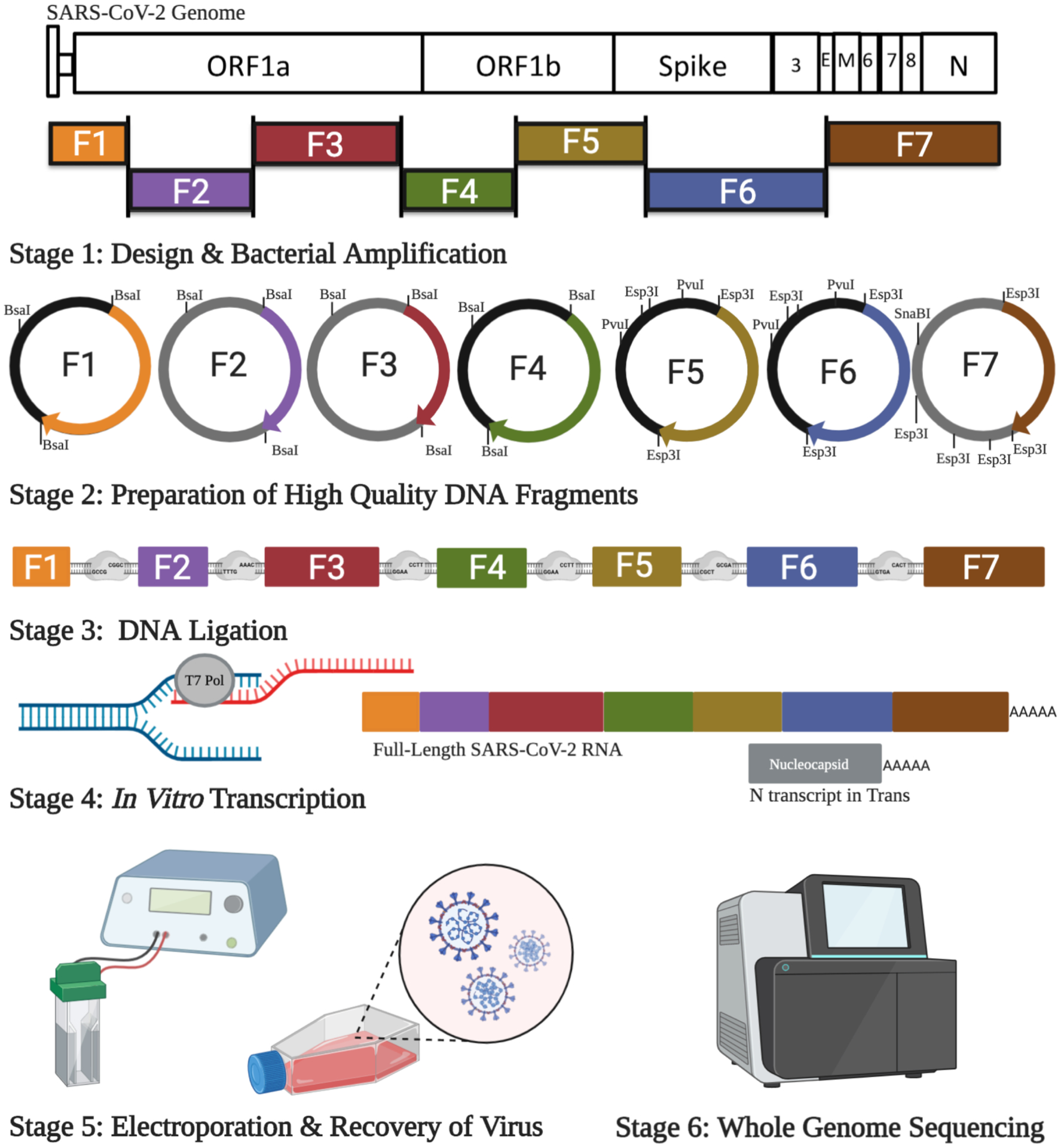
The SARS-CoV-2 infectious clone model contains seven cDNA fragments to cover the complete viral genome, to disrupt toxic elements, and to aid in genetic manipulation. The SARS-CoV-2 plasmids are amplified in E. coli and sequentially ligated following digestion with type II restriction enzymes to remove the plasmid backbone. The full-length viral DNA is then in vitro transcribed using T7 polymerase to generate full-length genomic SARS-CoV-2 RNA and electroporated into cells with N-protein transcript expressed in trans. Following electroporation, cells are seeded into cell culture flasks and virus recovered 2–5 days post electroporation.
Development of the protocol
With the rapid spread of SARS-CoV-2, researchers around the world initiated efforts to respond to the COVID-19 pandemic. The technical requirement for the assembly of full-genome coronavirus cDNA is challenging because of the large genomic size (~30,000 nucleotides), toxic genomic regions, and issues with mutations and deletions in the viral sequence11.
In response, our group developed a reverse genetic system that enables rapid synthesis of wild-type, mutant, and reporter SARS-CoV-2 strains to study viral infection, transmission, pathogenesis, therapeutics, and vaccines3. It applies the same principles from infectious clones developed for TGEV, MHV, SARS-CoV, MERS-CoV, and several bat coronaviruses5–7,12–15. Briefly, a contiguous panel of seven cDNA fragments was designed to span the entire genome of SARS-CoV-2 and were individually cloned into plasmids using type II restriction enzyme sites (Fig. 1). The type II restriction enzymes were chosen for cloning because they recognize asymmetric DNA sequences and generate unique cohesive overhangs that ensure one directional, seamless assembly of the seven DNA fragments into the genome-length cDNA. The assembled genome-length cDNA was used as a template for in vitro transcription. The resulting genome-length viral RNA was subsequently electroporated into cells to recover recombinant SARS-CoV-2. We originally described this method in our supporting Cell Host and Microbe paper3, which showed that the full-genome cDNA was highly infectious after electroporation into cells. The infectious-clone-derived SARS-CoV-2 (icSARS-CoV-2) exhibited similar plaque morphology, viral RNA profile, and replication kinetics to a clinical isolate. In addition, we generated a stable mNeonGreen SARS-CoV-2 (icSARS-CoV-2-mNG), which was successfully used to evaluate the antiviral activities of interferon and vaccine development2,3,16–18. In this protocol article, we provide more detailed information for using this method, including troubleshooting information.
Overview of the procedure
Stage 1 of the procedure (steps 1–33) is preparation of the seven plasmids that contain SARS-CoV-2 fragments F1–F7. The plasmids should be validated by restriction enzyme digestion and Sanger sequencing to exclude introduction of any undesired mutations into the plasmids prior to assembly of the full-length SARS-CoV-2 DNA. Stage 2 (steps 34–45) involves the preparation of high-quality DNA fragments for downstream experiments by restriction enzyme digestion of the Maxiprep plasmids. Stage 3 (steps 46–89) involves assembling the seven DNA fragments into a full-length SARS-CoV-2 DNA in vitro using a T4 DNA ligase. Two separate ligation steps increase the ligation efficiency of the full-length DNA and avoid nonspecific ligation between F3 and F7 fragments. Afterwards, the full-length ligation product is immediately purified by phenol-chloroform extraction and isopropanol precipitation. Stage 4 (steps 90–96) is in vitro transcription of full-length RNA and N gene RNA. Stage 5 (step 97) involves recovery of the SARS-CoV-2 recombinant virus from cell culture via RNA electroporation. Two different methods can be used for electroporation, using either Vero E6 cells only or BHK-21 and VeroE6 cells. Stage 6 (steps 98–108) involves whole-genome Sanger sequencing of the virus to verify the entire viral genome sequence. The procedures of stages 1–4 can be performed in general lab. The procedures of stages 5–6 involve manipulating the SARS-CoV-2 must be done in a biosafety laboratory level 3 (BSL-3) facility.
Comparison with alternative methods
Our seven-cDNA-fragment approach has several key advantages over alternative methods, including bacterial artificial chromosomes, a vaccinia virus, and a yeast recombination-based assembly11,19 (See details below). First, it permits rapid generation of mutant and reporter viruses by manipulation of a smaller plasmid (i.e., the plasmid that contains the targeted mutation fragment), reducing the risk of off-target mutations or deletions being inadvertently incorporated into the recombinant virus. Second, this approach allows simultaneous manipulation of multiple mutations from different cDNA fragments. More than one mutation from different cDNA fragments can be engineered in parallel to make combinatory mutant viruses. Such flexibility is important when characterizing a combinatory effect of multiple viral elements on host immune response or developing a live-attenuated vaccine platform, which often requires multiple mutation sites to be investigated at the same time20,21. In addition, the seven-fragment system allows quick insertion of mutations that arise from sequencing of new clinical isolates or swapping of regions from related coronaviruses found in animals13,22. Collectively, the reverse genetic system offers a wealth of opportunities to explore and study SARS-CoV-2 infection and pathogenesis.
Although the in vitro ligation approach allows rapid preparation of mutant and reporter viruses, the requirement to assemble and transcribe genome-length RNA requires technical expertise. Alternative coronavirus reverse genetic systems have used bacterial artificial chromosomes, a vaccinia virus, and a yeast recombination-based assembly11,19. These alternate systems offer less assembly requirements, but are more prone to potential off-target mutations due to the use of larger size of viral cDNA and the need for amplification in host cells. Besides our SARS-CoV-2 infectious cDNA clone3, a yeast-based platform and a similar multiple plasmid approach have been shown to produce recombinant SARS-CoV-219,23. The yeast platform required screening of several clones to identify virus equivalent to the original clinical isolate19. In contrast, both of the cDNA-fragment-based approaches yielded production of recombinant SARS-CoV-2 equivalent to the clinical isolate. These results are consistent with the previously characterized phenotypes of the epidemic SARS-CoV and MERS-CoV isolates as compared to their recombinant versions5,15. The fidelity to the clinical isolate of SARS-CoV-2 is an important advantage of these multiple plasmid infectious clone systems.
Applications
Limitations and experimental design considerations
Our experimental design is to clone seven cDNA fragments covering the entire genome of SARS-CoV-2 into plasmid vectors, resulting in seven plasmids. The seven viral cDNA fragments are engineered into plasmids based on the nucleotide sequences for Type IIS restriction enzymes. The Type IIS restriction enzymes was chosen because they recognize asymmetric DNA sequences and cleave outside of their recognition sequence, thus allowing directional assembly of multiple DNA fragments. There are two considerations for choosing the starting and ending nucleotide positions for each cDNA fragment: 1) to divide the entire viral genome to 7 fragments with reasonable DNA length for quick RT-PCR amplification and molecular cloning based on the BsaI and Esp3I restriction enzymes; 2) to minimize the nonspecific ligation of the 4-base overhangs generated by BsaI and Esp3I. This seven-plasmid approach allows simultaneous manipulation of different viral fragments of interest via standard molecular approaches (e.g., PCR or site-directed mutagenesis) to generate recombinant viruses with multiple changes.
Despite success across coronavirus platforms, several issues can potentially disrupt the efficacy of our reverse genetic system. We have found variability in the electroporation capacity of Vero E6 cell lineages. Although the electroporation buffer improves efficiency in Vero E6 cells, we also include an alternative approach utilizing BHK-21 cells, a Golden Syrian hamster fibroblast cell line, which can be used for virus generation in other CoV systems6,7. BHK-21 cells are not suitable for continued SARS-CoV-2 replication due to lack of ACE2 receptor expression; however, these cells tolerate electroporation well and allow sufficient SARS-CoV-2 production to seed co-cultured Vero E6 cells. Overall, electroporation efficiency is low in both BHK-21 and Vero E6 cells (<1% cells based on the mNeonGreen expression from cells electroporated with mNeonGreen-containing SARS-CoV-2 RNA) and co-culture with non-electroporated Vero E6 cells can improve viral yield for passage 0. Notably, we find that viral yields improve with the subsequent passage and these stocks are generally used for experiments.
Another key barrier to success with our reverse genetic system is deletions and mutations while propagating the cDNA plasmids. Despite their smaller size and our efforts to disrupt toxic elements, the SARS-CoV-2 plasmids are still prone to errors and deletions when amplified in E. coli. To reduce incorporation of these errors, we sequence to verify cDNA plasmids at each stage of amplification. To prevent continued mutations/deletions in certain SARS-CoV-2 plasmids, we have also included instructions for alternative growth conditions with lower temperatures (25°C or 30°C) for longer times (up to 48 hours) to facilitate generation of cDNA with fidelity to the original viral sequence.
In addition, we use a Dark Reader blue transilluminator for manipulation of SARS-CoV-2 plasmid DNA. We found that use of standard UV light boxes yields sequence mutations and poor virus recovery. We also note that each plasmid has prescribed competent cells (Top10 or EPI300), which are associated with lower mutation and deletion frequencies as well as improved plasmid yields. Overall, this reverse genetic system requires significant effort to prevent mutations/deletions from disrupting SARS-CoV-2 generation.
The conditions for assembly, ligation, and electroporation of the viral nucleic acid must be carefully considered when using this reverse genetic system. A key challenge is the requirement for a sufficient concentration of cDNA fragments for ligation. We have included calculations for the necessary amount of each fragment, as well as a visual image of the cDNA fragments (after gel purifications) to provide a reference for the amount of plasmid DNA needed for a successful in vitro assembly of full-length cDNA (Fig. 2a). We also include alternative growth conditions (larger cultures and longer culture time) to amplify low yield plasmids if necessary. In general, we find it necessary to complete a maxiprep (Qiagen) for each plasmid to have sufficient DNA concentrations to facilitate full-length cDNA assembly.
Figure 2. Gel extraction of SARS-CoV-2 fragments, full-length cDNA, and full-length viral RNA.
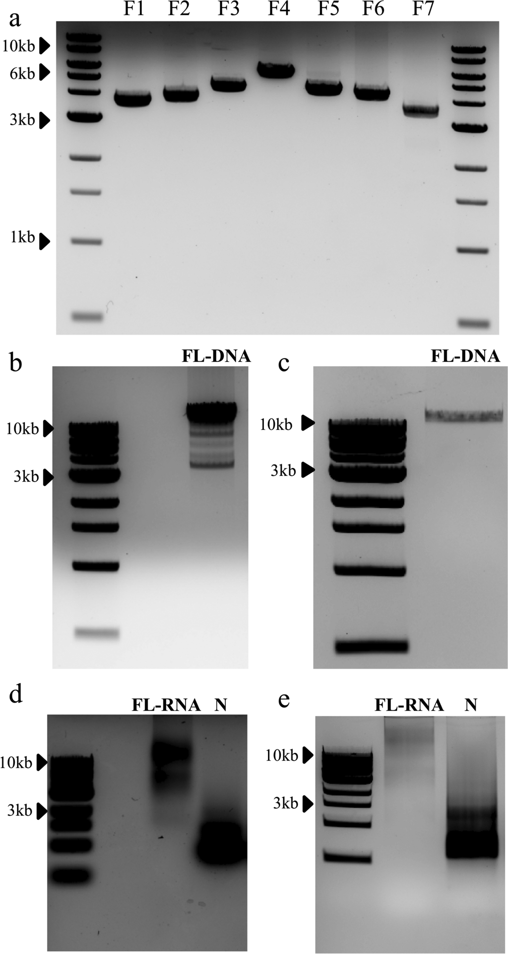
(a) Agarose gel showing each SARS-CoV-2 DNA fragment (1μl of each) isolated post restriction enzyne digestion and utilized for in vitro ligation. B&C) Representative gels from successful (b) and unsuccessful (c) attempts to generate full-length SARS-CoV-2 cDNA. Representative agarose gels from successful (d) and unsuccessful (e) attempts to generate in vitro transcribed full-length SARS-CoV-2 viral RNA prior to electroporation and virus recovery.
We have included gel images of full-length SARS-CoV-2 DNA after ligation and full-length RNA after in vitro transcription to show the required amount for effective versus ineffective electroporation and virus recovery (Fig. 2b–d). For poor full-length DNA yields, we offer alternative ligation conditions as well. Together, these tips and data should provide critical references for use and manipulation of the SARS-CoV-2 infectious clone.
Materials
Cells
EPI300 competent cells (Lucigen, cat. no. C300C105)
TOPO 10 chemically competent cells (Fisher Scientific, cat. no. C404010)
BHK-21 cells (cat. no. ATCC® CCL-10; https://scicrunch.org/resolver/RRID:CVCL_1915)
Vero E6 cells (Lab passaged derivative of ATCC® CRL-1586; https://scicrunch.org/resolver/RRID: CVCL_0574)
! CAUTION Periodically make sure the mammalian cells used are authentic and are not contaminated with mycoplasma.
Reagents
0.25% Trypsin-EDTA (1×) (ThermoFisher Scientific, cat.no. 25200–072)
0.4% Trypan blue (ThermoFisher Scientific, cat. no.15250–061)
1 kb DNA ladder (NEB, cat. No. N3232L)
10×Cutsmart buffer (NEB, cat. no. B7204S)
Absolute ethanol (EtOH; anhydrous, 200 proof/100% (vol/vol); VWR, cat. no. 89125–170)
Acid Phenol:Chloroform (pH 4.5; Ambion, cat. no. AM9722)
Agarose (BIO-RAD, cat. no.1613102)
Ampicillin sodium salt (Sigma-Aldrich, cat. no. A9518)
Chloramphenicol (Sigma-Aldrich, cat. no. 0378)
CopyControl™ Induction Solution (Lucigen, cat. no. CCIS125)
DMEM, high glucose (Life Technologies, cat. no. 11965–092)
DNA loading buffer (NEB, cat. no. B7024S)
EDTA (Sigma-Aldrich, cat. no. 324503)
Electroporation buffer (Mirus, cat. no. MIR 50117)
Ethidium bromide (EB; 10mg/ml; Bio-Rad, cat. no.161–0433)
FBS (Hyclone, cat.no. SH3007103HI)
Glycerol (≥99.5% (wt/vol); Sigma-Aldrich, cat. no. G9012)
Hydrogen Chloride (HCl; 36.5–38%; Sigma-Aldrich, cat. no. H1758–500ML)
illustra MicroSpin G-25 Columns (GE Healthcare, cat. no.27–5325-01)
Isopropanol (Sigma-Aldrich, cat. no. I9516)
LB Agar (ready-made powder; Fisher Scientific, cat.no. DF0401–17)
Luria broth (LB; ready-made powder; Fisher Scientific, cat.no. DF0402–08-0)
mMESSAGE mMACHINE™ T7 Transcription Kit (ThermoFisher Scientific, cat. no. AM1344)
PBS (Thermo Fisher Scientific, cat. no. 10010023)
Penicillin/Streptomycin (ThermoFisher Scientific, cat. no.15140–122)
Phenol:Chloroform:Isoamyl Alcohol 25:24:1 (pH 8.05; Invitrogen, cat. no. 15593–031)
Platinum™ SuperFi II DNA Polymerase (ThermoFisher Scientific, cat. No. 12361010)
QIAGEN Plasmid Maxi Kit (QIAGEN, cat. no. 12163)
QIAprep Spin Miniprep Kit (QIAGEN, cat. no. 27106)
QIAquick Gel Extraction Kit (QIAGEN, cat. no. 28706)
QIAquick® PCR Purification Kit (Qiagen, cat. no. 28106)
Restriction enzyme BsaI-HFv2 (NEB, cat. no. R3733)
Restriction enzyme Esp3I (NEB, cat. no. R0734L)
Restriction enzyme PvuI-HF (NEB, cat. no. R3150S)
Restriction enzyme SnaBI (NEB, cat. no. R0130L)
Ribonucleotide solution mix (rNTP solution mix; NEB, cat. no. N0466L)
SOC outgrowth medium (10 mL; Invitrogen, cat. no. 15544034)
Sodium acetate (Sigma-Aldrich, cat. no. S8750)
Sodium hydroxide pellets (NaOH; Sigma-Aldrich, cat. no. S8045)
SuperScript First-Strand Synthesis System (ThermoFisher Scientific, cat. No. 18091050)
T4 ligase and ligation buffer (NEB, cat. no. M0202L)
Tris-base (Sigma-Aldrich, cat. no. T1503)
TRIzol™ LS Reagent (ThermoFisher Scientific, cat. no. 10296028)
UltraPure DNase/RNase-free distilled water (ThermoFisher Scientific, cat. no. 10977015)
Equipment
1L glass bottle (Duran, cat. no. 21820545)
2 ml screw-top tube (VWR, cat. no. 101093–752)
250 ml glass bottle (Duran, cat. no. 21801365)
4 mm-Cuvettes (Bio-Rad, cat. no.1652088)
90-mm Petri dishes (Thermo Scientific, cat. no. 263991)
Automated cell counter (Bio-Rad, cat. no. 1450102)
C-fold paper towel (Scott)
CO2 incubator (NuAire)
Cooler (Coleman)
Counting slide (Bio-Rad, cat. no. 145–0011)
Darkreader transilluminators (DR89X model, Clare Chemical Research)
Eppendorf Benchtop centrifuge (models 5810R, 5424R, 5425)
Erlenmeyer flask, 1L (Pyrex, cat. no. 4446–1L)
Erlenmeyer flask, 250ml (Pyrex, cat. no. 4446–250)
Falcon 15ml conical tube (Corning, cat. no. 352096)
Falcon 50ml conical tube (Corning, cat. no. 352070)
Fisherbrand™ Isotemp™ Stirrer (Fisher Scientific)
Gel DOC™ EZ system (Bio-Rad, cat. no. 170827)
Gene Pulser Xcell Electroporation Systems (Bio-Rad, cat. no. 1652660)
Horizontal Electrophoresis Systems (BIO-RAD)
Incubator for bacteria culture (Fisher Scientific)
L-shaped cell spread (Fisher scientific, cat. no. 14–665-230)
Microcentrifuge tube, 1.7-ml (Axygen, cat. no. MCT-175-C)
Microwave (Oster)
Milli-Q™ Ultrapure Water Systems
New Brunswick Scientific™ Innova™ 43R Incubator Shakers (Eppendorf, cat. no. M1320–0000)
PCR tube, 0.2-ml (Axygen, cat. no. PCR-02-C)
pH meter (Sartorius)
Research plus pipettes (0.1–2.5, 2–20, 20–200, and 100–1,000 μL) (Eppendorf)
Secura® Balance (Sartorius, cat. no. ENTRIS 6202–1S)
S1 Pipet Fillers (Thermo Scientific, cat. no. 9501)
Spectrophotometer (DS-11 Series, DENOVIX)
T175 flask (Corning, cat. no. 431080)
T-75 flask (Corning, cat. no. 430641U)
Thermocycler (models C1000 Touch and T100, Bio-Rad)
VACUBOY (INTEGRA Biosciences)
Vortex (Fisher Scientific)
Water bath (Fisher Scientific)
Reagent setup
0.8% agarose gel
Weigh 0.8 g of agarose powder in a 250-ml conical flask and add 100 ml of 1×TAE buffer. Swirl the conical flask to blend the contents and cover the top of the conical flask with a plastic wrap to reduce evaporation. Microwave for 1–2 min to melt the agarose completely and do not over-boil. Cool down the agarose solution to 50–60°C and pour it into a gel dock slowly to avoid bubble formation. Add Ethidium Bromide (EB) to the agarose solution at a final concentration of 0.5 μg/ml, distribute the EB evenly by shaking the gel dock gently, and quickly insert a gel comb into the gel dock. The agarose gel will be ready to use once it solidifies.
Ampicillin stock (100 mg/ml)
Weigh out 10 g of ampicillin sodium salt in a clean 250mL glass bottle. Add 100 ml UltraPure deionized water and stir thoroughly until components are completely dissolved. Sterilize the ampicillin solution by passing through a 0.22 μm filter, and aliquot into sterile 1.7ml tubes (1 ml per tube) to avoid multiple freeze-thaws. Ampicillin aliquots may be stored at −20°C for at least 1 year. For frequent use, store the ampicillin stock at 4°C for no more than 1 month.
Cell culture media
Prepare all the cell culture medium in a biosafety cabinet. To make 10% FBS or 2% FBS media, add 55 ml or 10 ml FBS into 500 ml of high glucose DMEM supplemented with 1% penicillin-streptomycin solution, respectively. Use 10% FBS culture media for cell propagation and 2% FBS culture media for virus infection and propagation.
Chloramphenicol stock (25 mg/ml)
Add 2.5 g of Chloramphenicol into 100 ml of absolute ethanol and vortex vigorously to ensure all the chloramphenicol powders are fully dissolved. Filter sterilize is not necessary since it is in 100% ethanol. Dispense the chloramphenicol stock solution into aliquots (500 μl in 1.7 ml tube). Chloramphenicol aliquots may be kept in a −20°C freezer for at least 1 year.
! CAUTION Ethanol is a flammable. Keep ethanol and dissolved Chloramphenicol stocks away from fire.
EDTA, 0.5 M, pH 8.0
Add 148 g of EDTA into 1 liter of UltraPure deionized water and mix thoroughly on a magnetic stirrer. To improve EDTA solubility in water, adjust pH to 8.0 by adding NaOH (approximately 30 to 40 g) into the solution gradually. Keep stirring until all the components are fully dissolved and sterilize EDTA solution by autoclaving at 121°C for 30 min. The EDTA solution is stable at room temperature for up to 1 year.
70% ethanol
Mix 15 ml nuclease-free water with 35 ml absolute ethanol in a 50ml falcon tube and keep at - 20°C for long-term storage.
! CAUTION 70% ethanol is still flammable. Keep stocks away from fire.
50% glycerol buffer
Combine 50 ml of glycerol with 50 ml of UltraPure deionized water in a 250 ml glass bottle and shake up the solution. Autoclave the solution at 121°C for 20 min, and place in a 4°C freezer for storage.
! CAUTION Keep and store the 50% glycerol buffer in a sterile environment.
LB agar plates containing Ampicillin or Chloramphenicol
Add 28 g of LB Agar powder to 800 ml of UltraPure deionized water in a 1-L glass bottle and swirl to mix. Autoclave to sterilize at 121.0 °C for 30 min, and the components will be dissolved after autoclaving. Cool down the LB agar solution to 55 °C before adding ampicillin (to a final concentration of 100 μg/mL) or chloramphenicol (to a final solution of 12.5 μg/ml). On a sterile bench area, pour LB agar solution (approximately 20 ml per dish) into a 90-mm Petri dish. Usually, 800 ml LB agar solution is sufficient for casting 20–30 agar plates. Return the lids to the plates and cool the plates down at room temperature until the agar solidifies. Agar plates containing antibiotics can be stored in plastic bags or sealed with Parafilm at 4 °C in the dark for up to 3 months.
! CAUTION Keep the Chloramphenicol stocks away from fire.
LB media solution
Dissolve 20 g of LB powder into 1 L of UltraPure deionized water and autoclave at 121°C for 30 min for sterilization. After autoclaving, the LB media solution can be stored at room temperature for up to 4 months.
To prepare the LB media containing ampicillin or chloramphenicol for the bacteria selection, add 0.8 ml ampicillin stock (80 mg/ml) or 500 μl chloramphenicol stock (25 mg/ml) into 1 L LB media respectively in a sterilized environment. The final work concentrations are 100 μg/ml for ampicillin and 12.5 μg/ml for chloramphenicol. Store the LB media containing antibiotics at 4°C in dark.
! CAUTION Antibiotics degrade over time, so LB media containing antibiotics should be made up fresh or frequently.
Sodium acetate (3.0 M, pH 5.2)
Dissolve 246.1 g of sodium acetate in 500 ml of deionized water. Adjust the pH to 5.2 with glacial acetic acid. Allow the solution to cool overnight. Adjust the pH once more to 5.2 with glacial acetic acid. Adjust the final volume to 1 L with deionized water and filter-sterilize.
50× TAE buffer
Weigh out 484 mg of Tris-base in a clean 2-L glass bottle and add approximately 1500 ml of UltraPure deionized water. After the Tris-base has dissolved, carefully pour 114.2 ml of glacial acetic acid and 200 ml 0.5 M EDTA (pH 8.0) into solution and mix them up by agitating. Top up the TAE solution to the final volume of 2L with water and store the 50× TAE buffer at room temperature.
To prepare 1×TAE buffer (40 mM Tris, 20 mM acetate, and 1 mM EDTA), which is used for DNA electrophoresis, dilute 400 ml of 50×TAE buffer into 19.6 L of UltraPure deionized water.
Plasmids
We have successfully cloned seven different DNA fragments spanning the entire genome of SARS-CoV-2 into commercial pUC57 (GenScript, Piscataway, NJ) or pCC1 (Epicenter Biotechnologies, Madison, WI) vectors, resulting in seven plasmids: pUC57-CoV-2-F1, pCC1-CoV-2-F2, pCC1-CoV-2-F3, pUC57-CoV-2-F4, pUC57-CoV-2-F5, pUC57-CoV-2-F6 and pCC1-CoV-2-F7. The sequences of the seven plasmids are included in the Supplementary Fig.1. Type IIS restriction enzymes BsaI and Esp3I, which recognize asymmetric DNA sequences and cleave outside of their recognition sequence, have been widely used for Golden Gate Assembly to ensure directional assembly of multiple DNA fragments simultaneously. Additional restriction enzymes (like PvuI and SnaBI) are used to efficiently resolve the desired DNA fragments from other by-products in the same restriction reactions during electrophoresis. Table 1 outlines the restriction enzyme cleavage sites in the seven plasmids that would be used for preparing the fragments prior to ligation in this protocol. The seven plasmids can be used as templates for generating mutations of interest via standard molecular approaches, such as PCR or site-directed mutagenesis.
Table 1.
Restriction enzymes for validation of seven SARS-CoV-2 plasmids.
| Plasmid | Restriction enzyme(s) | DNA fragments with expected size |
|---|---|---|
| pUC57-CoV-2-F1 | BsaI | 3644 bp (F1)a + 1367 bp + 1366 bp |
| pCC1-CoV-2-F2 | BsaI | 6466 bp + 3886 bp (F2)a + 1344 bp |
| pCC1-CoV-2-F3 | BsaI | 6466 bp + 4480 bp (F3)a + 1344 bp |
| pUC57-CoV-2-F4 | BsaI | 5607 bp (F4)a + 1367 bp + 1366 bp |
| pUC57-CoV-2-F5 | Esp3I and PvuI | 4457 bp (F5)a + 1674 bp + 620 bp + 234 bp + 161 bp+ 42 bp |
| pUC57-CoV-2-F6 | Esp3I and PvuI | 4284 bp (F6)a + 1674 bp + 620 bp + 234 bp + 161 bp+ 42 bp |
| pCC1-CoV-2-F7 | Esp3I and SnaBI | 3563 bp (F7)a + 2522 bp + 2395 bp + 1689 bp + 651 bp + 553 bp |
pUC57
pUC57 is a high-copy-number (500–700 copies/cell) plasmid, which contains an ampicillin-resistant gene. It can be propagated in E. coli to produce a high yield of plasmids for downstream use. pUC57-F1, pUC57-F4, pUC57-F5 and pUC57-F6 are stable when they are propagated in the commercially available Top10 competent cells. Several times of attempts to clone the F2, F3 and F7 fragments into the pUC57 vector that can propagate in the Top10 competent cells failed, probably due to the toxicity of these fragments to the bacterial cells. Finally, F2, F3 and F7 were successfully cloned and in pCC1 vector and propagated stably in EPI300 competent cells (see details below).
! CRITICAL Top10 competent cells are recommended. Other cells must be verified for plasmid compatibility/stability prior to prepare large batch of those plasmids.
pCC1
pCC1 is a bacmid cloning vector with a controlled copy number. It is ideal for amplifying large, unstable, and bacteria-toxic DNA fragments in E. coli. Before induction, the copy number of pCC1 plasmid is one copy per cell. Upon induction by L-arabinose, the copy number of pcc1 can be 10–20 copies per cell in the bacteria cells (EPI300 competent cells are recommended for propagating the pCC1-derived plasmids).
Equipment setup
Electroporator setup
This protocol is based on the use of Gene Pulser Xcell Electroporation Systems using the exponential decay pulse for electroporating RNAs into mammalian cells. We optimized the parameter settings including voltages, capacitance, and pulse times for different cells. The conditions used in this study give us high and reproducible transformation efficiency with high viability of cells after electroporation. The parameter settings for electroporation of Vero cells are indicated below: voltage, 270 V; capacitance, 950 μF; resistance, ∞; cuvette size (mm): 4. One pulse is needed for electroporation of Vero cells. The parameter settings for electroporation of BHK-21 cells are: voltage, 850 V; capacitance, 25 μF; resistance, ∞; cuvette size (mm): 4. Three pulses with 3-second intervals between each pulse are needed for electroporation of BHK-21 cells. Alternative electroporation systems (such as 4D-Nucleofector™ X unit) with optimized settings can be also used.
Procedure
Stage 1:Propagation of plasmids containing SARS-CoV-2 fragments ●Timing 3.5 d
Chemical transformation ● Timing 2 h
▲CRITICAL STEP The transformation should be performed in a sterile environment to prevent contamination.
- 1
Before transformation, complete the following steps:- Quantify the concentration of each plasmid using a spectrophotometer.
- Warm up a water bath to 42°C.
- Disinfect the bench surface with 70% ethanol
- Light a Bunsen burner to provide a sterile environment for transformation.
▲CRITICAL STEP The water bath should be regularly calibrated for accurate temperature. Inappropriate temperature would decrease transformation efficiencies.
- 2
Thaw appropriate chemically competent cells on ice. Add 1–10 ng of plasmids to the thawed cells and mix by tapping the tube or swirling gently.- For pUC57-derived plasmids, including pUC57-CoV-2-F1, pUC57-CoV-2-F4, pUC57-CoV-2-F5 and pUC57-CoV-2-F6, use Top10 competent cells for transformation.
- For pCC1-derived plasmids, including pCC1-CoV-2-F2, pCC1-CoV-2-F3, pCC1-CoV-2-F7, use EPI300 competent cells for transformation.
▲CRITICAL STEP Competent cells are sensitive to temperature and salt/buffer conditions. Add the DNA solution at a volume <10% of the competent cell suspension. Keep the competent cells on ice prior to heat shock. The thawed competent cells should not be refrozen at −80°C.
- 3
After adding the plasmids, immerse the tube immediately into ice. Incubate the plasmid-cell mixtures for 20–30 min on ice. - 4
Incubate the tube containing the plasmid-cell mixtures in a 42°C water bath for 30 seconds. - 5
Immediately put the tube back on ice and incubate for 2 min. - 6
Add 250 μl SOC medium (pre-warmed at room temperature) into the tube containing transformed cells. - 7
Shake the cultures in a 37°C incubator at 230× rpm for 60 min to recover the cells. - 8
During the incubation period, warm LB agar plates supplemented with 100 μg/ml ampicillin (for pUC57-derived plasmids) or 12.5 μg/ml chloramphenicol (for pCC1-derived plasmids) in a 37°C incubator. - 9
After the 60 min incubation, inoculate 50 μl of the cultures onto a pre-warmed LB agar plate supplied with the appropriate antibiotics, and spread the cultures over the plate using L-shaped cell spreaders. - 10
Place the plates upright in a 37°C incubator for 30 min to allow the cultures to be fully absorbed. Afterwards, invert the plates and incubate at 37° overnight (16–20 hours).
? TROUBLESHOOTING
Colony screen (Timing 1 d or overnight).
- 11
After overnight incubation, pick up several well-separated colonies using sterile P10 tips and inoculate the colonies into LB broth containing proper antibiotics.- For pUC57 vector derived plasmids, inoculate the colonies into a 50-ml conical tube containing 10 ml LB broth containing 80 μg/ml Ampicillin.
- For pCC1 vector derived plasmids, inoculate the colonies into a 50-ml conical tube containing 10 ml LB broth containing 12.5 μg/ml Chloramphenicol.
▲CRITICAL STEP The colonies may vary in size due to potential cryptical foreign gene expression from viral genome, which is usually toxic to the E. coli. Pick up 4–6 medium-small colonies per plasmid for validation.
■ PAUSE POINT Store the LB agar plates at 4°C in dark for future use (for up to 1 month).
- 12
Incubate the bacterial cultures at 37°C with shaking at 230 × rpm overnight (12–16 h).
▲CRITICAL STEP Grow the bacterial cultures in tubes with volumes of more than three times that of the culture volume. The tubes should not be fully sealed to ensure enough air exchange. Do not extend the incubation time as more “toxin” accumulates over time. - 13
The next day, prior to miniprep, save a bacteria glycerol stock for each colony by mixing 0.5 ml overnight culture with 0.5 ml 50% glycerol in a sterile 1.7-ml Eppendorf tube. Store the glycerol stock in −80°C freezer for future use (stock should be stable for years). For pUC57-derived plasmids, the remaining overnight cultures are now ready for miniprep, so you can skip step 14 and proceed to step 15. For pCC1-derived plasmids, an extra induction step (step 14) is needed. - 14
OPTIONAL For pCC1-derived plasmids only, inoculate 1.5 ml of overnight cultures into a new 50 ml conical tube containing 13.5 ml LB broth supplemented with 12.5 μg/ml chloramphenicol and 15 μl induction solution (supplemented by the manufacturer). - 15
Shake the culture at 37°C 230× rpm for 5 hours, and then use the induced cultures for miniprep.
▲CRITICAL STEP The induction time should not exceed 5 hours. Longer culture time may increase the risk of mutation/deletion in the DNA fragments of the SARS-CoV-2.
Plasmid miniprep ●Timing 1–2 h
- 16
Harvest the bacterial culture by centrifuging at 3900× rpm for 10 min in a bench-top centrifuge (Eppendorf model 5810R). - 17
Dispense the media into a waste container containing 20% bleach. Invert the tubes and put them on absorbent papers to remove the residual liquid. - 18
Save the bacterial pellets to extract plasmids using a QIAGEN miniprep kit by following the manufacturer’s instructions.
■ PAUSE POINT The pellets can be either used immediately for Miniprep or preserved at −20°C until use. - 19
At the final step of Miniprep, add 50 μl nuclease-free water (prewarmed at 56°C) into the miniprep columns. Centrifuge at >12000× g for 2 min to elute the plasmids. - 20
Measure the yield and quality of the plasmids using a spectrophotometer.
■ PAUSE POINT The eluted plasmids can be either used immediately or stored at −20°C up to at least one year until use.
Plasmid validation by restriction enzyme digestion ●Timing 2 h
CRITICAL The plasmids prepared above are digested with appropriate restriction enzymes. The restriction enzymes used to validate the seven SARS-CoV-2 plasmids and the expected DNA fragments after restriction are indicated in Table 1.
- 21
Set up a 10-μl digestion reaction system for each plasmid in a 0.2-ml PCR tube. The setups are indicated in the table below.
Reaction systems for plasmid validation.Components Volume (μl) Nuclease-free water Up to 10 10×Cutsmart buffer 1 Restriction enzymes 0.5–1a DNA Plasmid TBD (200–300 ng) - 22
Incubate the reaction at 37°C for 1 h. During the incubation period, cast a 0.6% agarose gel containing Ethidium Bromide with 1mm-wide-well for DNA electrophoresis.
! CAUTION Ethidium Bromide (EB) is a mutagen. Wear proper PPE when handling EB-containing solutions and gels. - 23
After 1-hour incubation, reactions are stopped by adding 2 μl of 6× DNA loading buffer. Load the samples onto a 0.8% agarose gel using a P10 pipette. Pipet 5 μl of 1-kb DNA ladder into an independent well of the same gel. - 24
Resolve the DNA fragments by electrophoresis at 120×V for 25 min in 1× TAE buffer. - 25
Acquire the images of the gel using a Gel DOC-EZ imager. Evaluate the results for each plasmid, referring to Table 1 for expected fragment sizes. Figure 3 shows an example of the rection digestion results. For each plasmid, one validated colony is sufficient for proceeding to the next step.
? TROUBLESHOOTING
Figure 3. Representative gel images post restriction enzyme digestion.
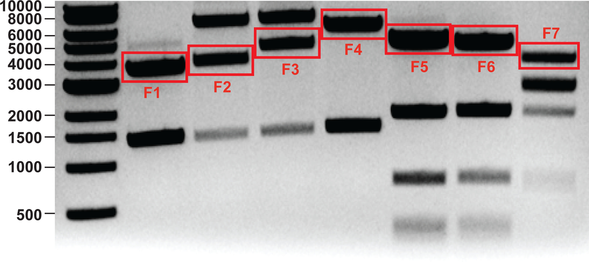
The DNA ladders in bp are indicated. The corresponding fragments of SARS-CoV-2 restricted from the plasmids are outlined.
Plasmid maxiprep ●Timing 24 h
CRITICAL The glycerol stocks of validated plasmids are used for preparing large batches of plasmids.
- 26
Inoculate 0.1 ml of glycerol stock into a 250-ml Erlenmeyer flask containing 50 ml of LB media supplemented with appropriate selective antibiotics. - 27
Shake the cultures in a 37°C incubator with speed of 230× rpm overnight (12–16h).For pUC57-derived plasmids, the overnight cultures are ready for maxiprep (50ml should be sufficient) so you can skip step 27 and proceed to step 28. - 28
OPTIONAL For pCC1-derived plasmids only, transfer 50 ml of the overnight cultures into a 2-L Erlenmeyer flask containing 450 ml of fresh LB broth supplemented with 12.5 μg/ml chloramphenicol and 500 μl induction solution. Incubate the culture at 37°C with shaking at 230 rpm for 5 h. The new cultures are used for maxiprep.
▲CRITICAL STEP The induction time should not exceed 5 hours. Longer culture time may increase the risk of mutation/deletion in the DNA fragments of the SARS-CoV-2. - 29
Pellet down the cultures by spinning at 7000 rpm for 5 min. - 30
Save the pellets for plasmid extraction using QIAGEN Plasmid Plus MAXI Kit according to the manufacturer’s instructions.
■ PAUSE POINT The pellets can be either used immediately or preserved at −20°C until use. - 31
At the last step of Maxiprep, add 100–200 μl nuclease-free water (prewarmed at 56°C) into the columns. - 32
Incubate at room temperature or 56°C incubator for 2 min. - 33
Elute the plasmids by centrifuging at >12000 rpm for 2 min. - 34
Determine the yield and quality of plasmids using a spectrophotometer. Note that the yields of plasmids may vary from 100 to 300 μg.
■ PAUSE POINT The eluted plasmids can be either used immediately or preserved at −20°C until use.
? TROUBLESHOOTING
Stage 2: Prepare DNA fragments by restriction enzyme digestion and purification ●Timing 1 d
Plasmid digesting ●Timing 4 h
CRITICAL Use 30 μg of plasmids for restriction enzyme digesting. The protocol described below will recover enough high-quality DNA fragments for more than two in vitro ligation reactions.
- 35
Set up a 50-μl digest reaction system with appropriate restriction enzymes for each plasmid according to the table below. Scale up the total digestion volume as needed. Table 1 shows the enzymes used for each plasmid.
Restriction enzyme digestion reactions for preparing large amounts of DNA fragments.Components Volume (μl) Nuclease-free water Up to 50 μl 10×Cutsmart buffer 5 μl Restriction enzymes 1 unit/μga Plasmids TBD (30 μg) - 36
Incubate the reaction mixtures in a 37°C incubator for 3–4 hours.
▲CRITICAL STEP The incubation time should not exceed 4 hours to prevent star activities of the restriction enzyme. - 37
During the incubation period, prepare a 5-cm-long 0.8% agarose gel with 3 mm-wide-well. The reaction of each plasmid will occupy 2–3 wells of the agarose gel. Prepare 7 gels for all seven reactions.
▲CRITICAL STEP Always clean the gel dock and the combs thoroughly before casting any new agarose gels. Before gel electrophoresis, always clean the electrophoresis chamber and use fresh gel running buffer to avoid contamination from other experiments.
DNA fragment extraction from gel ●Timing 2 h
- 38
After digestion, stop the reaction by adding 40 μl of 6× DNA loading buffer to each reaction tube. Mix thoroughly. - 39
Load ~34 μl of DNA samples to each well of the gel using a P100 pipette. Use one gel for loading one reaction of the DNA samples. Load 5 μl of 1 kb DNA ladder in a separate well.
▲CRITICAL STEP Load the sample slowly to prevent samples from flowing out of the wells. - 40
Resolve the DNA fragments by electrophoreses at 120× V for 30–40 min. - 41
Visualize the DNA fragments using a DarkReader transilluminator.
▲CRITICAL STEP Do not use UV light for visualizing the DNA because UV radiation cause DNA damage such as breaks in the sugar-phosphate backbone, pyrimidine dimerization, and inter-strand cross-links, which will result in the failure of downstream RNA transcription. Moreover, UV radiation may introduce undesired mutations into the final recovered viruses.
▲CRITICAL STEP Always clean the DarkReader before placing the gel on the device to avoid contamination from other experiments.
! CAUTION Minimize your own exposure to the lights from the DarkReader. - 42
Excise the target bands (referring to Table 1 and Figure 3 for guidance on expected fragment size and expected results) using a clean blade. Place the gel slices containing the targeted DNA fragments into a 15-ml falcon tube. - 43
Extract DNA fragments from the gel slice using a QIAquick Gel Extraction Kit according to the manufacturer’s instructions. Use one column for each DNA fragment. - 44
At the end of the Gel extraction procedures, add 20 μl nuclease-free water (prewarmed at 56°C) to the column. - 45
Elute the DNA by centrifuging at 14000 rpm for 2 min. - 46
Measure the concentration of DNA fragments using a spectrophotometer. Usually, the concentration of DNA fragments is 50–200 ng/μl.
■ PAUSE POINT The DNA fragments can be used immediately or stored at 4°C until use. The purified DNA fragment can be kept at 4°C for up to 1 month or stored at −20°C for longer term storage.
? TROUBLESHOOTING
Stage 3: In vitro ligation ●Timing 2.5 d
Set up in vitro ligation reactions ●Timing 2 d
- 47
Prior to reaction setup, calculate the required volume of DNA fragments needed for assembling the full-length clone. Equal molar concentrations of each fragment should be used for the ligation in this protocol. The table below shows how to calculate the volume of DNA fragments for assembling 5 μg of SARS-CoV-2 full-length DNA.
Calculation of volume of each DNA fragment required for ligationFragment Size (kb) Mass of each fragment for assembling 1 μg full-length DNA Mass of each fragment for assembling 5 μg of full-length DNA (μg) F1 3644 0.12179 0.61 F2 3886 0.12988 0.65 F3 4480 0.14973 0.75 F4 5607 0.18739 0.935 F5 4457 0.14896 0.745 F6 4284 0.14318 0.715 F7 3565 0.11915 0.595 Total 29921 1 5 - 48
Prepare the first-step ligation reactions. Set up two separation ligations (A and B) according to the table below. Ligate F1, F2, F3, F4 in a 0.2-ml PCR tube to produce F1–4 DNA, and ligate F5, F6, F7 in a separate PCR tube to produce F5–7 DNA. Set up a 40 ul ligation reaction for individual ligation reactions as below.
Reaction system for the first-step ligation.Components Volume Nuclease-free water Top up to 40 μl 10×T4 ligation buffer 4 μl T4 DNA ligase 4 μl DNA fragmentsa TBD - 49
Incubate both ligation reactions at 4°C for 16–20 h. - 50
Set up the second step of ligation. Combine the above two ligation reactions in a new 1.7 ml tube. Top up the reactions with 16 μl of nuclease-free water, 2 μl of T4 DNA ligase, and 2 μl of 10× ligation buffer to 100 μl. Mix the reactions thoroughly by gently taping the tube.
Incubate the ligation reaction at 4°C for another 16–20 h to produce the full-length SARS-CoV-2 DNA.
Purify the ligation products. ● Timing 1.5 h
- 51
After ligation, add 100 μl of Phenol:Chloroform:Isoamyl Alcohol to the ligation reaction, and mix the solution by tapping the tube several times.
! CAUTION Organic reagents such as Phenol:Chloroform:Isoamyl Alcohol, chloroform, and isopropanol are toxic. Chemical fume hood is required for handling such chemicals. - 52
Centrifuge the mixture at maximal speed for 1 min in a bench-top centrifuge. - 53
After centrifugation, check that two layers have formed (the aqueous phase on the top and the organic phase at the bottom). Carefully transfer the aqueous phase (80 μl) containing the DNAs to a separate 1.7-ml Eppendorf tube.
▲CRITICAL STEP Be careful not to transfer the organic phase, which would deteriorate downstream RNA transcription. - 54
Add 100 μl of nuclease-free water to the previous tube containing the organic phase. Mix the solution by tapping the tube several times. Repeat steps 52–54. - 55
Combine the aqueous phase collected from the above two extractions. Add an equal volume of chloroform (~200μl) to the aqueous phase containing DNAs. - 56
Mix gently by tapping the tube several times. Centrifuge the tube in a bench-top centrifuge at maximum speed for 1 min. - 57
Transfer the upper aqueous phase (~200 μl) and transfer it to a new 1.7-ml tube. - 58
Add sodium acetate (3.0 M, pH 5.2) to the DNA solution to a final concentration of 0.3 M. - 59
Add an equal volume of isopropanol and mix gently by inverting the tube several times.
▲CRITICAL STEP It is important to precipitate DNA at room temperature using isopropanol instead of ethanol to minimize co-precipitation of ATPs in the T4 ligation buffer. High presence of ATPs would interfere with DNA quantification and downstream in vitro transcription. - 60
Incubate the mixtures at room temperature for 15–30 min to precipitate DNA. - 61
Pellet down the DNA by centrifuging at maximum speed for 15 min in a bench-top centrifuge to pellet DNA. - 62
Carefully decant the supernatant.
▲CRITICAL STEP The pellet may come out with the supernatant. To prevent loss of the pellet during decanting, save the supernatant in a new tube until you are sure that the precipitated DNA has been recovered. - 63
Add 1 ml 70% ethanol to the pellet. Centrifuge at maximum speed for 5 min and carefully decant the supernatant.
▲CRITICAL STEP The pellet may come out with the supernatant. To prevent loss of the pellet during decanting, save the supernatant in a new tube until you are sure that the precipitated DNA has been recovered. - 64
Add 1 ml >95% ethanol to the pellet. Centrifuge at maximum speed for 2 min and carefully decant the supernatant.
▲CRITICAL STEP The pellet may come out with the supernatant. To prevent loss of the pellet during decanting, save the supernatant in a new tube until you are sure that the precipitated DNA has been recovered. - 65
Carefully remove the residual liquid completely with a P100 pipet loaded with 100 μl tips, and air-dry the pellet for <5 min.
▲CRITICAL STEP Do not touch the pellets. The pellet may stick to the tip.
▲CRITICAL STEP Do not over dry the pellet, as this might cause low recovery of the DNA. - 66
Add 10 μl nuclease-free water (prewarmed at 56°C) to resuspend the DNA. - 67
Wait for 1 min until the DNAs are completely dissolved. Gently tape the tube to mix the DNA. Quickly spin the tube to bring all the liquid down to the bottom. - 68
Use 0.5–1 μl of samples to measure the DNA quantity and quality with a spectrometer. Transfer the recovered DNA samples to a new tube containing DNA loading dye. The expected total yield of DNA products is approximately 3 μg. - 69
Load the recovered DNA samples for agarose gel electrophoresis to examine the full-length DNA. The ligated full-length SARS-CoV-2 DNA should be separated from other DNA fragments in a 0.8% agarose gel, as shown in Figure 2.
■ PAUSE POINT The purified DNAs can be used immediately or stored at 4°C until use.
? TROUBLESHOOTING
Preparation of N-gene DNA ● Timing 1d
CRITICAL Co-electroporation with N-gene RNA is used in this protocol because N protein can enhance the infectivity of coronavirus RNA5,6,24. The SARS-CoV-2 N-gene cDNA is prepared from the plasmid pCC1-CoV-2-F7 via PCR with a pair of primers CoV-T7-N-F (tactgTAATACGACTCACTATAGGatgtctgataatggaccccaaaatc and polyT-N-R [(t)37aggcctgagttgagtcagcac].
- 70
Prepare 50-μl PCR reactions according to the instructions of the Platinum™ SuperFI™ II PCR master mix. Reactions can be prepared as a 200 μl-master solution and aliquot the solution into 4 PCR tubes as 50 μl/tube, as per the table below.
PCR reactions for amplifying N-geneComponents Volume (μl) Nuclease-free water Top up to 50 2× Platinum™ SuperFI™ II PCR master mix 25 Forward primer CoV-T7-N-F 2.5 Reverse primer polyT-N-R 2.5 Template DNA (Plasmid pCC1-CoV-2-F7) TBD (1–10 ng) - 71
Incubate reactions in a thermal cycler according to the thermal cycling program outlined in the table below.
Thermal cycling program for PCRCycle step Temperature Time Cycles Initial denaturation 98°C 30 seconds 1 Denaturation 98°C 10 seconds Annealing 60°C 10 seconds 35 cycles Extention 72°C 20 seconds Final extension 72°C 5 minutes 1 12°C Hold -- - 72
Load 1 μl of PCR product (mixed with 6× DNA loading dye) onto a 0.8% agarose gel and check the PCR products by gel electrophoresis. Usually, PCR will yield a single DNA band with size of 1319-bp. - 73
Clarify the PCR products by using the Microspin™ G-25 columns according to the manufacturer’s instructions to remove extra dNTPs from the PCR reactions. - 74
Combine all four clarified PCR reactions (~200 μl) in a 1.5-ml Eppendorf tube. - 75
Add 200 μl of Phenol:Chloroform:Isoamyl Alcohol 25:24:1. Mix the reactions by vortexing for 5 s. - 76
Centrifuge at >12000× rpm for 1 min. - 77
Transfer the top aqueous layer to a new 1.7-ml Eppendorf tube. - 78
Add 100 μl of nuclease-free water to the tube containing phenol. Repeat steps 76–78. - 79
Combine the aqueous phase collected from the above two extractions. Add an equal volume of chloroform (~250 μl) to the aqueous phase containing DNAs. Mix the reactions by vortexing for 5 s. - 80
Centrifuge at >12000× rpm for 2 min. Collect the top layer (~250 μl) in another 1.7-ml Eppendorf tube. - 81
Add ~30 μl of sodium acetate (3.0 M, pH 5.2) to the DNA solution to a final concentration of 0.3 M. - 82
Add 900 μl of pure ethanol and 1.2 μl of glycogen. Mix by vortexing for 5 s. - 83
Incubate the tube at −20°C for >30 minutes. - 84
Centrifuge the tube at 4°C at 14000× for 15 min to pellet the PCR product. - 85
Wash the pellet once with 70% ethanol. - 86
Carefully remove the residual liquid completely using a P100 pipet loaded with 100 μl tips, and air-dry the pellet for <5 min. - 87
Add 15 μl nuclease-free water (prewarmed at 56°C) to resuspend the DNA. - 88
Wait for 1 min until the DNAs are completely dissolved. Gently tape the tube to mix the DNA. Quickly spin the tube to bring all the liquid down to the bottom. - 89
Use 0.5–1 μl of samples to measure the DNA quantity and quality using a spectrometer. Transfer the recovered DNA samples to a new tube containing DNA loading dye. - 90
Load the recovered DNA samples for agarose gel electrophoresis to examine the quality of the purified DNA.
■ PAUSE POINT The purified DNAs can be used immediately or stored at −20°C until use.
? TROUBLESHOOTING
Stage 4: Prepare full-length RNA and N gene RNA by in vitro transcription
In Vitro transcription ● Timing 1d
CRITICAL Use mMESSAGE mMACHINE™ T7 Transcription Kit to generate SARS-CoV-2 and N-protein RNA
- 91
Set up the in vitro transcription reactions as shown in the table below.
in vitro transcription reaction setupComponents For transcribing full-length RNA For transcribing N RNA 2×NTP/CAP 25 μl 10 μl GTP 7.5 μl 0.75 μl 10× reaction buffer 5 μl 2 μl DNA template Up to 7.5 μl (use 1–2 μg) TBD (use 1 μg) Enzyme Mix 5 μl 2 μl Nuclease-free water Top up to 50 μl Top up to 20 μl - 92
Incubate the reactions for transcribing SARS-CoV-2 full-length RNA at 32°C for 8 h. Incubate the reactions for transcribing SARS-CoV-2 N RNA at 37°C for 3 h. - 93
Add 1–2 μl DNase to digest the DNA templates for 15 min at 37°C. - 94
Extract and purify the RNA using acid Phenol:Chloroform according to the instructions of the mMESSAGE mMACHINE™ T7 Transcription Kit. - 95
After isopropanol precipitation and 70% ethanol wash, resuspend the pellet with 20–50 μl nuclease-free water - 96
Measure the DNA quantity and quality using a spectrometer and load 1 μl RNA samples to a 0.8% agarose gel to examine the RNA quality (Fig. 2b). - 97
Aliquot SARS-CoV-2 full-length RNA and N RNA into PCR tubes (20 μg per tube) and store RNA samples at −80°C.
? TROUBLESHOOTING
Stage 5: Electroporation and virus production ●Timing 1h for electroporation and 2–4 d for recovering viruses
CRITICAL The section describes how to recover the SARS-CoV-2 recombinant virus from cell culture via RNA electroporation. Two different methods using either Vero E6 cells alone (option A) or BHK-21 cells and Vero E6 cells (option B) are described separately. In option B, RNA transcripts are electroporated into BHK-21 cells and the electroporated BHK cells are seeded onto a monolayer of Vero E6 cells. The steps involving cell culture should be performed in a sterile environment in a biosafety cabinet.
- 98
Recover the SARS-CoV-2 recombinant virus from cell culture via RNA electroporation using either Vero E6 cells (option A) or BHK-21 cells (option B).
A). Electroporation using Vero E6 cells only
- Split Vero E6 cells one day before electroporation to ensure 80–90% confluent the next day. Seed cells in a T-175 flask and grow cells in a 37°C incubator with 5% CO2. Usually, one T-175 flask of cells is enough to perform electroporation of two samples.
▲CRITICAL Cell maintained in BSL-2 lab should be checked without mycoplasma contamination prior to electroporation. The electroporation and cell culture steps must be strictly performed in BSL-3 laboratories. Use fresh cells with 80–90% of confluence prior to electroporation to ensure cells are at the exponential growth stage. Using cells that are too confluent and/or old could result in low transfection efficiency and low cell viability after electroporation. - Before electroporation, get the following reagents and equipment ready.
- Pre-warm PBS, 0.25% trypsin-EDTA, and cell growth medium in a 37°C water bath.
- Pre-chill a 4-mm cuvette and a bottle of PBS on ice.
- Thaw 20 μg of SARS-CoV-2 and 20 μg N RNA on ice.
- Cool down the centrifuge to 4°C.
- Remove cell culture media from the T175-flask using VACUBOY.
- Add 12 ml warm PBS to wash the cell monolayer twice.
▲CRITICAL STEP Vero E6 cells are easily detached. Do not pipet PBS against cell monolayer. - Discard PBS and add 4 ml warm 0.25% trypsin-EDTA to the flask.
- Incubate the flask at 37°C for 1 minute to detach cells from the flask.
- Add 12 ml culture medium supplemented with 10%FBS to neutralize the activities of trypsin.
- Pipet the cell suspension gently several times to make a single cell suspension.
- Transfer the cell suspension into a 50-ml falcon tube.
- Wash the flask one more time with 12 ml culture medium. Collect as many of the cells as possible.
- Pellet down the cells by centrifuging at 420× g for 5 minutes at 4°C.
- Discard the supernatant and resuspend the cells in 20 ml chilled PBS.
▲CRITICAL STEP keep the cells on ice before electroporation. - Take 30 μl of cell suspension for cell counting by mixing cells with equal volume of Trypan blue in a 1.7-ml EP tube.
- Count the cell numbers using Bio-Rad automated cell counter.
- Calculate the total number of cells for electroporation (8 million cells per electroporation) and discard any extra cells.
- Pellet down the cells by centrifuging at 420× g for 5 minutes at 4°C.
- Resuspend the cell pellet with 0.8 ml chilled (4°C) electroporation buffer. The concentration of the cells should be around 107 cells/ml.
- In BSL-3, add 20 μg of SARS-CoV-2 full-length RNA and 20 μg of N-protein RNA into the chilled 4-mm cuvette.
▲CRITICAL STEP Electroporation should be performed in a biosafety cabinet in BSL-3. - Add 800 μl of the cell suspension and mix gently by pipetting up and down.
▲CRITICAL STEP Try to prevent bubbles from forming in the cuvette when mixing cells with RNAs. - Place the cuvette into Gene Pulser Xcell Electroporation System quickly and apply a single electrical pulse with a setting of 270V at 950 μF (see equipment setup for more details). Keep the Shockpod in the hood and the rest of the electroporator outside the hood.
- After electroporation, place the cuvette at room temperature for 5 min to recover the cells.
- Gently aspirate the cells out of the cuvette and transfer cells in a new T75 flask containing 15 ml culture medium supplemented with 10%FBS.
- Gently tilt the flasks left and right to distribute cells evenly.
- Incubate the cells in a 37°C incubator with 5% CO2.
- The next day, change the culture media to fresh medium supplemented with 2% FBS.
- Monitor the cells daily for virus-mediated cytopathic effect (CPE). For recombinant wild-type (WT) SARS-CoV-2, minor CPE will be expected to occur at 24–48 hours post-electroporation. Severe CPEs occur at 48–72 h post-transfection. WT SARS-CoV-2 from electroporation (defined as P0 virus) is usually harvested around 40–60 hours post-transfection.
- Harvest P0 virus by centrifuging at 1000 g for 10 min at 4°C. Aliquot the P0 virus as 500 μl per tube and store the viruses in −80°C freezer for future use.
- Seed 50–100 μl of P0 stock virus into a T175 flask of Vero E6 monolayers.
- Harvest the supernatants at 48 h post-infection (Defined as P1) by centrifuging at 1000× g for 10 min at 4°C. Aliquot the P1 virus as 500–1000 μl per tube. Store the viruses in −80°C freezer up to at least one year for future use.
B: Electroporation using BHK-21 cells and Vero E6 cells
CRITICAL Using this approach, RNA transcripts are electroporated into BHK-21 cells and the electroporated BHK cells are seeded onto a monolayer of Vero E6 cells. This has the advantage of higher transfection efficiency and better cell viability post-electroporation over using Vero E6 cells alone.
- Prepare two T75 flasks of Vero E6 cells and eight T75 flasks of BHK-21 cells to be 80–90% confluent at electroporation. BHK-21 cells are maintained in MEM Alpha (1×) supplemented with Glutamax, 5% FBS and 1% antibiotics. Vero E6 cells are grown in DMEM media containing 10% FBS and 1% antibiotics.
- Before electroporation, replace the culture media of Vero E6 flasks with 8 ml of fresh media containing 5% FBS and 1% antibiotics.
- Harvest BHK-21 cells from all 8 flasks using the same procedures described in option A (using Vero E6 cells only).
- Resuspend the BHK-21 cell pellet in 2 ml cold PBS. CRITICAL Keep BHK-21 cells and RNAs on ice always.
- In a BSL3 lab, mix 20 μg of N-protein RNA, 20 μg of SARS-CoV-2 RNA with 800 μl of BHK-21 cells in the chilled cuvette. For the control sample, mix 20 μg of N-protein RNA with 800 μl of BHK-21 cells in a separate cuvette.
- Setup the exponential protocol in the electroporator with the following parameters: voltage (V): 850; capacitance: 25 μF; resistance: ∞; cuvette (mm): 4.
- Keep the Shockpod in the hood and the rest of the electroporator outside the hood. Insert the control cuvette into the Shockpod and apply 3 pulses with a 3-second interval between each pulse.
- Incubate the electroporated cells at room temperature for 5 min.
- Gently aspirate the cells out of the cuvette and transfer to a 15-ml tube containing 2 ml Vero E6 culture media.
- Finally, transfer the cell suspension to each Vero E6 flask.xi) Tilt the flasks to distribute cells and incubate at 37°C with CO2 until CPE appear (usually on day 3–4).
? TROUBLESHOOTING
Stage 6: Viral whole genome sequencing ●Timing 2–3 d
Viral RNA extraction ●Timing 2 h
- 99
In a BSL-3 lab, add 200 μl virus sample into a 2-ml O-ring capped tube containing 1000 μl Trizol LS reagent.
▲CRITICAL STEP Tubes used to collect viral RNA must be equipped with screw-top lids and sealing rings to prevent leakage of the virus. - 100
Screw down the lid tightly and mix the culture fluid and Trizol LS thoroughly. Place the tube at room temperature for 5 min to permit disruption of the virus.
■ PAUSE POINT The Trizol LS inactivated samples can be stored at −80°C for 1 year without RNA degradation. - 101
After careful surface decontamination, bring the samples down to the BSL-2 lab for downstream processing. - 102
Isolate RNA by following the instructions of Trizol LS reagent. - 103
Finally, dissolve the extracted RNA in 15 μl nuclease-free water.
■ PAUSE POINT The extracted RNA can be used immediately or stored at −80°C for up to months until use.
RT-PCR and Sanger sequence ●Timing 5 h
- 104
Synthesize the first-strand cDNA from 11 μl isolated RNA using a SuperScript IV reverse transcriptase (according to the manufacturer ‘ s instructions). Random hexamers supplied in the kit are used as primers for the reverse transcription. A total of 20 μl first-strand cDNA should be obtained.
■ PAUSE POINT The cDNA can be used immediately or stored at −20°C for future use. - 105
Nine DNA fragments (gF1 to gF9) spanning the entire genome of SARS-CoV-2 are PCR amplified using the Platium™ SuperFi™ II DNA Polymerase master solutions. For each fragment amplified by PCR, prepare a 50-μl reaction containing 2 μl cDNA template and a pair of specific primers. The 9 primer pairs used for each fragment amplification are indicated in the table below.
Primer and annealing temperature for PCRFragment gF1 gF2 gF3 gF4 gF5 gF6 gF7 gF8 gF9 Forward primer (in 10 μM) cov-1V cov-3225V cov-7382V cov-11707V cov-14618V cov-18037V cov-21521V cov-25068V cov-27875V Reverse primer (in 10 μM) pncov-R1 pncov-F2m-R pncov-R3 cov-14995R cov-18377R pncov-R5 cov-25238R cov-28099R pncov-R7 Annealing temperature 58°C 58°C 59°C 59°C 54°C 55°C 55°C 55°C 58°C Amplicon size in base-pair 3.6 kb 4.3 kb 4.6 kb 3.2 kb 3.8 kb 4.02 kb 3.7kb 3.0 kb 2.0 kb The nucleic acid sequences of primers are shown in the following table. Sequence of 9 pairs of primers for PCR Primer name Sequence (5’–3’) ----------- ---------------------------------------- cov-1V ATTAAAGGTTTATACCTTCCCAGG pncov-R1 GGGCCGACAACATGAAGACAGTG cov-3225V CTGTTGGTCAACAAGACGG pncov-F2m-R CTATTACGTTTGTAACACATCATACATGTAGATGAATTAC cov-7382V CAAATGGCCCCGATTTCAG pncov-R3 CAAAGGCTTCAGTAGTATCTTTAGC cov-11707V AGTTTCTACACAGGAGTTTAG cov-14995R TGGAAAACCAGCTGATTTGTC cov-14618V CTACGTGCTTTTCAGTAG cov-18377R GTAGAAAAACCTAGCTGTAAAGG cov-18037V AAGCTGAAAATGTAACAGG pncov-R5 TCGCACTAGAATAAACTCTGAACTC cov-21521V TGTTATTTCTAGTGATGTTCTTG cov-25238R CAATCAAGCCAGCTATAAAACC cov-25068V TCTCTGGCATTAATGCTTC cov-28099R GATTTAGAACCAGCCTCATCC cov-27875V TTGTCACGCCTAAACGAAC pncov-R7 TTTTTTTTTTTTTGTCATTCTCCTAAGAAGC ▲CRITICAL STEP A DNA polymerase with a high-fidelity is used to avoid errors occurring during DNA amplification. - 106
To check the size of PCR products, run 1 μl of each fragment in a 0.8% agarose gel.
Image the gel using a Gel DOC-EZ imager. The expected bands are shown in Fig.4. - 107
Purify the PCR products using the QIAquick® PCR Purification Kit according to the manufacturer’s instruction. - 108
Elute final DNA in 60 μl nuclease-free water (prewarmed at 56°C), and determine the concentration using spectrometer.
■ PAUSE POINT The cDNA can be used immediately or stored at −20°C for future use. - 109
Prepare DNA and primers for Sanger sequencing (outsourced or performed in house). The table below lists the primers for Sanger sequencing of corresponding DNA fragments.Step Problem Possible reason Possible solution 10 No colonies occur after transformation Use wrong competent cells. Use wrong antibiotics Plasmids are degraded or badly preserved. Use the correct competent cells for corresponding plasmids. Other competent cells than the recommended cells used in this protocol are not guaranteed. Check the concentration and shelf life of the antibiotics. The antibiotics should be not expired. Prepare new plasmids with concentration between 1–10 ng/μl. 24 Incorrect sizes of fragments occur after restriction enzyme digestion Wrong colonies are picked up. Culture are grown too long. Potential mutations are introduced into the plasmids during the PCR or mutagenesis. Discard the colonies and pick up new colonies as recommended above. Grow and induce cultures as recommended above. Pick up more new colonies for screening. Grow the culture at 25°C or 30°C for 48 h instead of 37°C for overnight. 33 Plasmid yield from maxiprep is low Bacterial cultures are not well prepared. Re-prepare the bacterial cultures. Check carefully of the LB media, antibodies, preservation conditions of the glycerol stocks, and shaker settings. If possible, scale up culture to >1 liter. 45 Low DNA recovery after gel extraction Low quality of the Maxprep plasmids due to bacterial RNA and/or RNA contamination in the plasmid preparation. Incomplete digestion by the restriction enzymes. Follow the Maxprep procedures. Check the expiration date of the restriction enzymes. 68 No full-length of SARS-CoV-2 DNA fragments recovered Ligation fails. DNA breaks down. Wrong or expired phenol used during the purification. Re-set up the ligation. Always run a portion of purified full-length DNA (1 μl) on a gel to check the ligation efficiency. Large blobs of full-length genomic DNA are needed for success. If full-length DNA appeared as a thin band, do DNA ligation at 16°C for 18 h. Do not store the DNA fragments at −80°C. Use the phenol as recommended. Do not use acidified phenol. 89 Low yield of N-gene DNA PCR of N-gene did not work. Nonspecific amplification occurs during the PCR. Re-set up the PCR as recommended. Optimize the PCR conditions if needed. Run a portion of PCR product (1 μl) on a gel to check the quality of the PCR product. It should be only one DNA band after gel electrophoresis. 96 RNA recovery is low Poor quality of DNA template. Low input of DNA template. Contamination of residual phenol. Too much residual ATP carried over from the ligation. Re-prepare the ligation and purify high quality DNA. Increase the in vitro transcription time or the amounts of input template. Avoid transferring the organic phase during purification. Follow the protocol as recommended. Do not use alcohol for precipitation. Good quality of purified DNA (A260/A230>1, A260/280 around 1.8) is needed for success. 97 No CPE/virus at given time Poor RNA quality. Low amount of full-length RNA transcribed. Engineered mutations significantly attenuate viral replication. Extend monitoring time after electroporation. Increase the amount of RNA transcripts (up to 60 μg) for transfection. Use other approaches (RT-PCR or immunofluorescence) to detect the production of viruses. 97 Significant cell death after electroporation Cells are too overgrown prior to transfection. Always use fresh cells with 80–90% of confluence prior to electroporation 108 Yield of DNA fragments for sequencing is low Virus titer is low. Increase the input of cDNA template for PCR or pool more viruses for RNA extraction. 108 Undesired mutations occur in the viral genome Mutations occur when plasmid propagation in E. coli. Random cell-adaptive mutations occur after electroporation. Sequence the plasmids from Maxiprep. Use correct ones to recover the virus. Prepare a new batch of RNA transcripts and redo the electroporation. Use the WT stain as described in this protocol as a control may help figure it out.
Figure 4. Nine PCR amplicons prepared for Sanger sequencing.
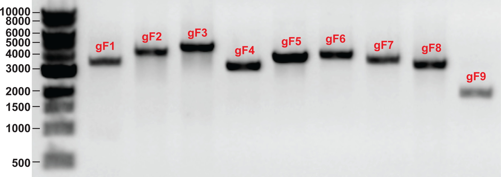
The DNA ladders in bp are shown.
Anticipated results
This protocol efficiently produces recombinant SARS-CoV-2 viruses. The recovered virus can cause significant CPE on Vero E6 (Figure 5a). The engineered molecular markers with no other mutations are retained in the recombinant SARS-CoV-2 genome (Figure 5b). The recombinant virus can generate similar plaque morphologies and replication kinetics as the clinical isolate strain WA1 on Vero E6 cells3.
Figure 5. Characterization of recombinant SARS-CoV-2.
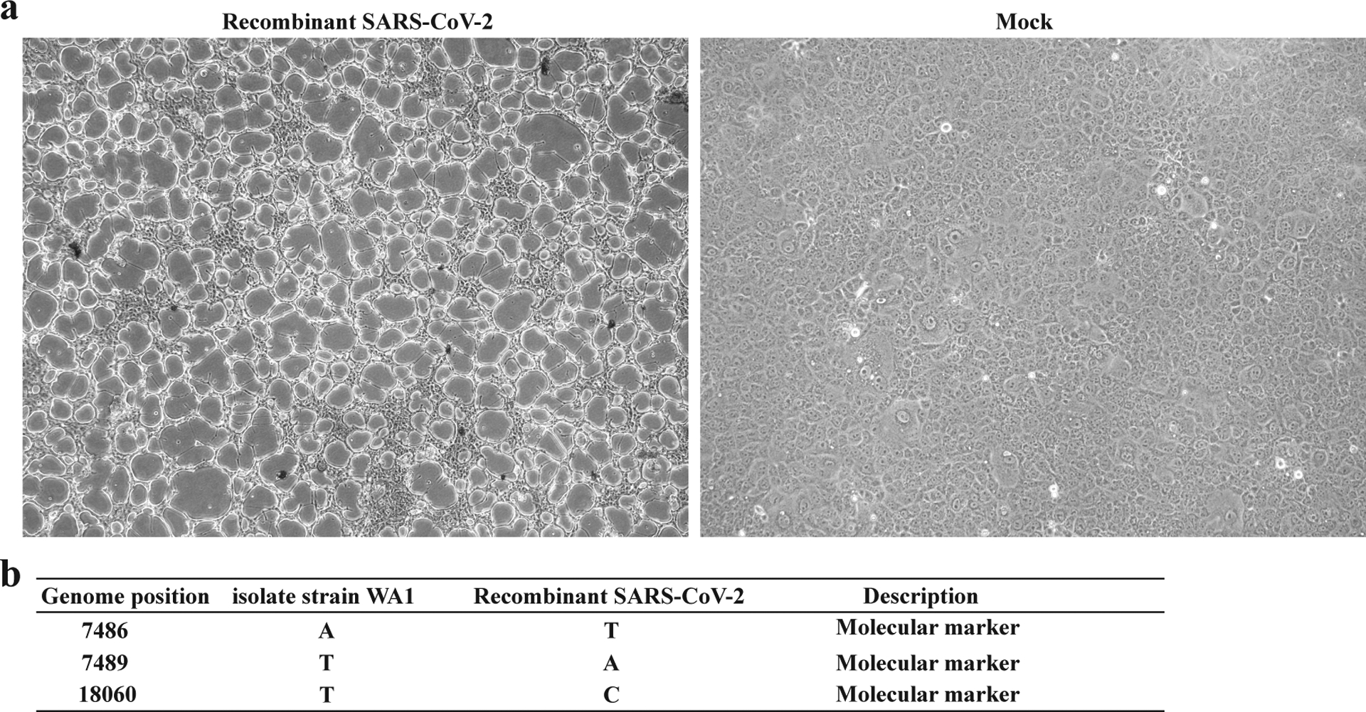
(a) Bright-field images of the recombinant SARS-CoV-2 infected Vero E6 cells using EVOS M5000 Imaging System with 10x objective. Cytopathic effects appeared on day 2 after cells were inoculated with recovered SARS-CoV-2 P0 virus. (b) Sequence results of the recombinant SARS-CoV-2.
Supplementary Material
supplemental material
Table 2.
Sequencing primer list
| Fragments | Primer name | Sequence (5’–3’) |
|---|---|---|
| gF1 | cov-1V | ATTAAAGGTTTATACCTTCCCAGG |
| cov-655V | AGCTGGTGGCCATAGTTAC | |
| cov-1321V | AGGTGCCACTACTTGTGG | |
| cov-1925V | CTGCTCAAAATTCTGTGCG | |
| cov-2572V | CTACTAGTGAAGCTGTTGAAGC | |
| cov-3225V | CTGTTGGTCAACAAGACGG | |
| cov-528R | AGCTCAACCATAACATGACC | |
| gF2 | cov-3225V | CTGTTGGTCAACAAGACGG |
| cov-3824V | GTTTCAAGCTTTTTGGAAATG | |
| cov-4431V | TGCCTGTCTGTGTGGAAAC | |
| cov-4990V | CAACATTAACCTCCACACGC | |
| cov-5525V | ACTTGTGGACAACAGCAG | |
| cov-6109V | GAAACCTGCTTCAAGAGAG | |
| cov-6737V | ACACGGTGTTTAAACCGTG | |
| cov-7382V | CAAATGGCCCCGATTTCAG | |
| gF3 | cov-7382V | CAAATGGCCCCGATTTCAG |
| cov-7930V | TCAGCGTCTGTTTACTACAG | |
| cov-8481V | CTTTTAAGTTGACATGTGCAAC | |
| cov-8995V | ATCAGCTTGTGTTTTGGC | |
| cov-9534V | CTGTACTCTGTTTAACACC | |
| cov-10094V | GAGGGTTGTATGGTACAAG | |
| cov-10680V | ACGCTGCTGTTATAAATGG | |
| cov-11188V | ACCTTCTCTTGCCACTG | |
| cov-11707V | AGTTTCTACACAGGAGTTTAG | |
| gF4 | cov-11707V | AGTTTCTACACAGGAGTTTAG |
| cov-12205V | GAAGAAGTCTTTGAATGTGG | |
| cov-12806V | GTACTTGCACTGTTATCCG | |
| cov-13441V | GTCAGCTGATGCACAATCG | |
| cov-14062V | GATAATCAAGATCTCAATGG | |
| cov-14618V | CTACGTGCTTTTCAGTAG | |
| gF5 | cov-14618V | CTACGTGCTTTTCAGTAG |
| cov-15170V | ATCAATAGCCGCCACTAG | |
| cov-15677V | ACGCATATTTGCGTAAAC | |
| cov-16273V | TCATTAAGATGTGGTGCTTG | |
| cov-16853V | GTGATGCTGTTGTTTACCG | |
| cov-17444V | CTCAATTACCTGCACCAC | |
| cov-18037V | AAGCTGAAAATGTAACAGG | |
| gF6 | cov-18037V | AAGCTGAAAATGTAACAGG |
| cov-18588V | TGTCTTATGGGCACATGG | |
| cov-19211V | GATATCCTGCTAATTCCATTG | |
| cov-19840V | ATTTGGGTGTGGACATTG | |
| cov-20459V | AACAGATGCGCAAACAGG | |
| cov-20934V | TACGCTGCTTGTCGATTC | |
| cov-21521V | TGTTATTTCTAGTGATGTTCTTG | |
| gF7 | cov-21521V | TGTTATTTCTAGTGATGTTCTTG |
| cov-22092V | TGGACCTTGAAGGAAAAC | |
| cov-22685V | TCCACTTTTAAGTGTTATGGAG | |
| cov-23203V | AGGCACAGGTGTTCTTAC | |
| cov-23840V | GTACACAATTAAACCGTGC | |
| cov-24428V | CACAAGCTTTAAACACGC | |
| cov-25068V | TCTCTGGCATTAATGCTTC | |
| gF8 | cov-25068V | TCTCTGGCATTAATGCTTC |
| cov-25624V | CACTTTGTTTGCAACTTGC | |
| cov-26245V | CATTCGTTTCGGAAGAGAC | |
| cov-26778V | GTCTTGTAGGCTTGATGTG | |
| cov-27372V | ATGGAGATTGATTAAACGAAC | |
| cov-27875V | TTGTCACGCCTAAACGAAC | |
| cov-25068V | TCTCTGGCATTAATGCTTC | |
| gF9 | cov-27875V | TTGTCACGCCTAAACGAAC |
| cov-28404V | GTTTACCCAATAATACTGCG | |
| cov-28994V | CAACAAGGCCAAACTGTC | |
| cov-29611V | GTGCAGAATGAATTCTCG | |
| F7-AvrII-R | GAAGTCCAGCTTCTGGCC |
Acknowledgements
X.X. was partially supported by NIH5UC7AI094660. V.D.M was supported by NIH and NIAID grants AI153602 and AG049042, and STARs Award provided by the University of Texas System. P.-Y.S. was supported by NIH grants AI142759, AI134907, AI145617, and UL1TR001439, and awards from the Sealy & Smith Foundation, Kleberg Foundation, the John S. Dunn Foundation, the Amon G. Carter Foundation, the Gilson Longenbaugh Foundation, and the Summerfield Robert Foundation.
Footnotes
Data Availability Statement
Similar data that support the this study are reported in previous publications (ref.3). The seven plasmids of SARS-CoV-2 has been deposited to the World Reference Center for Emerging Viruses and Arboviruses (https://www.utmb.edu/wrceva) at UTMB for distribution.
Competing interests
X.X., V.D.M, and P.-Y.S. have filed a patent on the reverse genetic system of SARS-CoV-2 and reporter SARS-CoV-2. Other authors declare no competing interests.
References
- 1.Gralinski LE & Menachery VD Return of the Coronavirus: 2019-nCoV. Viruses 12, doi: 10.3390/v12020135 (2020). [DOI] [PMC free article] [PubMed] [Google Scholar]
- 2.Muruato AE et al. A high-throughput neutralizing antibody assay for COVID-19 diagnosis and vaccine evaluation. Nature Communications 11, 4059, doi: 10.1038/s41467-020-17892-0 (2020). [DOI] [PMC free article] [PubMed] [Google Scholar]
- 3.Xie X et al. An Infectious cDNA Clone of SARS-CoV-2. Cell Host Microbe 27, 841–848 e843, doi: 10.1016/j.chom.2020.04.004 (2020). [DOI] [PMC free article] [PubMed] [Google Scholar]
- 4.Xie X et al. A nanoluciferase SARS-CoV-2 for rapid neutralization testing and screening of anti-infective drugs for COVID-19. Nature Communications 11, 5214, doi: 10.1038/s41467-020-19055-7 (2020). [DOI] [PMC free article] [PubMed] [Google Scholar]
- 5.Yount B et al. Reverse genetics with a full-length infectious cDNA of severe acute respiratory syndrome coronavirus. Proc Natl Acad Sci U S A 100, 12995–13000, doi: 10.1073/pnas.1735582100 (2003). [DOI] [PMC free article] [PubMed] [Google Scholar]
- 6.Yount B, Denison MR, Weiss SR & Baric RS Systematic assembly of a full-length infectious cDNA of mouse hepatitis virus strain A59. J Virol 76, 11065–11078, doi: 10.1128/jvi.76.21.11065-11078.2002 (2002). [DOI] [PMC free article] [PubMed] [Google Scholar]
- 7.Yount B, Curtis KM & Baric RS Strategy for systematic assembly of large RNA and DNA genomes: transmissible gastroenteritis virus model. J Virol 74, 10600–10611, doi: 10.1128/jvi.74.22.10600-10611.2000 (2000). [DOI] [PMC free article] [PubMed] [Google Scholar]
- 8.Harcourt J et al. Severe Acute Respiratory Syndrome Coronavirus 2 from Patient with 2019 Novel Coronavirus Disease, United States. Emerg Infect Dis 26, doi: 10.3201/eid2606.200516 (2020). [DOI] [PMC free article] [PubMed] [Google Scholar]
- 9.Johnson BA et al. Furin Cleavage Site Is Key to SARS-CoV-2 Pathogenesis. bioRxiv, doi: 10.1101/2020.08.26.268854 (2020). [DOI] [Google Scholar]
- 10.Plante JA et al. Spike mutation D614G alters SARS-CoV-2 fitness. Nature, doi: 10.1038/s41586-020-2895-3 (2020). [DOI] [PMC free article] [PubMed] [Google Scholar]
- 11.Almazan F et al. Coronavirus reverse genetic systems: infectious clones and replicons. Virus Res 189, 262–270, doi: 10.1016/j.virusres.2014.05.026 (2014). [DOI] [PMC free article] [PubMed] [Google Scholar]
- 12.Becker MM et al. Synthetic recombinant bat SARS-like coronavirus is infectious in cultured cells and in mice. Proc Natl Acad Sci U S A 105, 19944–19949, doi: 10.1073/pnas.0808116105 (2008). [DOI] [PMC free article] [PubMed] [Google Scholar]
- 13.Menachery VD et al. A SARS-like cluster of circulating bat coronaviruses shows potential for human emergence. Nat Med 21, 1508–1513, doi: 10.1038/nm.3985 (2015). [DOI] [PMC free article] [PubMed] [Google Scholar]
- 14.Menachery VD et al. SARS-like WIV1-CoV poised for human emergence. Proc Natl Acad Sci U S A 113, 3048–3053, doi: 10.1073/pnas.1517719113 (2016). [DOI] [PMC free article] [PubMed] [Google Scholar]
- 15.Scobey T et al. Reverse genetics with a full-length infectious cDNA of the Middle East respiratory syndrome coronavirus. Proc Natl Acad Sci U S A 110, 16157–16162, doi: 10.1073/pnas.1311542110 (2013). [DOI] [PMC free article] [PubMed] [Google Scholar]
- 16.Sahin U et al. COVID-19 vaccine BNT162b1 elicits human antibody and TH1 T cell responses. Nature 586, 594–599, doi: 10.1038/s41586-020-2814-7 (2020). [DOI] [PubMed] [Google Scholar]
- 17.Mulligan MJ et al. Phase I/II study of COVID-19 RNA vaccine BNT162b1 in adults. Nature 586, 589–593, doi: 10.1038/s41586-020-2639-4 (2020). [DOI] [PubMed] [Google Scholar]
- 18.Walsh EE et al. Safety and Immunogenicity of Two RNA-Based Covid-19 Vaccine Candidates. New England Journal of Medicine, doi: 10.1056/NEJMoa2027906 (2020). [DOI] [PMC free article] [PubMed] [Google Scholar]
- 19.Thi Nhu Thao T et al. Rapid reconstruction of SARS-CoV-2 using a synthetic genomics platform. Nature 582, 561–565, doi: 10.1038/s41586-020-2294-9 (2020). [DOI] [PubMed] [Google Scholar]
- 20.Menachery VD et al. Combination Attenuation Offers Strategy for Live Attenuated Coronavirus Vaccines. J Virol 92, doi: 10.1128/JVI.00710-18 (2018). [DOI] [PMC free article] [PubMed] [Google Scholar]
- 21.Menachery VD et al. MERS-CoV Accessory ORFs Play Key Role for Infection and Pathogenesis. mBio 8, doi: 10.1128/mBio.00665-17 (2017). [DOI] [PMC free article] [PubMed] [Google Scholar]
- 22.Menachery VD et al. Trypsin Treatment Unlocks Barrier for Zoonotic Bat Coronavirus Infection. J Virol 94, doi: 10.1128/JVI.01774-19 (2020). [DOI] [PMC free article] [PubMed] [Google Scholar]
- 23.Hou YJ et al. SARS-CoV-2 Reverse Genetics Reveals a Variable Infection Gradient in the Respiratory Tract. Cell 182, 1–18 (2020). [DOI] [PMC free article] [PubMed] [Google Scholar]
- 24.Curtis KM, Yount B & Baric RS Heterologous gene expression from transmissible gastroenteritis virus replicon particles. J Virol 76, 1422–1434, doi: 10.1128/jvi.76.3.1422-1434.2002 (2002). [DOI] [PMC free article] [PubMed] [Google Scholar]
Key references using this protocol
- 1.Xie X et al. An Infectious cDNA Clone of SARS-CoV-2. Cell Host Microbe 27, 841–848 e843, doi: 10.1016/j.chom.2020.04.004 (2020). [DOI] [PMC free article] [PubMed] [Google Scholar]
- 2.Johnson BA et al. Furin Cleavage Site Is Key to SARS-CoV-2 Pathogenesis. bioRxiv, doi: 10.1101/2020.08.26.268854 (2020). [DOI] [Google Scholar]
- 3.Plante JA et al. Spike mutation D614G alters SARS-CoV-2 fitness. Nature, doi: 10.1038/s41586-020-2895-3 (2020). [DOI] [PMC free article] [PubMed] [Google Scholar]
Associated Data
This section collects any data citations, data availability statements, or supplementary materials included in this article.
Supplementary Materials
supplemental material