Human Slug is a repressor that localizes to sites of active transcription - PubMed (original) (raw)
Human Slug is a repressor that localizes to sites of active transcription
K Hemavathy et al. Mol Cell Biol. 2000 Jul.
Abstract
Snail/Slug family proteins have been identified in diverse species of both vertebrates and invertebrates. The proteins contain four to six zinc fingers and function as DNA-binding transcriptional regulators. Various members of the family have been demonstrated to regulate cell movement, neural cell fate, left-right asymmetry, cell cycle, and apoptosis. However, the molecular mechanisms of how these regulators function and the target genes involved are largely unknown. In this report, we demonstrate that human Slug (hSlug) is a repressor and modulates both activator-dependent and basal transcription. The repression depends on the C-terminal DNA-binding zinc fingers and on a separable repression domain located in the N terminus. This domain may recruit histone deacetylases to modify the chromatin and effect repression. Protein localization study demonstrates that hSlug is present in discrete foci in the nucleus. This subnuclear pattern does not colocalize with the PML foci or the coiled bodies. Instead, the hSlug foci overlap extensively with areas of the SC-35 staining, some of which have been suggested to be sites of active splicing or transcription. These results lead us to postulate that hSlug localizes to target promoters, where activation occurs, to repress basal and activator-mediated transcription.
Figures
FIG. 1
Structural relationship of Snail family proteins. The three proteins shown on the top, Snail, Worniu, and Escargot, are from Drosophila. mSnail is from the mouse, and the three Slug proteins are from the mouse, chicken, and human, respectively. The other members of the family, including two zebra fish Snails, frog Snail and Slug, protochordate Snails, chicken Snail, human Snail, and Drosophila Scratch, are not shown here. The N termini of all three Drosophila proteins are highly divergent among themselves; these regions are also highly divergent among vertebrate and invertebrate proteins. The Drosophila N-terminal (NT) box and the vertebrate SNAG domain are different motifs, but both contain highly basic amino acid residues. The C-terminal binding protein (CtBP) interaction motif has the sequence related to P-DLS-K/R (41, 42, 47). The DBD contains four to six highly conserved zinc fingers.
FIG. 2
mRNA expression of hSlug in adult tissues. The hSlug full-length cDNA was used to prepare a radioactive probe, which was then used to hybridize with mRNA from various human tissues. The analysis reveals a single mRNA species of approximately 2.2 kb that hybridized with the probe. Expression is relatively high in the ovary and almost undetectable in peripheral blood leukocytes. All other tissues tested have detectable and variable levels of expression. The lower panel shows hybridization of the same blots after stripping using the rat 18S rRNA probe, which cross-hybridized with the homologous human transcripts.
FIG. 3
hSlug is a sequence-specific DNA-binding protein. Bacterial extracts that contained full-length or zinc finger domain-truncated hSlug proteins were incubated with a double-stranded oligonucleotide probe that contained the consensus SBS. The mixture was then analyzed on an acrylamide gel. A prominent protein-DNA complex was detected that has slower mobility than the free DNA (lanes 2 to 4, increasing amount of extract). This complex was not seen in extract that contained the zinc finger domain-deleted hSlug (lanes 5 to 7). The competition assay (lane 8 to 14) demonstrates that the wild-type, unlabeled oligonucleotide (SBS) is an efficient competitor, while the oligonucleotide that contained mutations in the recognition core (SBSmut) could not compete. One microliter each of two antisera (ab1 and ab2) raised against full-length hSlug protein was added in similar assays (lane 15 to 21). The antisera abolished complex formation, while the preimmune sera (preimm1 and 2) did not. Weak bands (*) with slower mobility were also formed when the antisera were added to the mixture, indicating the formation of complexes that contained the antibodies for hSlug.
FIG. 4
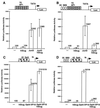
Repression of basal and activated transcription by hSlug. Human 293T cells were transfected with different combinations of expression and reporter plasmids. The specific DNA-binding sites on the promoter of the reporter plasmids used are illustrated at the top of each panel. Luciferase reporter activities were measured 48 h after transfection. In the presence of appropriate binding sites, JunD (A) and Gal4-VP16 (C) increased the reporter activity substantially, indicating activation of transcription. The addition of hSlug did not repress transcription if the binding sites for hSlug were not present (A and C). In the presence of the SBS, hSlug repressed the activated transcription by both JunD and Gal4-VP16 (B and D). Furthermore, the basal transcription was also repressed significantly by hSlug (B and D). Panel E shows that a fusion construct, the hSlug N terminus fused with the Gal4 DBD, repressed basal transcription. The reporter contained the Gal4 binding sites and was modestly activated by the GAL4 DBD alone. Therefore, occupation of the binding sites by the hSlug fusion reduces the activity to a level much lower than the basal level, suggesting active repression.
FIG. 5
The N terminus of hSlug contains both repression and activation modules. A transfection assay using the M1 and M2 constructs and SBS-containing reporter demonstrates that neither the N or C terminus of hSlug changes the reporter activity. Various portions of the N terminus of hSlug were then fused in frame with the Gal4 DBD (A). The constructs were cotransfected with a luciferase reporter that contained both four Gal4 binding sites (for hSlug-Gal4 repression) and seven AP1 binding sites (for JunD activation) (B). The N-terminal hSlug-Gal4 DBD fusion (M3) repressed activation efficiently, demonstrating the presence of the repression domain in the N terminus. Serial deletion shows that the first 32 aa contain the most potent repression domain. This domain is dominant over the central activation domain (aa 33 to 94). The region from aa 95 to 129 contains a helper domain for repression (compare M3 and M4 with M14 and M15), since the construct M14 presents the best repressor activity. However, this helper domain is not sufficient to override the activation by the central domain (M8). Full-length hSlug, M1, and M2 were transfected with a different reporter that contained the SBS target.
FIG. 6
hSlug repression is affected by an HDAC inhibitor. The cultured cells were cotransfected with expression plasmids as indicated. The reporter plasmids contain the corresponding binding sites for both protein expression plasmids. The HDAC inhibitor TSA was added 24 h after transfection; the cells were harvested 24 h later. The presence of dimethyl sulfoxide solvent (−TSA) did not result in any relief of repression. Basal transcription and activated transcription could be elevated to some extent by the addition of TSA, demonstrating some nonspecific increase of transcription. However, the addition of TSA caused more significant relief of the hSlug-mediated repression (5-fold versus 2-fold in panel A; 18-fold versus 5-fold in panel B). These suggest that HDACs may mediate part of the repressor function of hSlug.
FIG. 7
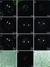
Colocalization of hSlug with SC-35 domains. The antibodies raised against hSlug were affinity purified and used for immunofluorescence staining. The staining for hSlug is predominantly nuclear and punctate in HeLa cells (A to J) as well as in other cell types tested (data not shown). Furthermore, the transfected hSlug-HA fusion protein in 293T cells (K and L) also exhibited punctate nuclear staining. Double stainings were performed together with the anti-SC-35 (A to C), anti-PML (E to G), and anti-coilin (H to J) antibodies. There are fewer foci for PML, and the pattern does not overlap with that of hSlug (PML staining is indicated by arrows in panels F and G). Similar double staining with SC-35 shows that the two patterns overlap extensively (two examples are indicated by the arrows in panels A to C). The coiled bodies also contain splicing factors, but the double staining with coilin reveals that hSlug and coilin do not colocalize (coilin staining is indicated by arrows in panels I and J). All panels are images obtained from confocal microscopy.
Similar articles
- Identification of Daxx interacting with p73, one of the p53 family, and its regulation of p53 activity by competitive interaction with PML.
Kim EJ, Park JS, Um SJ. Kim EJ, et al. Nucleic Acids Res. 2003 Sep 15;31(18):5356-67. doi: 10.1093/nar/gkg741. Nucleic Acids Res. 2003. PMID: 12954772 Free PMC article. - Interaction with members of the heterochromatin protein 1 (HP1) family and histone deacetylation are differentially involved in transcriptional silencing by members of the TIF1 family.
Nielsen AL, Ortiz JA, You J, Oulad-Abdelghani M, Khechumian R, Gansmuller A, Chambon P, Losson R. Nielsen AL, et al. EMBO J. 1999 Nov 15;18(22):6385-95. doi: 10.1093/emboj/18.22.6385. EMBO J. 1999. PMID: 10562550 Free PMC article. - Fusion proteins of the retinoic acid receptor-alpha recruit histone deacetylase in promyelocytic leukaemia.
Grignani F, De Matteis S, Nervi C, Tomassoni L, Gelmetti V, Cioce M, Fanelli M, Ruthardt M, Ferrara FF, Zamir I, Seiser C, Grignani F, Lazar MA, Minucci S, Pelicci PG. Grignani F, et al. Nature. 1998 Feb 19;391(6669):815-8. doi: 10.1038/35901. Nature. 1998. PMID: 9486655 - Modulation of thyroid hormone receptor silencing function by co-repressors and a synergizing transcription factor.
Lutz M, Baniahmad A, Renkawitz R. Lutz M, et al. Biochem Soc Trans. 2000;28(4):386-9. Biochem Soc Trans. 2000. PMID: 10961925 Review. - Snail/Gfi-1 (SNAG) family zinc finger proteins in transcription regulation, chromatin dynamics, cell signaling, development, and disease.
Chiang C, Ayyanathan K. Chiang C, et al. Cytokine Growth Factor Rev. 2013 Apr;24(2):123-31. doi: 10.1016/j.cytogfr.2012.09.002. Epub 2012 Oct 25. Cytokine Growth Factor Rev. 2013. PMID: 23102646 Free PMC article. Review.
Cited by
- Characterization of the SNAG and SLUG domains of Snail2 in the repression of E-cadherin and EMT induction: modulation by serine 4 phosphorylation.
Molina-Ortiz P, Villarejo A, MacPherson M, Santos V, Montes A, Souchelnytskyi S, Portillo F, Cano A. Molina-Ortiz P, et al. PLoS One. 2012;7(5):e36132. doi: 10.1371/journal.pone.0036132. Epub 2012 May 2. PLoS One. 2012. PMID: 22567133 Free PMC article. - Epigenetic polypharmacology: A new frontier for epi-drug discovery.
Tomaselli D, Lucidi A, Rotili D, Mai A. Tomaselli D, et al. Med Res Rev. 2020 Jan;40(1):190-244. doi: 10.1002/med.21600. Epub 2019 Jun 20. Med Res Rev. 2020. PMID: 31218726 Free PMC article. Review. - The mouse snail gene encodes a key regulator of the epithelial-mesenchymal transition.
Carver EA, Jiang R, Lan Y, Oram KF, Gridley T. Carver EA, et al. Mol Cell Biol. 2001 Dec;21(23):8184-8. doi: 10.1128/MCB.21.23.8184-8188.2001. Mol Cell Biol. 2001. PMID: 11689706 Free PMC article. - Phosphorylation regulates the subcellular location and activity of the snail transcriptional repressor.
Domínguez D, Montserrat-Sentís B, Virgós-Soler A, Guaita S, Grueso J, Porta M, Puig I, Baulida J, Francí C, García de Herreros A. Domínguez D, et al. Mol Cell Biol. 2003 Jul;23(14):5078-89. doi: 10.1128/MCB.23.14.5078-5089.2003. Mol Cell Biol. 2003. PMID: 12832491 Free PMC article. - Snail family regulation and epithelial mesenchymal transitions in breast cancer progression.
de Herreros AG, Peiró S, Nassour M, Savagner P. de Herreros AG, et al. J Mammary Gland Biol Neoplasia. 2010 Jun;15(2):135-47. doi: 10.1007/s10911-010-9179-8. Epub 2010 May 9. J Mammary Gland Biol Neoplasia. 2010. PMID: 20455012 Free PMC article. Review.
References
- Alberga A, Boulay J-L, Kempe E, Dennefeld C, Haenlin M. The snail gene required for mesoderm formation is expressed dynamically in derivatives of all three germ layers. Development. 1991;111:983–992. - PubMed
- Ashraf S I, Ip Y T. Transcriptional control: repression by local chromatin modification. Curr Biol. 1998;8:R683–686. - PubMed
- Ayer D E. Histone deacetylases: transcriptional repression with SINers and NuRDs. Trends Cell Biol. 1999;9:193–198. - PubMed
- Bohmann K, Ferreira J, Santama N, Weis K, Lamond A I. Molecular analysis of the coiled body. J Cell Sci Suppl. 1995;19:107–113. - PubMed
Publication types
MeSH terms
Substances
LinkOut - more resources
Full Text Sources
Other Literature Sources
Medical
Molecular Biology Databases
Research Materials


