Dominant-negative C/EBP disrupts mitotic clonal expansion and differentiation of 3T3-L1 preadipocytes - PubMed (original) (raw)
Dominant-negative C/EBP disrupts mitotic clonal expansion and differentiation of 3T3-L1 preadipocytes
Jiang-Wen Zhang et al. Proc Natl Acad Sci U S A. 2004.
Abstract
Hormonal induction of growth-arrested 3T3-L1 preadipocytes rapidly activates expression of CCAAT/enhancer-binding protein (C/EBP) beta. Acquisition of DNA-binding activity by C/EBPbeta, however, is delayed until the cells synchronously enter the S phase of mitotic clonal expansion (MCE). After MCE, C/EBPbeta activates expression of C/EBPalpha and peroxisome proliferator-activated receptor gamma, which then transcriptionally activate genes that give rise to the adipocyte phenotype. A-C/EBP, which possesses a leucine zipper but lacks functional DNA-binding and transactivation domains, forms stable inactive heterodimers with C/EBPbeta in vitro. Infection of 3T3-L1 preadipocytes with an adenovirus A-C/EBP expression vector interferes with C/EBPbeta function after induction of differentiation. A-C/EBP inhibited events associated with hormone-induced entry of S-phase of the cell cycle, including the turnover of p27/Kip1, a key cyclin-dependent kinase inhibitor, expression of cyclin A and cyclin-dependent kinase 2, DNA replication, MCE, and, subsequently, adipogenesis. Although A-C/EBP blocked cell proliferation associated with MCE, it did not inhibit normal proliferation of 3T3-L1 preadipocytes. Immunofluorescent staining of C/EBPbeta revealed that A-C/EBP prevented the normal punctate nuclear staining of centromeres, an indicator of C/EBPbeta binding to C/EBP regulatory elements in centromeric satellite DNA. The inhibitory effects of A-C/EBP appear to be due primarily to interference with nuclear import of C/EBPbeta caused by obscuring its nuclear localization signal. These findings show that both MCE and adipogenesis are dependent on C/EBPbeta.
Figures
Fig. 1.
Effect of dominant-negative A-C/EBP on the differentiation of 3T3-L1 preadipocytes. Nearly confluent 3T3-L1 preadipocytes were infected with adenoviruses either lacking an insert (control) or containing an insert that encodes A-C/EBP, a dominant-negative C/EBP. Exposure to the viruses was continued for 2 days, at which time (2 days postconfluent) the preadipocytes were induced to differentiate by using the standard protocol. (A) Eight days after induction, the cell monolayers were stained with Oil red O. (B) Four days after induction, cell lysates were immunoblotted with antibody specific against C/EBPα, PPARγ, and 422/aP2.
Fig. 2.
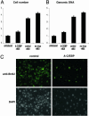
Effect of A-C/EBP on MCE. 3T3-L1 preadipocytes were infected with A-C/EBP or control adenoviruses and induced as in Fig. 1. Three days after induction of differentiation, cell number was determined (A), genomic DNA was quantified (B), and incorporation of BrdUrd into cellular DNA was determined by using a monoclonal anti-BrdUrd antibody and FITC-conjugated secondary antibody (C). Cells were then stained with DAPI and photographed with a fluorescence microscope.
Fig. 3.
Effect of A-C/EBP on the expression of cell-cycle markers. 3T3-L1 preadipocytes were infected with control or A-C/EBP adenoviruses and induced to differentiate as in Fig. 1. Cell lysates were prepared every 4 h after induction differentiation. Cell lysates were subjected to SDS/PAGE and immunoblotted with antibodies against cyclin D1, cyclin B1, p27/Kip1, cyclin A, Cdk2, C/EBPβ, and Flag (recognizing Flag-A-C/EBP fusion protein).
Fig. 4.
Interaction of endogenous C/EBPβ with A-C/EBP. 3T3-L1 preadipocytes were infected with A-C/EBP or control adenoviruses and induced as in Fig. 1. Twenty hours after induction, cell lysates were prepared and subjected to immunoprecipitation by using anti-Flag agarose beads. After three washes, supernates and precipitates were separated by SDS/PAGE and immunoblotted with anti-C/EBPβ and anti-flag antibodies.
Fig. 5.
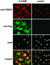
A-C/EBP prevents nuclear localization and centromeric localization of C/EBPβ. 3T3-L1 preadipocytes on coverslips were infected with control and A-C/EBP (Flag-tagged) adenoviruses as in Fig. 1 and then induced to differentiate. Cells were immunostained with antibodies to C/EBPβ and Flag and were stained with DAPI.
References
- Shepherd, P. R., Gnudi, L., Tozzo, E., Yang, H., Leach, F. & Kahn, B. B. (1993) J. Biol. Chem. 268, 22243–22246. - PubMed
- Gnudi, L., Shepherd, P. R. & Kahn, B. B. (1996) Proc. Nutr. Soc. 55, 191–199. - PubMed
- Bernlohr, D. A., Bolanowski, M. A., Kelly, T. J. & Lane, M. D. (1985) J. Biol. Chem. 260, 5563–5567. - PubMed
- Cornelius, P., MacDougald, O. A. & Lane, M. D. (1994) Annu. Rev. Nutr. 14, 99–129. - PubMed
Publication types
MeSH terms
Substances
Grants and funding
- K01 DK061355/DK/NIDDK NIH HHS/United States
- R01 DK038418/DK/NIDDK NIH HHS/United States
- R01 DK060787/DK/NIDDK NIH HHS/United States
- DK060787/DK/NIDDK NIH HHS/United States
- DK38418/DK/NIDDK NIH HHS/United States
- K01-DK61355/DK/NIDDK NIH HHS/United States
LinkOut - more resources
Full Text Sources
Molecular Biology Databases
Miscellaneous


