Retroviral vectors pseudotyped with severe acute respiratory syndrome coronavirus S protein - PubMed (original) (raw)
Retroviral vectors pseudotyped with severe acute respiratory syndrome coronavirus S protein
Tsanan Giroglou et al. J Virol. 2004 Sep.
Abstract
The worldwide outbreak of severe acute respiratory syndrome (SARS) was shown to be associated with a novel coronavirus (CoV) now called SARS CoV. We report here the generation of SARS CoV S protein-pseudotyped murine leukemia virus (MLV) vector particles. The wild-type S protein pseudotyped MLV vectors, although at a low efficiency. Partial deletion of the cytoplasmic tail of S dramatically increased infectivity of pseudotypes, with titers only two- to threefold lower than those of pseudotypes generated in parallel with the vesicular stomatitis virus G protein. S-pseudotyped MLV particles were used to analyze viral tropism. MLV(SARS) pseudotypes and wild-type SARS CoV displayed similar cell types and tissue and host restrictions, indicating that the expression of a functional receptor is the major restraint in permissiveness to SARS CoV infection. Efficient gene transfer could be detected in Vero and CaCo2 cells, whereas the level of gene marking of 293T, HeLa, and HepG2 cells was only slightly above background levels. A cat cell line and a dog cell line were not susceptible. Interestingly, PK-15, a porcine kidney cell line, and primary porcine kidney cells were also highly permissive for SARS S pseudotypes and wild-type SARS CoV. This finding suggests that swine may be susceptible to SARS infection and may be a source for infection of humans. Taken together, these results indicate that MLV(SARS) pseudotypes are highly valuable for functional studies of viral tropism and entry and, in addition, can be a powerful tool for the development of therapeutic entry inhibitors without posing a biohazard to human beings.
Figures
FIG. 1.
Schematic illustration of SARS S expression constructs. The extracellular, membrane-spanning, and cytoplasmic domains of the SARS S expression constructs as determined by computational prediction (
) are shown. Deletions of the cytoplasmic domains were performed via PCR. Amino acid residues of the cytoplasmic domains of truncated S proteins are indicated.
FIG. 2.
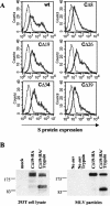
Expression of S protein constructs. (A) Cell surface expression of S mutants in 293T cells. Cells were transfected with DNA encoding either full-length S (wt) or cytoplasmically truncated S protein (CΔ8 to CΔ39). Mock-transfected 293T cells (thin line) were used as a negative control. FACS analysis was performed with SARS antiserum (1:50) and anti-human IgG antibody conjugated with phycoerythrin. (B) Immunoblot of transfected 293T cells (left panel) and purified viral particles (right panel). At 48 h after transfection with HA-tagged CΔ19 S expression construct, cells were lysed either directly (CΔ19-HA) or after being detached from the wells with trypsin-EDTA (CΔ19-HA/Trypsin) and subjected to Western blot analysis with monoclonal anti-HA antibody. Lysates of mock-transfected 293T cells were used as a control (mock). Viral particles with or without trypsin treatment were analyzed likewise. Supernatants produced in the absence of a viral envelope (no env) served as a control. Molecular size standards (in kilodaltons) are indicated.
FIG. 3.
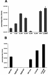
Infectivities of S pseudotypes. (A) Pseudotype titers for different S expression constructs. Vector titers were calculated from end point dilutions on Vero cells and are displayed as TU per milliliter of supernatant (mean ± standard deviation from up to four independent experiments). Envelope proteins of VSV and LCMV were used as positive controls. (B) Infectivities of CΔ19 pseudotypes in the presence of 1, 10, and 25 μM AZT, with HIV-1 core proteins, and with an HA tag without concentration and after 60× concentration by ultracentrifugation. Infectious titers were determined on Vero cells.
FIG. 4.
Incorporation of SARS S protein into MLV particles. Pelleted virus particles were layered on top of a 20 to 60% sucrose gradient and centrifuged to equilibrium. The gradient was fractionated from the top, and individual aliquots were trichloroacetic acid pelleted and then analyzed for the presence of HA-tagged CΔ19 (arrowhead in upper panel) and MLV Gag/p30 (arrowhead in lower panel) by Western blotting with monoclonal anti-HA or polyclonal anti-Gag antibody. The density of each fraction, in grams per milliliter, is shown. Molecular sizes of marker proteins (in kilodaltons) are indicated on the left.
FIG. 5.
Neutralization of SARS pseudotypes. Prior to infection of Vero cells, pseudotype particles were incubated with antisera from four different SARS patients or with a control serum for 1 h at room temperature at the indicated dilutions. The infectivity of pseudotypes in absence of antiserum was set as 100%. Patient antisera were named according to their origin: BNI, Bernhard Nocht Institute, Hamburg, Germany; FFM, Frankfurt, Germany; HK, Hong Kong.
FIG. 6.
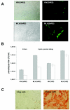
Susceptibility of primary porcine cells to MLV/HIV(SARS) pseudotypes and to SARS CoV. (A) Primary cells of porcine kidney were grown on 48-well plates and transduced with 5.4 × 10² TU of HIV(SARS) pseudotype per ml (upper panel) and with 1.6 × 10⁴ TU of MLV(SARS) pseudotype per ml (lower panel). The phase-contrast and eGFP fluorescences are shown (magnification, ×200). (B) Supernatants containing either HIV(SARS) or MLV(SARS) pseudotypes were used for transduction of Vero cells and primary porcine kidney cells. Pseudotype titers on each cell type were determined by FACS analysis as described in Materials and Methods. Means and standard deviations from duplicates are shown. VSV G-pseudotyped lentiviral and oncoretroviral vectors served as controls. (C) Primary cells of porcine lung and kidney were grown on six-well plates and infected with SARS CoV strain Frankfurt at a multiplicity of infection of 5. Virus replication was assessed by immunostaining with reconvalescent-phase serum from a SARS patient.
Similar articles
- S protein of severe acute respiratory syndrome-associated coronavirus mediates entry into hepatoma cell lines and is targeted by neutralizing antibodies in infected patients.
Hofmann H, Hattermann K, Marzi A, Gramberg T, Geier M, Krumbiegel M, Kuate S, Uberla K, Niedrig M, Pöhlmann S. Hofmann H, et al. J Virol. 2004 Jun;78(12):6134-42. doi: 10.1128/JVI.78.12.6134-6142.2004. J Virol. 2004. PMID: 15163706 Free PMC article. - Contributions of the structural proteins of severe acute respiratory syndrome coronavirus to protective immunity.
Buchholz UJ, Bukreyev A, Yang L, Lamirande EW, Murphy BR, Subbarao K, Collins PL. Buchholz UJ, et al. Proc Natl Acad Sci U S A. 2004 Jun 29;101(26):9804-9. doi: 10.1073/pnas.0403492101. Epub 2004 Jun 21. Proc Natl Acad Sci U S A. 2004. PMID: 15210961 Free PMC article. - Longitudinally profiling neutralizing antibody response to SARS coronavirus with pseudotypes.
Temperton NJ, Chan PK, Simmons G, Zambon MC, Tedder RS, Takeuchi Y, Weiss RA. Temperton NJ, et al. Emerg Infect Dis. 2005 Mar;11(3):411-6. doi: 10.3201/eid1103.040906. Emerg Infect Dis. 2005. PMID: 15757556 Free PMC article. - Vaccine design for severe acute respiratory syndrome coronavirus.
He Y, Jiang S. He Y, et al. Viral Immunol. 2005;18(2):327-32. doi: 10.1089/vim.2005.18.327. Viral Immunol. 2005. PMID: 16035944 Review. - SARS-CoV and emergent coronaviruses: viral determinants of interspecies transmission.
Bolles M, Donaldson E, Baric R. Bolles M, et al. Curr Opin Virol. 2011 Dec;1(6):624-34. doi: 10.1016/j.coviro.2011.10.012. Curr Opin Virol. 2011. PMID: 22180768 Free PMC article. Review.
Cited by
- Identification of SARS-CoV-2 viral entry inhibitors using machine learning and cell-based pseudotyped particle assay.
Sun H, Wang Y, Chen CZ, Xu M, Guo H, Itkin M, Zheng W, Shen M. Sun H, et al. Bioorg Med Chem. 2021 May 15;38:116119. doi: 10.1016/j.bmc.2021.116119. Epub 2021 Mar 26. Bioorg Med Chem. 2021. PMID: 33831697 Free PMC article. - Enhanced protective efficacy of H5 subtype influenza vaccine with modification of the multibasic cleavage site of hemagglutinin in retroviral pseudotypes.
Tao L, Chen J, Meng J, Chen Y, Li H, Liu Y, Zheng Z, Wang H. Tao L, et al. Virol Sin. 2013 Jun;28(3):136-45. doi: 10.1007/s12250-013-3326-5. Epub 2013 May 10. Virol Sin. 2013. PMID: 23728771 Free PMC article. - In Silico Prediction and Experimental Confirmation of HA Residues Conferring Enhanced Human Receptor Specificity of H5N1 Influenza A Viruses.
Schmier S, Mostafa A, Haarmann T, Bannert N, Ziebuhr J, Veljkovic V, Dietrich U, Pleschka S. Schmier S, et al. Sci Rep. 2015 Jun 19;5:11434. doi: 10.1038/srep11434. Sci Rep. 2015. PMID: 26091504 Free PMC article. - Characterization of the spike protein of human coronavirus NL63 in receptor binding and pseudotype virus entry.
Lin HX, Feng Y, Tu X, Zhao X, Hsieh CH, Griffin L, Junop M, Zhang C. Lin HX, et al. Virus Res. 2011 Sep;160(1-2):283-93. doi: 10.1016/j.virusres.2011.06.029. Epub 2011 Jul 20. Virus Res. 2011. PMID: 21798295 Free PMC article. - Exosomal vaccines containing the S protein of the SARS coronavirus induce high levels of neutralizing antibodies.
Kuate S, Cinatl J, Doerr HW, Uberla K. Kuate S, et al. Virology. 2007 May 25;362(1):26-37. doi: 10.1016/j.virol.2006.12.011. Epub 2007 Jan 26. Virology. 2007. PMID: 17258782 Free PMC article.
References
MeSH terms
Substances
LinkOut - more resources
Full Text Sources
Other Literature Sources
Miscellaneous


