The Drosha-DGCR8 complex in primary microRNA processing - PubMed (original) (raw)
Comparative Study
. 2004 Dec 15;18(24):3016-27.
doi: 10.1101/gad.1262504. Epub 2004 Dec 1.
Affiliations
- PMID: 15574589
- PMCID: PMC535913
- DOI: 10.1101/gad.1262504
Comparative Study
The Drosha-DGCR8 complex in primary microRNA processing
Jinju Han et al. Genes Dev. 2004.
Abstract
RNase III proteins play key roles in microRNA (miRNA) biogenesis. The nuclear RNase III Drosha cleaves primary miRNAs (pri-miRNAs) to release hairpin-shaped pre-miRNAs that are subsequently cut by the cytoplasmic RNase III Dicer to generate mature miRNAs. While Dicer (class III) and other simple RNase III proteins (class I) have been studied intensively, the class II enzyme Drosha remains to be characterized. Here we dissected the action mechanism of human Drosha by generating mutants and by characterizing its new interacting partner, DGCR8. The basic action mechanism of Drosha was found to be similar to that of human Dicer; the RNase III domains A and B form an intramolecular dimer and cleave the 3' and 5' strands of the stem, respectively. Human Drosha fractionates at approximately 650 kDa, indicating that Drosha functions as a large complex. In this complex, Drosha interacts with DGCR8, which contains two double-stranded RNA (dsRNA)-binding domains. By RNAi and biochemical reconstitution, we show that DGCR8 may be an essential component of the pri-miRNA processing complex, along with Drosha. Based on these results, we propose a model for the action mechanism of class II RNase III proteins.
Figures
Figure 1.
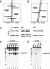
Site-directed mutagenesis of human Drosha. (A) A model for the cleavage mechanism of RNase III (Blaszczyk et al. 2001). Two processing centers are formed between two RIIIDs. Each center contains two catalytic sites that cleave two nearby phosphodiester bonds on the opposite RNA strands of dsRNA (W-C, Watson-Crick base pairs). The 5′ strand indicates the strand that contains the 5′ phosphate group at the terminus of stem. The 3′ strand means the strand that possesses the 3′-hydroxyl group at the 3′ end. (B) The “single processing center” model where only two catalytic sites are formed in a single processing center (Zhang et al. 2004). (C) Naming of the single amino acid mutants in the RIIIDs of human Drosha. (D) In vitro processing of pri-let-7a-1. The substrate was incubated with E64aA, E64aQ, E64bA, E64bQ, or wild-type (WT) Drosha-Flag proteins that were expressed in HEK293T cells and prepared by immunoprecipitation with anti-Flag antibody. The RNA size markers (Decade Marker, Ambion) are indicated on the left side of the gel. (E) In vitro processing of pri-let-7a-1. The substrate was incubated with E110aQ, E110bQ, or wild-type (WT) Drosha-Flag proteins that were immobilized on anti-Flag beads. The asterisks indicate fragments that accumulate when the particular mutant was used in the assay.
Figure 2.
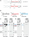
Processing of the “minimal” pri-miRNA with E110 mutants. (A) Sequences and secondary structure of the short pri-miRNAs derived from pri-miR-16-1 and pri-miR-30a. These RNAs are 111 nt and 105 nt, respectively. The cleavage sites (the 5′ and 3′ ends of pre-miRNAs) are indicated with arrows. Actual cleavage sites of pri-miR-16-1 were determined by directional cloning of pre-miR-16-1. Pre-miR-16-1 was prepared by in vitro processing, ligated to the 5′ and 3′ adapters, amplified by RT-PCR, and subcloned into pGEM-T-easy vector. Ten clones were randomly chosen and sequenced. All the clones were identical at the 5′ end. Five of the 10 clones had the same 3′ end shown here. Three other clones were 1 nt shorter and two other clones were 2 nt shorter at the 3′ end, which is likely to be due to exonucleolytic trimming. The cleavage sites at miR-30a were reported (Lee et al. 2003). Pre-miR-16-1 shown here is 65 nt; pre-miR-30a is 63 nt. (B) Schematic representation of the processing products from in vitro processing by wild-type (WT), E110aQ, and E110bQ Drosha. Wild-type Drosha generates F1, F2, and F3 E110aQ produces F1 and F4 and E110bQ generates F3 and F5. (C) In vitro processing of pri-miR-16-1. The short pri-miR-16-1 was incubated with wild-type (WT) Drosha, E110aQ, or E110bQ proteins that were expressed in HEK293T cells and immobilized on anti-Flag affinity gel. The substrate was either internally labeled during transcription (lanes _1_-4) or labeled at the 5′ end after transcription (lanes _5_-8). The size of each fragment is indicated in the bracket next to the arrow. The RNA size markers (Ambion) are indicated on the side of the gel. (D)In vitro processing of pri-miR-30a. The short pri-miR-30a was incubated with wild-type Drosha, E110aQ, or E110bQ proteins as in C. The substrate was either internally labeled during transcription (lanes _1_-4) or labeled at the 5′ end after transcription (lanes _5_-8).
Figure 3.
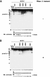
Gel exclusion chromatography of Drosha. (A) Nuclear extract was prepared from twenty-five 10-cm plates of HEK293T cells and fractionated through a Sephacryl-S300 HR column. (Top panel) Each fraction was 1.75 mL in volume, from which 20 μL was taken for in vitro processing of pri-let-7a-1. The fraction number and the protein molecular mass standards (Sigma) are indicated at the top of the gel. “Mock” means that lysis buffer was used instead of protein fractions for the in vitro processing assay. (Bottom panel) The fractions were also analyzed by SDS-PAGE, and Drosha proteins were detected by Western blotting using anti-Drosha antibody. (B) Nuclear extract was treated with 50 μg/mL of RNase A for 30 min at 4°C before loading to the column.
Figure 4.
Role of human DGCR8 in pri-miRNA processing. (A) Immunoprecipitation of the Drosha complex with antibody against Flag. Drosha-Flag protein was expressed in HEK293T cells by transfection of pCK-Drosha-Flag. Total cell extract was prepared from four 10-cm dishes and used for immunoprecipitation with anti-Flag antibody-conjugated agarose beads. (Lane 1) Extract prepared from the same amount of “mock” transfected cells was used as a control. Proteins were separated on 7.5% denaturing polyacrylamide gels and visualized by silver staining. The specific bands were analyzed by MALDI-TOF mass spectrometry. Two forms of truncated Drosha proteins were marked with one or two asterisks (Drosha★ and Drosha★★). Heavy and light chains of anti-Flag antibody are abbreviated as h.c. and l.c. The position of the molecular mass markers is indicated on the left in kilodaltons. (B) Coimmunoprecipitation of DGCR8 and Drosha. V5-tagged DGCR8 and Flag-tagged Drosha proteins were coexpressed in HEK293T cells. Flag-pCK is the backbone for Drosha expression and was used here to match the total amount of plasmids in each transfection. Immunoprecipitation was carried out by incubating total cell extract with anti-Flag antibody-conjugated agarose beads in buffer D-K′250. For Western blot analysis, anti-V5 antibody was used to visualize the V5-DGCR8 protein. (C) In vitro processing of pri-let-7a-1 using Drosha-Flag or Flag-DGCR8 immunoprecipitates prepared in buffer D-K′100. (D) Reconstitution of pri-miRNA processing activity using Drosha-Flag immunoprecipitate and recombinant GST-DGCR8 protein. Pri-miRNA processing activity was measured by in vitro processing of pri-let-7a-1. Drosha-Flag immunoprecipitate was prepared by washing with either buffer A (0.5 M NaCl) or buffer B (2.5 M NaCl); 1.5 μg of recombinant proteins (GST or GST-DGCR8) was added to 1.5 μg of the Drosha-Flag immunoprecipitate. GST-DGCR8 (ΔN275) protein contains 276-773 amino acids of human DGCR8. (E) RT-PCR of pri-miRNA. The siRNA duplex against luciferase, Drosha, or DGCR8 was transfected to HeLa cells. After 72 h, total RNA was prepared and used for RT-PCR. (F) Northern blot analysis of miR-21. RNAi was carried out by transfection of siRNA duplexes to HeLa cells as in E. 5S rRNA bands stained with ethidium bromide are presented as a loading control.
Figure 5.
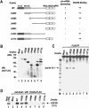
Domain mapping of human Drosha. (A) Schematic representation of Drosha mutants. Results from the pri-miRNA processing assay and the DGCR8 binding assay are summarized at the right. (B) Western blot analysis of the mutant proteins. Expression plasmids were transfected into HEK293T cells. Forty-eight hours post-transfection, total cell extract was analyzed by Western blot analysis using anti-Flag antibody. (C) In vitro processing of pri-let-7a-1. The substrate was incubated with wild-type (WT) Drosha or mutant proteins that were prepared by immunoprecipitation in Buffer D-K′100 from transiently transfected HEK293T cells. (D) In vitro binding assay. Drosha protein (wild type [WT] or mutants) were immobilized on anti-Flag beads and incubated with radiolabeled DGCR8 that had been prepared in TnT-coupled reticulocyte lysate system (Promega). Drosha and DGCR8 were incubated with rotation in buffer D-K′250 for 90 min at 4°C.
Figure 6.
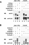
Homodimerization of Drosha and DGCR8. (A) Flag-tagged and V5-tagged Drosha proteins were coexpressed in HEK293T cells by transient transfection of the indicated plasmids. Forty-eight hours post-transfection, total cell extract was prepared and used for immunoprecipitation with anti-Flag affinity gel. Proteins were analyzed by Western blotting with anti-V5 antibody (upper panel) or anti-Flag antibody (lower panel). V5-Dicer was used as a control. (B) Flag-tagged and V5-tagged DGCR8 proteins were coprecipitated. Immunoprecipitation and Western blot analysis were done as in A.
Figure 7.
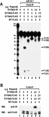
Intramolecular dimerization between the RIIIDa and RIIIDb of human Drosha. (A) Flag-tagged or V5-tagged Drosha proteins were expressed in HEK293T cells by transient transfection of the indicated plasmids. Forty-eight hours post-transfection, immunoprecipitation was carried out using anti-Flag antibody from total cell extract. Processing activity was determined by incubating the immunoprecipitates with internally labeled pri-miR-16-1. Wild-type (WT) enzyme produces F1 (the 5′ flanking fragment), F2 (pre-miRNA), and F3 (the 3′ flanking fragment). Fragment F4 corresponds to F2 and F3 combined, whereas fragment F5 corresponds to F1 and F2 combined. Refer to Figure 2B for graphical explanation. (B) Proteins were analyzed by immunoprecipitation with anti-Flag antibody and Western blotting with anti-V5 antibody (upper panel) or anti-Flag antibody (lower panel).
Similar articles
- Characterization of DGCR8/Pasha, the essential cofactor for Drosha in primary miRNA processing.
Yeom KH, Lee Y, Han J, Suh MR, Kim VN. Yeom KH, et al. Nucleic Acids Res. 2006;34(16):4622-9. doi: 10.1093/nar/gkl458. Epub 2006 Sep 8. Nucleic Acids Res. 2006. PMID: 16963499 Free PMC article. - Molecular basis for the recognition of primary microRNAs by the Drosha-DGCR8 complex.
Han J, Lee Y, Yeom KH, Nam JW, Heo I, Rhee JK, Sohn SY, Cho Y, Zhang BT, Kim VN. Han J, et al. Cell. 2006 Jun 2;125(5):887-901. doi: 10.1016/j.cell.2006.03.043. Cell. 2006. PMID: 16751099 - MicroRNA biogenesis: isolation and characterization of the microprocessor complex.
Gregory RI, Chendrimada TP, Shiekhattar R. Gregory RI, et al. Methods Mol Biol. 2006;342:33-47. doi: 10.1385/1-59745-123-1:33. Methods Mol Biol. 2006. PMID: 16957365 Review. - In vitro and in vivo assays for the activity of Drosha complex.
Lee Y, Kim VN. Lee Y, et al. Methods Enzymol. 2007;427:89-106. doi: 10.1016/S0076-6879(07)27005-3. Methods Enzymol. 2007. PMID: 17720480 - Rethinking the microprocessor.
Seitz H, Zamore PD. Seitz H, et al. Cell. 2006 Jun 2;125(5):827-9. doi: 10.1016/j.cell.2006.05.018. Cell. 2006. PMID: 16751089 Review.
Cited by
- Trans-acting regulators of ribonuclease activity.
Lee J, Lee M, Lee K. Lee J, et al. J Microbiol. 2021 Apr;59(4):341-359. doi: 10.1007/s12275-021-0650-6. Epub 2021 Mar 29. J Microbiol. 2021. PMID: 33779951 Review. - Regulating miRNA by natural agents as a new strategy for cancer treatment.
Sethi S, Li Y, Sarkar FH. Sethi S, et al. Curr Drug Targets. 2013 Sep;14(10):1167-74. doi: 10.2174/13894501113149990189. Curr Drug Targets. 2013. PMID: 23834152 Free PMC article. Review. - Crosstalk between breast cancer-derived microRNAs and brain microenvironmental cells in breast cancer brain metastasis.
Khan MS, Wong GL, Zhuang C, Najjar MK, Lo HW. Khan MS, et al. Front Oncol. 2024 Aug 8;14:1436942. doi: 10.3389/fonc.2024.1436942. eCollection 2024. Front Oncol. 2024. PMID: 39175471 Free PMC article. Review. - Cell-type-specific profiling of loaded miRNAs from Caenorhabditis elegans reveals spatial and temporal flexibility in Argonaute loading.
Brosnan CA, Palmer AJ, Zuryn S. Brosnan CA, et al. Nat Commun. 2021 Apr 13;12(1):2194. doi: 10.1038/s41467-021-22503-7. Nat Commun. 2021. PMID: 33850152 Free PMC article. - AntiVIRmiR: A repository of host antiviral miRNAs and their expression along with experimentally validated viral miRNAs and their targets.
Thakur A, Kumar M. Thakur A, et al. Front Genet. 2022 Sep 8;13:971852. doi: 10.3389/fgene.2022.971852. eCollection 2022. Front Genet. 2022. PMID: 36159991 Free PMC article.
References
- Bartel D.P. 2004. MicroRNAs: Genomics, biogenesis, mechanism, and function. Cell 116: 281-297. - PubMed
- Bernstein E., Caudy, A.A., Hammond, S.M., and Hannon, G.J. 2001. Role for a bidentate ribonuclease in the initiation step of RNA interference. Nature 409: 363-366. - PubMed
- Blaszczyk J., Tropea, J.E., Bubunenko, M., Routzahn, K.M., Waugh, D.S., Court, D.L., and Ji, X. 2001. Crystallographic and modeling studies of RNase III suggest a mechanism for double-stranded RNA cleavage. Structure (Camb) 9: 1225-1236. - PubMed
Publication types
MeSH terms
Substances
LinkOut - more resources
Full Text Sources
Other Literature Sources
Molecular Biology Databases
