Variants in a novel epidermal collagen gene (COL29A1) are associated with atopic dermatitis - PubMed (original) (raw)
doi: 10.1371/journal.pbio.0050242.
Ingo Marenholz, Tamara Kerscher, Franz Rüschendorf, Jorge Esparza-Gordillo, Margitta Worm, Christoph Gruber, Gabriele Mayr, Mario Albrecht, Klaus Rohde, Herbert Schulz, Ulrich Wahn, Norbert Hubner, Young-Ae Lee
Affiliations
- PMID: 17850181
- PMCID: PMC1971127
- DOI: 10.1371/journal.pbio.0050242
Variants in a novel epidermal collagen gene (COL29A1) are associated with atopic dermatitis
Cilla Söderhäll et al. PLoS Biol. 2007 Sep.
Abstract
Atopic dermatitis (AD) is a common chronic inflammatory skin disorder and a major manifestation of allergic disease. AD typically presents in early childhood often preceding the onset of an allergic airway disease, such as asthma or hay fever. We previously mapped a susceptibility locus for AD on Chromosome 3q21. To identify the underlying disease gene, we used a dense map of microsatellite markers and single nucleotide polymorphisms, and we detected association with AD. In concordance with the linkage results, we found a maternal transmission pattern. Furthermore, we demonstrated that the same families contribute to linkage and association. We replicated the association and the maternal effect in a large independent family cohort. A common haplotype showed strong association with AD (p = 0.000059). The associated region contained a single gene, COL29A1, which encodes a novel epidermal collagen. COL29A1 shows a specific gene expression pattern with the highest transcript levels in skin, lung, and the gastrointestinal tract, which are the major sites of allergic disease manifestation. Lack of COL29A1 expression in the outer epidermis of AD patients points to a role of collagen XXIX in epidermal integrity and function, the breakdown of which is a clinical hallmark of AD.
Conflict of interest statement
Competing interests. The authors have declared that no competing interests exist.
Figures
Figure 1. Positional Cloning Strategy for the AD Disease Gene on Chromosome 3q21
(A) The candidate region spanned 12.75 cM between markers D3S1303 and D3S1292. The _y_-axis depicts the GENEHUNTER nonparametric Zall as previously reported [16]. (B) Fine mapping with 96 microsatellite markers narrowed the interval to 5.4 Mb between markers M3CS075 and M3CS233. An association scan using 212 SNPs of the region revealed association of AD with two adjacent SNPs, rs5852593 and rs1497309. Genotyping of 16 additional SNPs refined the associated region. (C) Genomic positions of the 42 exons of COL29A1 are shown. The gene entirely encompasses the associated region. (D) The COL29A1 mRNA consists of 9226 bp. Translation start site and stop codon are indicated. (E) The predicted open reading frame encodes a protein of 2614 amino acids including a secretion peptide (SP), six N-terminal and three C-terminal vWAs, flanking a short collagen triple helix.
Figure 2. Pairwise LD Values (D′) Between 28 SNPs Based on Genotypes of the Founders in the Discovery Cohort
Boxes contain the LD values (D′) between the respective markers indicated on top. Higher LD values correspond to a darker shade of red. Positions on Chromosome 3 are given in Mb; 131.547 denotes the start and 131.686 the end of COL29A1 on the genomic sequence. Boxes on the horizontal bar represent the 42 exons of COL29A1.
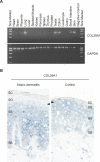
Figure 3. Gene Expression Analysis of COL29A1
(A) RT-PCR analysis of collagen XXIX in human tissues. (B) In situ gene expression analysis of COL29A1 in AD skin. In situ hybridization results of a _COL29A1_-specific antisense probe on cryostat sections (5 μm thick) of an AD skin biopsy (left) and a normal human control (right) are shown. COL29A1 mRNA detected by the digoxigenin-labelled probe is stained in blue (BCIP/NBT staining). The arrows point the different gene expression in the upper spinous and granular layer of the epidermis of an AD patient and a normal human control. Stratum basale (SB), stratum spinosum (SS), stratum granulosum (SG), and stratum corneum (SC) are indicated.
Figure 4. Immunohistochemical Analysis of Collagen XXIX Expression in AD Skin
Cryostat sections (5μm thick) of an AD skin biopsy (A) and a normal human control (B) are shown. In normal, human skin, collagen XXIX is expressed in the epidermis. In patients with atopic dermatitis, a striking lack of collagen XXIX staining was observed in the viable outermost spinous and granular layers of the epidermis (arrow). Stratum basale (SB), stratum spinosum (SS), stratum granulosum (SG), and stratum corneum (SC) are indicated. Collagen XXIX was stained with fuchsin (red). Sections were counterstained with hematoxylin (blue).
Similar articles
- A comprehensive analysis of the COL29A1 gene does not support a role in eczema.
Naumann A, Söderhäll C, Fölster-Holst R, Baurecht H, Harde V, Müller-Wehling K, Rodríguez E, Ruether A, Franke A, Wagenpfeil S, Novak N, Mempel M, Kalali BN, Allgaeuer M, Koch J, Gerhard M, Melén E, Wahlgren CF, Kull I, Stahl C, Pershagen G, Lauener R, Riedler J, Doekes G, Scheynius A, Illig T, von Mutius E, Schreiber S, Kere J, Kabesch M, Weidinger S. Naumann A, et al. J Allergy Clin Immunol. 2011 May;127(5):1187-94.e7. doi: 10.1016/j.jaci.2010.12.1123. Epub 2011 Feb 25. J Allergy Clin Immunol. 2011. PMID: 21353297 - A major susceptibility locus for atopic dermatitis maps to chromosome 3q21.
Lee YA, Wahn U, Kehrt R, Tarani L, Businco L, Gustafsson D, Andersson F, Oranje AP, Wolkertstorfer A, v Berg A, Hoffmann U, Küster W, Wienker T, Rüschendorf F, Reis A. Lee YA, et al. Nat Genet. 2000 Dec;26(4):470-3. doi: 10.1038/82625. Nat Genet. 2000. PMID: 11101848 - Identification of novel candidate variants including COL6A6 polymorphisms in early-onset atopic dermatitis using whole-exome sequencing.
Heo WI, Park KY, Jin T, Lee MK, Kim M, Choi EH, Kim HS, Bae JM, Moon NJ, Seo SJ. Heo WI, et al. BMC Med Genet. 2017 Jan 26;18(1):8. doi: 10.1186/s12881-017-0368-9. BMC Med Genet. 2017. PMID: 28125976 Free PMC article. - Unravelling the complex genetic background of atopic dermatitis: from genetic association results towards novel therapeutic strategies.
Hoffjan S, Stemmler S. Hoffjan S, et al. Arch Dermatol Res. 2015 Oct;307(8):659-70. doi: 10.1007/s00403-015-1550-6. Epub 2015 Feb 19. Arch Dermatol Res. 2015. PMID: 25693656 Review. - On the role of the epidermal differentiation complex in ichthyosis vulgaris, atopic dermatitis and psoriasis.
Hoffjan S, Stemmler S. Hoffjan S, et al. Br J Dermatol. 2007 Sep;157(3):441-9. doi: 10.1111/j.1365-2133.2007.07999.x. Epub 2007 Jun 15. Br J Dermatol. 2007. PMID: 17573887 Review.
Cited by
- The expanded collagen VI family: new chains and new questions.
Fitzgerald J, Holden P, Hansen U. Fitzgerald J, et al. Connect Tissue Res. 2013;54(6):345-50. doi: 10.3109/03008207.2013.822865. Epub 2013 Aug 23. Connect Tissue Res. 2013. PMID: 23869615 Free PMC article. Review. - EMR-linked GWAS study: investigation of variation landscape of loci for body mass index in children.
Namjou B, Keddache M, Marsolo K, Wagner M, Lingren T, Cobb B, Perry C, Kennebeck S, Holm IA, Li R, Crimmins NA, Martin L, Solti I, Kohane IS, Harley JB. Namjou B, et al. Front Genet. 2013 Dec 3;4:268. doi: 10.3389/fgene.2013.00268. eCollection 2013. Front Genet. 2013. PMID: 24348519 Free PMC article. - Meta-analysis of 20 genome-wide linkage studies evidenced new regions linked to asthma and atopy.
Bouzigon E, Forabosco P, Koppelman GH, Cookson WO, Dizier MH, Duffy DL, Evans DM, Ferreira MA, Kere J, Laitinen T, Malerba G, Meyers DA, Moffatt M, Martin NG, Ng MY, Pignatti PF, Wjst M, Kauffmann F, Demenais F, Lewis CM. Bouzigon E, et al. Eur J Hum Genet. 2010 Jun;18(6):700-6. doi: 10.1038/ejhg.2009.224. Epub 2010 Jan 13. Eur J Hum Genet. 2010. PMID: 20068594 Free PMC article. - From Structure to Phenotype: Impact of Collagen Alterations on Human Health.
Arseni L, Lombardi A, Orioli D. Arseni L, et al. Int J Mol Sci. 2018 May 8;19(5):1407. doi: 10.3390/ijms19051407. Int J Mol Sci. 2018. PMID: 29738498 Free PMC article. Review. - LILRA6 copy number variation correlates with susceptibility to atopic dermatitis.
López-Álvarez MR, Jiang W, Jones DC, Jayaraman J, Johnson C, Cookson WO, Moffatt MF, Trowsdale J, Traherne JA. López-Álvarez MR, et al. Immunogenetics. 2016 Oct;68(9):743-7. doi: 10.1007/s00251-016-0924-z. Epub 2016 Jun 22. Immunogenetics. 2016. PMID: 27333811 Free PMC article.
References
- The International Study of Asthma and Allergies in Childhood (ISAAC) Steering Committee. Worldwide variation in prevalence of symptoms of asthma, allergic rhinoconjunctivitis, and atopic eczema: ISAAC. Lancet. 1998;351:1225–1232. - PubMed
- Kay J, Gawkrodger DJ, Mortimer MJ, Jaron AG. The prevalence of childhood atopic eczema in a general population. J Am Acad Dermatol. 1994;30:35–39. - PubMed
- Taylor B, Wadsworth J, Wadsworth M, Peckham C. Changes in the reported prevalence of childhood eczema since the 1939–45 war. Lancet. 1984;2:1255–1257. - PubMed
- Rajka G. Prurigo Besnier (atopic dermatitis) with special reference to the role of allergic factors. Acta Derm Venereol (Stockh) 1960;40:285–306. - PubMed
- Wahn U. What drives the allergic march? Allergy. 2000;55:591–599. - PubMed
Publication types
MeSH terms
Substances
LinkOut - more resources
Full Text Sources
Molecular Biology Databases


