Association between crystals and cartilage degeneration in the ankle - PubMed (original) (raw)
. 2008 Jun;35(6):1108-17.
Epub 2008 Apr 15.
Affiliations
- PMID: 18412302
- PMCID: PMC6240447
Association between crystals and cartilage degeneration in the ankle
Carol Muehleman et al. J Rheumatol. 2008 Jun.
Abstract
Objective: Monosodium urate (MSU) and calcium pyrophosphate dihydrate (CPPD) crystals have been observed in synovial joints both before and after the onset of osteoarthritis (OA). The relationship between crystals and OA, however, remains controversial. We compared histologic and immunohistochemical patterns in articular cartilage of ankle joints with and without crystals.
Methods: A sample of 7,855 human cadaveric tali was examined for the presence of surface and beneath-the-surface crystals. A random subsample of tali with and without crystals underwent crystal analysis by Fourier transform infrared spectroscopy (FTIR), histologic examination, and immunohistochemistry for S100 protein, superficial zone protein, collagen X, cSRC.
Results: The prevalence of grossly visible crystals in the pool of donors over 18 years of age was 4.7% and correlated with advanced age, male sex, and obesity. Crystals were strongly associated with cartilage lesions and these lesions appeared to be biomechanically induced, being located where opposing articular surfaces might not be in congruence with each other. Thirty-four percent of the random subsamples of crystals upon which FTIR was performed contained CPPD, and the remainder were MSU crystals. Both crystal types were associated with higher levels of superficial zone protein and collagen X.
Conclusion: We show that the presence of surface crystals of either MSU or CPPD is strongly correlated with cartilage lesions in the talus. The histologic similarities in cartilage from joints with CPPD crystals compared to those with MSU crystals, together with what is known about the dramatically different etiologic factors producing these crystals, strongly suggest that these lesions are biomechanically induced.
Figures
Figure 1.
The process of selecting tali used for histology and immunohistochemistry.
Figure 2.
Crystals (white arrows) associated with cartilage lesions (black arrows). The crystals surround the lesion in more severe cases. Lesions are located in regions that could be subject to biomechanically-induced damage from the opposing articular surface, particularly in joint instability. Note that in regions with a smooth normal appearance, without lesions, no crystals are present.
Figure 3.
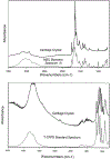
FTIR curves for the tested material (MSU, top; CPPD, bottom) on histological sections of tali compared to the sodium urate standard.
Figure 4.
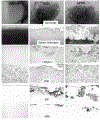
Representative macroscopic (top row) and microscopic views of normal, MSU crystal, and CPPD crystal tali (left, middle, and right columns, respectively). Note the middle zone CPPD crystal cysts that are often in continuity with the articular surface.
Comment in
- Crystal deposition in joints: prevalence and relevance for arthritis.
Pritzker KP. Pritzker KP. J Rheumatol. 2008 Jun;35(6):958-9. J Rheumatol. 2008. PMID: 18528950 No abstract available.
Similar articles
- The prevalence of and factors related to calcium pyrophosphate dihydrate crystal deposition in the knee joint.
Ryu K, Iriuchishima T, Oshida M, Kato Y, Saito A, Imada M, Aizawa S, Tokuhashi Y, Ryu J. Ryu K, et al. Osteoarthritis Cartilage. 2014 Jul;22(7):975-9. doi: 10.1016/j.joca.2014.04.022. Epub 2014 May 9. Osteoarthritis Cartilage. 2014. PMID: 24814686 - Characterization of lesions of the talus and description of tram-track lesions.
Li J, Jadin K, Masuda K, Sah R, Muehleman C. Li J, et al. Foot Ankle Int. 2006 May;27(5):344-55. doi: 10.1177/107110070602700506. Foot Ankle Int. 2006. PMID: 16701055 - Upregulation of ANK protein expression in joint tissue in calcium pyrophosphate dihydrate crystal deposition disease.
Uzuki M, Sawai T, Ryan LM, Rosenthal AK, Masuda I. Uzuki M, et al. J Rheumatol. 2014 Jan;41(1):65-74. doi: 10.3899/jrheum.111476. Epub 2013 Dec 1. J Rheumatol. 2014. PMID: 24293574 Free PMC article. - [Chondrocalcinosis due to calcium pyrophosphate deposition (CPPD). From incidental radiographic findings to CPPD crystal arthritis].
Tausche AK, Aringer M. Tausche AK, et al. Z Rheumatol. 2014 May;73(4):349-57; quiz 358-9. doi: 10.1007/s00393-014-1364-5. Z Rheumatol. 2014. PMID: 24811359 Review. German. - Metabolism of crystals within the joint.
Oliviero F, Scanu A, Punzi L. Oliviero F, et al. Reumatismo. 2012 Jan 19;63(4):221-9. doi: 10.4081/reumatismo.2011.221. Reumatismo. 2012. PMID: 22303528 Review.
Cited by
- Mechanisms of joint damage in gout: evidence from cellular and imaging studies.
McQueen FM, Chhana A, Dalbeth N. McQueen FM, et al. Nat Rev Rheumatol. 2012 Jan 10;8(3):173-81. doi: 10.1038/nrrheum.2011.207. Nat Rev Rheumatol. 2012. PMID: 22231231 Review. - Gout and Osteoarthritis: Associations, Pathophysiology, and Therapeutic Implications.
Yokose C, Chen M, Berhanu A, Pillinger MH, Krasnokutsky S. Yokose C, et al. Curr Rheumatol Rep. 2016 Oct;18(10):65. doi: 10.1007/s11926-016-0613-9. Curr Rheumatol Rep. 2016. PMID: 27686950 Review. - Crystal arthropathies and osteoarthritis-where is the link?
Jarraya M, Roemer F, Kwoh CK, Guermazi A. Jarraya M, et al. Skeletal Radiol. 2023 Nov;52(11):2037-2043. doi: 10.1007/s00256-022-04246-8. Epub 2022 Dec 20. Skeletal Radiol. 2023. PMID: 36538066 Review. - Association between serum zinc and copper concentrations and copper/zinc ratio with the prevalence of knee chondrocalcinosis: a cross-sectional study.
He H, Wang Y, Yang Z, Ding X, Yang T, Lei G, Li H, Xie D. He H, et al. BMC Musculoskelet Disord. 2020 Feb 12;21(1):97. doi: 10.1186/s12891-020-3121-z. BMC Musculoskelet Disord. 2020. PMID: 32050963 Free PMC article. - Unraveling the pathological biomineralization of monosodium urate crystals in gout patients.
Rodriguez-Navarro C, Elert K, Ibañez-Velasco A, Monasterio-Guillot L, Andres M, Sivera F, Pascual E, Ruiz-Agudo E. Rodriguez-Navarro C, et al. Commun Biol. 2024 Jul 7;7(1):828. doi: 10.1038/s42003-024-06534-6. Commun Biol. 2024. PMID: 38972919 Free PMC article.
References
- Neogi T, Nevitt M, Niu J, et al. Lack of association between chondrocalcinosis and increased risk of cartilage loss in knees with osteoarthritis. Arthritis Rheum 2006;54:1822–8. - PubMed
- Lally EV, Ho G Jr, Kaplan SR. The clinical spectrum of gouty arthritis in women. Arch Intern Med 1986;146:2221–5. - PubMed
- Simkin PA, Campbell PM, Larson EB. Gout in Heberden’s nodes. Arthritis Rheum 1985;26:461–8. - PubMed
Publication types
MeSH terms
Substances
LinkOut - more resources
Full Text Sources


