The biology of intestinal immunoglobulin A responses - PubMed (original) (raw)
Review
The biology of intestinal immunoglobulin A responses
Andrea Cerutti et al. Immunity. 2008 Jun.
Abstract
The gut mucosa is exposed to a large community of commensal bacteria that are required for the processing of nutrients and the education of the local immune system. Conversely, the gut immune system generates innate and adaptive responses that shape the composition of the local microbiota. One striking feature of intestinal adaptive immunity is its ability to generate massive amounts of noninflammatory immunoglobulin A (IgA) antibodies through multiple follicular and extrafollicular pathways that operate in the presence or absence of cognate T-B cell interactions. Here we discuss the role of intestinal IgA in host-commensal mutualism, immune protection, and tolerance and summarize recent advances on the role of innate immune cells in intestinal IgA production.
Figures
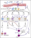
Figure 1. Intestinal IgA Responses in Mice and Humans
In mice (left model), DCs lodged in the subepithelial dome of Peyer’s patches capture bacteria or antigen internalized by M cells or by epithelial cells (ECs) via receptor-mediated endocytosis. These DCs migrate to the interfollicular region (IFR) of Peyer’s patches, where they present antigen to CD4+ T cells. Antigen-activated CD4+ T cells elicit IgA class switching by stimulating IgM+IgD+ B cells through CD40L and TGF. A subset of Peyer’s patch DCs, TNF-α+iNOS+ DCs, enhance IgA class switching by upregulating the expression of the TGF-β receptor on B cells through nitric oxide (NO). In the presence of retinoic acid (RA), IgA+ B cells upregulate the expression of CCR9 and α4β7 and thereafter migrate to the lamina propria, where they differentiate into plasma cells that release dimeric IgA antibodies. This T cell-dependent pathway yields high-affinity, monoreactive IgA antibodies that preferentially target pathogens and toxins. IgA class switching can also take place in the lamina propria via a T independent mechanism that involves activation of B-1 cells and possibly other IgM+IgD+ B cell subsets by DCs, including TNF-α+iNOS+ DCs. These DCs release innate IgA class-switch-inducing factors, such as BAFF, APRIL, TGF-β, and NO, as well as IgA secretion-inducing factors, such as IL-6 and RA, after sensing bacteria through TLRs. NO amplifies IgA class switching by enhancing BAFF and APRIL production by DCs. This T cell-independent pathway preferentially yields low-affinity, polyreactive IgA antibodies to commensal bacteria. In humans (right model), CD4+ T cells elicit IgA1 class switching by activating Peyer’s patch IgM+IgD+ B cells through CD40L and TGF-β. The resulting IgA1+ B cells migrate to the lamina propria through a mechanism presumably similar to that utilized by mouse IgA+ B cells. In the lamina propria, IgA1+ B cells sequentially switch to IgA2 in response to APRIL and IL-10 released by TLR-activated ECs. Also, DCs can release these cytokines in response to TSLP produced by ECs. In the lamina propria, additional IgM+IgD+ B cells can undergo direct class switching from IgM to IgA1 or IgA2 in response to BAFF or APRIL and IL-10. In general, IgA2 is more resistant to bacterial proteases than IgA1 and may therefore have a longer half-life in the lumen of the distal intestinal tract.
Figure 2. Putative Role of IgA in Intestinal Tolerance and Homeostasis
Intestinal M cells transfer IgA-bound antigen from the lumen to DCs. In the presence of TSLP and other epithelial cell (EC) products, possibly including retinoic acid (RA), TGF-β, and IL-10, multiple subsets of Peyer’s patch DCs initiate noninflammatory CD4+ T cell responses. By blocking DC production of IL-12 and inducing DC production of IL-10, TSLP prevents intestinal DCs from initiating proinflammatory Th1 responses, including IFN-γ-dependent activation of macrophages and cytotoxic T lymphocytes (CTLs). The resulting Th2 response triggers IgA (and IgG) class switching and production by activating B cells via CD40L (not shown) as well as IL-4 and IL-10. By upregulating DC release of TGF-β, IL-6, IL-27, and RA, TSLP alone or combined with other epithelial factors might also initiate Treg, Tr1, and Th17 cell responses. Treg cells dampen Th1-Th2 immunity through contact-dependent mechanisms and TGF-β, whereas Tr1 cells and regulatory-stage Th17 cells attenuate Th1-Th2 immunity via IL-10. Treg, Tr1, and Th17 cells might also trigger IgA (but not IgG) class switching and production by activating B cells via CD40L (not shown) as well as TGF-β and IL-10. Intestinal Treg, Tr1, and Th17 cell responses might be further amplified by TGF-β, IL-10, IL-6, and IgA derived from B cells.
Similar articles
- Induction of protective IgA by intestinal dendritic cells carrying commensal bacteria.
Macpherson AJ, Uhr T. Macpherson AJ, et al. Science. 2004 Mar 12;303(5664):1662-5. doi: 10.1126/science.1091334. Science. 2004. PMID: 15016999 - Diverse regulatory pathways for IgA synthesis in the gut.
Suzuki K, Fagarasan S. Suzuki K, et al. Mucosal Immunol. 2009 Nov;2(6):468-71. doi: 10.1038/mi.2009.107. Epub 2009 Sep 9. Mucosal Immunol. 2009. PMID: 19741602 Review. - Alternate mucosal immune system: organized Peyer's patches are not required for IgA responses in the gastrointestinal tract.
Yamamoto M, Rennert P, McGhee JR, Kweon MN, Yamamoto S, Dohi T, Otake S, Bluethmann H, Fujihashi K, Kiyono H. Yamamoto M, et al. J Immunol. 2000 May 15;164(10):5184-91. doi: 10.4049/jimmunol.164.10.5184. J Immunol. 2000. PMID: 10799877 - Metabolic changes during B cell differentiation for the production of intestinal IgA antibody.
Kunisawa J. Kunisawa J. Cell Mol Life Sci. 2017 Apr;74(8):1503-1509. doi: 10.1007/s00018-016-2414-8. Epub 2016 Nov 12. Cell Mol Life Sci. 2017. PMID: 27838744 Free PMC article. Review. - Effect of the administration of Lactobacillus paracasei subsp. paracasei NTU 101 on Peyer's patch-mediated mucosal immunity.
Tsai YT, Cheng PC, Liao JW, Pan TM. Tsai YT, et al. Int Immunopharmacol. 2010 Jul;10(7):791-8. doi: 10.1016/j.intimp.2010.04.012. Epub 2010 Apr 24. Int Immunopharmacol. 2010. PMID: 20417727
Cited by
- Immunoglobulin class-switch DNA recombination: induction, targeting and beyond.
Xu Z, Zan H, Pone EJ, Mai T, Casali P. Xu Z, et al. Nat Rev Immunol. 2012 Jun 25;12(7):517-31. doi: 10.1038/nri3216. Nat Rev Immunol. 2012. PMID: 22728528 Free PMC article. Review. - Decreased IgA+ B cells population and IgA, IgG, IgM contents of the cecal tonsil induced by dietary high fluorine in broilers.
Liu J, Cui H, Peng X, Fang J, Zuo Z, Deng J, Wang H, Wu B, Deng Y, Wang K. Liu J, et al. Int J Environ Res Public Health. 2013 May 2;10(5):1775-85. doi: 10.3390/ijerph10051775. Int J Environ Res Public Health. 2013. PMID: 23644827 Free PMC article. - Characterization of the Probiotic Yeast Saccharomyces boulardii in the Healthy Mucosal Immune System.
Hudson LE, McDermott CD, Stewart TP, Hudson WH, Rios D, Fasken MB, Corbett AH, Lamb TJ. Hudson LE, et al. PLoS One. 2016 Apr 11;11(4):e0153351. doi: 10.1371/journal.pone.0153351. eCollection 2016. PLoS One. 2016. PMID: 27064405 Free PMC article. - Polysaccharide from Atractylodes macrocephala Koidz Binding with Zinc Oxide Nanoparticles as a Novel Mucosal Immune Adjuvant for H9N2 Inactivated Vaccine.
Liu X, Lin X, Hong H, Wang J, Tao Y, Huai Y, Pang H, Liu M, Li J, Bo R. Liu X, et al. Int J Mol Sci. 2024 Feb 9;25(4):2132. doi: 10.3390/ijms25042132. Int J Mol Sci. 2024. PMID: 38396809 Free PMC article. - Interleukin (IL)-21 promotes intestinal IgA response to microbiota.
Cao AT, Yao S, Gong B, Nurieva RI, Elson CO, Cong Y. Cao AT, et al. Mucosal Immunol. 2015 Sep;8(5):1072-82. doi: 10.1038/mi.2014.134. Epub 2015 Jan 14. Mucosal Immunol. 2015. PMID: 25586558 Free PMC article.
References
- Andrew DP, Rott LS, Kilshaw PJ, Butcher EC. Distribution of alpha 4 beta 7 and alpha E beta 7 integrins on thymocytes, intestinal epithelial lymphocytes and peripheral lymphocytes. Eur. J. Immunol. 1996;26:897–905. - PubMed
- Babyatsky MW, Rossiter G, Podolsky DK. Expression of transforming growth factors alpha and beta in colonic mucosa in inflammatory bowel disease. Gastroenterology. 1996;110:975–984. - PubMed
- Bergqvist P, Gardby E, Stensson A, Bemark M, Lycke NY. Gut IgA class switch recombination in the absence of CD40 does not occur in the lamina propria and is independent of germinal centers. J. Immunol. 2006;177:7772–7783. - PubMed
- Bergtold A, Desai DD, Gavhane A, Clynes R. Cell surface recycling of internalized antigen permits dendritic cell priming of B cells. Immunity. 2005;23:503–514. - PubMed
Publication types
MeSH terms
Substances
Grants and funding
- R01 AI057653-03/AI/NIAID NIH HHS/United States
- R01 AI057653-S1/AI/NIAID NIH HHS/United States
- R01 AI057653-02/AI/NIAID NIH HHS/United States
- R01 AI074378-03/AI/NIAID NIH HHS/United States
- R01 AI074378/AI/NIAID NIH HHS/United States
- R01 AI074378-02/AI/NIAID NIH HHS/United States
- R01 AI057653/AI/NIAID NIH HHS/United States
- R01 AI057653-02S1/AI/NIAID NIH HHS/United States
- R01 AI074378-01A1/AI/NIAID NIH HHS/United States
LinkOut - more resources
Full Text Sources
Other Literature Sources
Miscellaneous

