The role of microRNA-221 and microRNA-222 in androgen-independent prostate cancer cell lines - PubMed (original) (raw)
The role of microRNA-221 and microRNA-222 in androgen-independent prostate cancer cell lines
Tong Sun et al. Cancer Res. 2009.
Abstract
Androgen-dependent prostate cancer typically progresses to castration-resistant prostate cancer (CRPC) after the androgen deprivation therapy. MicroRNAs (miR) are noncoding small RNAs (19-25nt) that play an important role in the regulation of gene expression. Recent studies have shown that miR expression patterns are significantly different in normal and neoplastic prostate epithelial cells. However, the importance of miRs in the development of CRPC has not yet been explored. By performing genome-wide expression profiling of miRs, we found that expression levels of several miRs, in particular miR-221 and miR-222, were significantly increased in CRPC cells (the LNCaP-derived cell line LNCaP-Abl), compared with those in the androgen-dependent prostate cancer cell line (LNCaP). Overexpression of miR-221 or miR-222 in LNCaP or another androgen-dependent cell line, LAPC-4, significantly reduced the level of the dihydrotestosterone (DHT) induced up-regulation of prostate-specific antigen (PSA) expression and increased androgen-independent growth of LNCaP cells. Knocking down the expression level of miR-221 and miR-222 with antagonist miRs in the LNCaP-Abl cell line restored the response to the DHT induction of PSA transcription and also increased the growth response of the LNCaP-Abl cells to the androgen treatment. Changing the expression level of p27/kip1, a known target of miR-221 and miR-222, alone in LNCaP cells affected the DHT-independent cell growth but did not significantly influence the response of PSA transcription to the DHT treatment. In conclusion, our data suggest the involvement of miR-221 and miR-222 in the development or maintenance of the CRPC phenotype.
Conflict of interest statement
The authors declare no conflict of interests.
Figures
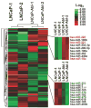
Figure 1. Comparison of miR expression patterns in LNCaP and LNCaP-Abl
Fold changes (LNCaP-Abl versus LNCaP) of the miRs are presented. LNCaP-1/LNCaP-Abl-1 and LNCaP-2 /LNCaP-Abl-2 represent two independently performed experiments. The tree graphs display the log2 transformation of the average fold changes. Arrays were mean centered and normalized by Gene Cluster 2.0. Average linkage clustering was performed by using uncensored correlation metric. The scale bar on the right upper corner displays the color range and the corresponding log2 transformation of average fold changes.
Figure 2. The relative expression of selected miRs in LNCaP and LNCaP derived CRPC cell lines
25 ng of total RNA from each cell line was used to measure miR expression levels by qRT-PCR. All of the data was normalized by the U6 expression level and presented as the relative expression level. The relative expression level of each miR in LNCaP was arbitrarily set as 1.0.
Figure 3. The impact of miR-221 and -222 expression levels on the AR mediated transcription in response to the DHT treatment
(A, B) Quantitative analysis of the expression level of PSA and AR in LNCaP (A) and LNCaP-Abl (B) with (+) or without androgen, DHT (-). Total RNAs were isolated from LNCaP that were mock-transfected (−), and transfected with pre-miR-221 (221), pre-miR-222 (222), pre-miR-221&-222 (221&222), or pre-miR-Neg RNA (NegRNA), and from LNCaP-Abl that were mock-transfected (−) and transfected with anti-miR-221 (221), anti-miR-222 (222), anti-miR221&-222 (221&222) or anti-miR-Neg RNA (Neg RNA). Left panels confirm the expression levels of miR-221 and -222 by RT-PCR in each transfected LNCaP (A) and LNCaP-Abl (B) cell lines. The expression of U6 was used as a control. Middle panels show the PSA expression levels analyzed by qRT-PCR. Right panels show the AR expression in each transfected cell line measured by qRT-PCR. (C) Comparison of the DHT induced PSA expression in LNCaP and LAPC-4. Cells were transfected with (+) or without (−) pre-miR-221 or pre-miR-Neg. (D) Effect of anti-androgen treatment on the PSA expression inducted by DHT. LNCaP and LNCaP-Abl were first transfected with Pre-miR-Negative (Neg RNA) or Pre-miR-221 and Anti-miR-Negative (Neg RNA) or Anti-miR-221 or without transfection (Mock) as indicated respectively, and then treated with (+) or without (−) DHT, Casodex and Flutamide. In all experiments, the relative expression levels of PSA and AR in each sample were normalized by the expression level of GAPDH. Values represent the fold differences relative to those in cells without any drug treatment or transfection (Mock), which were set as 1.0. *s indicate that the fold changes of those tranfected samples compared with their corresponding negative controls show a _P_-value < 0.05 in one-way ANOVA.
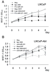
Figure 4. Effect of the expression level of miR-221 on the growth of LNCaP and LNCaP-Abl
(A) WST-1 analysis of the growth of LNCaP cells that were transfected with Pre-miR-221, miRNA-precursor-negative control (Neg), mocked transfected (lipo 2000) and kept in medium with (+DHT, solid lines) or without androgen (broken lines). (B) WST-1 analysis of the growth of LNCaP-Abl cells that were transfected with Anti-miR-221, Anti-miRNA inhibitors negative control (Neg), mocked transfected (lipo 2000) and kept in medium with (+DHT, solid lines) or without androgen (broken lines). Triplicate experiments were performed for each set. The data represents mean ± S.D. (n = 3).
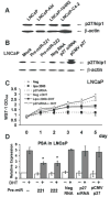
Figure 5. Effect of p27/kip1 expression on the DHT induction of the PSA expression in LNCaP
(A). Western blot analysis of p27/kip1 in LNCaP and LNCaP derived CRPC cell lines. The amount of β-actin in each lane is used as a loading control. (B). Western blot analysis of p27/kip1 in LNCaP cells that were mock-transfected (mock) or transfected with Pre-miR-221 (Pre-221), Pre-miR-222 (Pre-222), miRNA precursor-negative control (Neg RNA), p27/kip1 siRNA (p27 siRNA) or pCMV-SOPRT 6-p27/kip1 vector (pCMV-p27). Total proteins were extracted from cells 48 hours after transfection. (C). The impact of p27/kip1 siRNA on the growth of LNCaP. WST-1 assay was used to measure the cell growth. LNCaP cells that were transfected with p27/kip1 siRNA (p27siRNA, open squares), anti-miRNA inhibitors negative control (Neg, triangles), and mocked transfected (lipo 2000, black squares) were cultured in medium with (+DHT, solid lines) or without DHT (broken lines). Triplicate experiments were performed for each set. The data represents mean ± S.D. (n = 3). (D). QRT-PCR of the relative PSA expression level in transfected LNCaP cells as those described in (B). The relative expression level of PSA in each sample was normalized by the expression level of GAPDH. Values represent the fold differences relative to that in mock transfected cells without any drug treatment, which was arbitrarily set as 1.0. *s indicate that the fold changes of those transfected samples compared with their corresponding negative control exhibited a _P_-value < 0.05 in one-way ANOVA analysis.
Similar articles
- Androgen regulation of micro-RNAs in prostate cancer.
Waltering KK, Porkka KP, Jalava SE, Urbanucci A, Kohonen PJ, Latonen LM, Kallioniemi OP, Jenster G, Visakorpi T. Waltering KK, et al. Prostate. 2011 May;71(6):604-14. doi: 10.1002/pros.21276. Epub 2010 Oct 13. Prostate. 2011. PMID: 20945501 - Src promotes castration-recurrent prostate cancer through androgen receptor-dependent canonical and non-canonical transcriptional signatures.
Chattopadhyay I, Wang J, Qin M, Gao L, Holtz R, Vessella RL, Leach RW, Gelman IH. Chattopadhyay I, et al. Oncotarget. 2017 Feb 7;8(6):10324-10347. doi: 10.18632/oncotarget.14401. Oncotarget. 2017. PMID: 28055971 Free PMC article. - Changes in androgen receptor nongenotropic signaling correlate with transition of LNCaP cells to androgen independence.
Unni E, Sun S, Nan B, McPhaul MJ, Cheskis B, Mancini MA, Marcelli M. Unni E, et al. Cancer Res. 2004 Oct 1;64(19):7156-68. doi: 10.1158/0008-5472.CAN-04-1121. Cancer Res. 2004. PMID: 15466214 - MiR-221 promotes the development of androgen independence in prostate cancer cells via downregulation of HECTD2 and RAB1A.
Sun T, Wang X, He HH, Sweeney CJ, Liu SX, Brown M, Balk S, Lee GS, Kantoff PW. Sun T, et al. Oncogene. 2014 May 22;33(21):2790-800. doi: 10.1038/onc.2013.230. Epub 2013 Jun 17. Oncogene. 2014. PMID: 23770851 Free PMC article. - Interplay between steroid signalling and microRNAs: implications for hormone-dependent cancers.
Fletcher CE, Dart DA, Bevan CL. Fletcher CE, et al. Endocr Relat Cancer. 2014 Oct;21(5):R409-29. doi: 10.1530/ERC-14-0208. Epub 2014 Jul 25. Endocr Relat Cancer. 2014. PMID: 25062737 Review.
Cited by
- MicroRNAs as putative mediators of treatment response in prostate cancer.
O'Kelly F, Marignol L, Meunier A, Lynch TH, Perry AS, Hollywood D. O'Kelly F, et al. Nat Rev Urol. 2012 May 22;9(7):397-407. doi: 10.1038/nrurol.2012.104. Nat Rev Urol. 2012. PMID: 22613932 Review. - Epigenetic and miRNAs Dysregulation in Prostate Cancer: The role of Nutraceuticals.
Bosutti A, Zanconati F, Grassi G, Dapas B, Passamonti S, Scaggiante B. Bosutti A, et al. Anticancer Agents Med Chem. 2016;16(11):1385-1402. doi: 10.2174/1871520616666160425105257. Anticancer Agents Med Chem. 2016. PMID: 27109021 Free PMC article. Review. - Serum microRNA expression patterns that predict early treatment failure in prostate cancer patients.
Singh PK, Preus L, Hu Q, Yan L, Long MD, Morrison CD, Nesline M, Johnson CS, Koochekpour S, Kohli M, Liu S, Trump DL, Sucheston-Campbell LE, Campbell MJ. Singh PK, et al. Oncotarget. 2014 Feb 15;5(3):824-40. doi: 10.18632/oncotarget.1776. Oncotarget. 2014. PMID: 24583788 Free PMC article. - The Interactions of microRNA and Epigenetic Modifications in Prostate Cancer.
Singh PK, Campbell MJ. Singh PK, et al. Cancers (Basel). 2013 Aug 9;5(3):998-1019. doi: 10.3390/cancers5030998. Cancers (Basel). 2013. PMID: 24202331 Free PMC article. - Uncovering the roles of miRNAs and their relationship with androgen receptor in prostate cancer.
ChunJiao S, Huan C, ChaoYang X, GuoMei R. ChunJiao S, et al. IUBMB Life. 2014 Jun;66(6):379-86. doi: 10.1002/iub.1281. Epub 2014 Jun 30. IUBMB Life. 2014. PMID: 24979663 Free PMC article. Review.
References
- Huggins CB, Hodges CV. Studies on prostatic cancer. 1. The effect of castration, of estrogen and of androgen injections on serum phosphatases in metastatic carcinoma of the prostate. Cancer Res. 1941;1:293–7. - PubMed
- Feldman BJ, Feldman D. The development of androgen-independent prostate cancer. Nat Rev Cancer. 2001;1:34–45. - PubMed
- Rana TM. Illuminating the silence: understanding the structure and function of small RNAs. Nat Rev Mol Cell Biol. 2007;8:23–36. - PubMed
- Glinsky GV. An SNP-guided microRNA map of fifteen common human disorders identifies a consensus disease phenocode aiming at principal components of the nuclear import pathway. Cell Cycle. 2008;7:2570–83. - PubMed
Publication types
MeSH terms
Substances
LinkOut - more resources
Full Text Sources
Other Literature Sources
Medical
Research Materials
Miscellaneous


