GATA3 inhibits lysyl oxidase-mediated metastases of human basal triple-negative breast cancer cells - PubMed (original) (raw)
GATA3 inhibits lysyl oxidase-mediated metastases of human basal triple-negative breast cancer cells
I M Chu et al. Oncogene. 2012.
Abstract
Discovery of mechanisms that impede the aggressive and metastatic phenotype of human basal triple-negative-type breast cancers (BTNBCs) could provide novel targets for therapy for this form of breast cancer that has a relatively poor prognosis. Previous studies have demonstrated that expression of GATA3, the master transcriptional regulator of mammary luminal differentiation, can reduce the tumorigenicity and metastatic propensity of the human BTNBC MDA-MB-231 cell line (MB231), although the mechanism for reduced metastases was not elucidated. We demonstrate through gene expression profiling that GATA3 expression in 231 cells resulted in the dramatic reduction in the expression of lysyl oxidase (LOX), a metastasis-promoting, matrix-remodeling protein, in part, through methylation of the LOX promoter. Suppression of LOX expression by GATA3 was further confirmed in the BTNBC Hs578T cell line. Conversely, reduction of GATA3 expression by small interfering RNA in luminal BT474 cells increased LOX expression. Reconstitution of LOX expression in 231-GATA3 cells restored metastatic propensity. A strong inverse association between LOX and GATA3 expression was confirmed in a panel of 51 human breast cancer cell lines. Similarly, human breast cancer microarray data demonstrated that high LOX/low GATA3 expression is associated with the BTNBC subtype of breast cancer and poor patient prognosis. Expression of GATA3 reprograms BTNBCs to a less aggressive phenotype and inhibits a major mechanism of metastasis through inhibition of LOX. Induction of GATA3 in BTNBC cells or novel approaches that inhibit LOX expression or activity could be important strategies for treating BTNBCs.
Conflict of interest statement
Conflict of Interest. The authors report no conflict of interest.
Figures
Figure 1
GATA3 over-expression reduces proliferation in 3D culture and experimental metastasis in mice. (a) 231-Empty and 231-GATA3 cells were seeded on 3D Cultrex® for 12 days. 231-GATA3 cells show reduced proliferation as measured by MTS (mean +/- SEM). (b) Top panels, bright field images; lower panel, confocal microscopy of cells on 3D Cultrex® fixed and stained with DAPI (blue) for nuclear localization and phalloidin (green) for f-actin. (c) Lung lesions of mice injected by tail vein with 231-Emtpy and 231-GATA3 cells. Lungs were imaged by fluorescent microscopy with total metastatic burden calculated per lung.
Figure 2
GATA3 regulates LOX expression in breast cancer cells in part through LOX promoter methylation. (a) Relative LOX expression by Q-RT-PCR. Samples were normalized to cyclophilin B. Over-expression of GATA3 in MB231 and Hs578T cells reduces LOX mRNA expression. (b) Immunohistochemical staining of cell pellets confirmed positive staining for GATA3 in only 231-GATA3 cells and positive staining for LOX only in 231-Empty cells. (c) Relative LOX activity in the media of 231-Empty and 231-GATA3 cells measured as the increase in fluorescence over BAPN containing controls. Relative activity measured at 2400 seconds (40 min). (d) Relative LOX and GATA3 mRNA expression measured by Q-RT-PCR. BT474 cells were transfected with GATA3 siRNA for 72 hrs prior to RNA isolation. (e) Cells were treated with 5-AZA for 4 days prior to DNA or mRNA isolation. Top panel, PCR of the LOX promoter using LOX unmethylated (U) or methylated (M) specific primers. Lower panel, relative LOX expression by Q-RT-PCR. Treatment of 231-GATA3 cells with 5-AZA increased LOX mRNA expression.
Figure 3
GATA3 reduces macrophage recruitment to the lung (a) ELISA of media collected from 231-Empty and 231-GATA3 cells. 231-GATA3 cells showed reduced secretion of CSF-1 and GM-CSF. (b) Flow cytometric analyses of immune cells collected from lungs of tail-vein injected mice (n=4). Cells were labeled with anti CD45, F4/80, Gr1 or CD11b antibodies. Lungs collected from mice injected with 231-GATA3 cells showed reduced F4/80+/Gr1- recruitment.
Figure 4
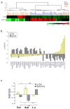
Analysis of the Neve et al. 51 breast cancer cell line microarray database for LOX and GATA expression (Neve et al., 2006). (a) Heatmap of LOX and GATA3 expression in breast cancer cell lines. The displayed expression of each gene was standardized with Z-score. The hierarchical clustering used 1-uncentered correlation distance metric and average linkage. (b) Relative GATA3 and LOX expression in breast cancer cell lines arranged in order of increasing LOX expression (Pearson's correlation coefficient r=-0.53, P<0.001). (c) Relative expression of GATA3 as represented by Z-score (see Supplementary Materials and Methods). GATA3 is enriched in luminal breast cancer cells whereas LOX is enriched in Basal B cells.
Figure 5
Re-expression of LOX in 231-GATA3 cells increased metastatic potential of 231-GATA3 cells. (a) Lentiviral transduction of 231-GATA3 cells with LOX increases LOX expression in 231-GATA3 cells. Relative LOX expression by Q-RT-PCR. (b) Immunohistochemical staining of cell pellets confirmed positive staining for GATA3 in 231-GATA3-Empty and 231-GATA3-LOX cells and positive staining for LOX in only 231-GATA3-LOX cells. (c) Relative LOX enzymatic activity measured at 2400 seconds (40 min). (d) Mice tail-vein injected with 231-GATA3-Empty and 231-GATA3-LOX cells with lungs collected after 2 months. Lungs imaged by fluorescent microscopy with total metastatic burden calculated per lung.
Figure 6
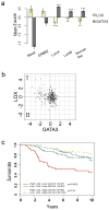
Retrospective microarray analysis of breast cancer patient microarray data from Van de Vijver et al, (van de Vijver et al., 2002). (a) GATA3 is associated with the Luminal A and luminal B subtype whereas LOX is enriched in the Basal subtype. (b) Correlation between LOX (y-axis) and GATA3 (x-axis) among breast cancer patients (n=295). GATA3 and LOX are inversely correlated (Pearson's correlation coefficient r=-0.30, P<0.001). (c) Kaplan-Meier survival curves showing that patients with high LOX and reduced GATA3 expression (quadrant I in (b) above) had significantly reduced overall survival (HR=2.65, p<0.01) compared to patients with low LOX and Low GATA3 (quadrant II in (b) above). High and low are defined as above or below median expression as depicted in (b). The log-rank test p-values are indicated. The interaction between LOX and GATA3 was statistically significant (P<0.05).
Similar articles
- Human breast cancer cell metastasis is attenuated by lysyl oxidase inhibitors through down-regulation of focal adhesion kinase and the paxillin-signaling pathway.
Chen LC, Tu SH, Huang CS, Chen CS, Ho CT, Lin HW, Lee CH, Chang HW, Chang CH, Wu CH, Lee WS, Ho YS. Chen LC, et al. Breast Cancer Res Treat. 2012 Aug;134(3):989-1004. doi: 10.1007/s10549-012-1986-8. Epub 2012 Mar 21. Breast Cancer Res Treat. 2012. PMID: 22434522 - A molecular role for lysyl oxidase in breast cancer invasion.
Kirschmann DA, Seftor EA, Fong SF, Nieva DR, Sullivan CM, Edwards EM, Sommer P, Csiszar K, Hendrix MJ. Kirschmann DA, et al. Cancer Res. 2002 Aug 1;62(15):4478-83. Cancer Res. 2002. PMID: 12154058 - ELK3-GATA3 axis modulates MDA-MB-231 metastasis by regulating cell-cell adhesion-related genes.
Kim KS, Kim J, Oh N, Kim MY, Park KS. Kim KS, et al. Biochem Biophys Res Commun. 2018 Apr 6;498(3):509-515. doi: 10.1016/j.bbrc.2018.03.011. Epub 2018 Mar 3. Biochem Biophys Res Commun. 2018. PMID: 29510139 - GATA3 in development and cancer differentiation: cells GATA have it!
Chou J, Provot S, Werb Z. Chou J, et al. J Cell Physiol. 2010 Jan;222(1):42-9. doi: 10.1002/jcp.21943. J Cell Physiol. 2010. PMID: 19798694 Free PMC article. Review. - Lysyl oxidase mediates hypoxic control of metastasis.
Erler JT, Giaccia AJ. Erler JT, et al. Cancer Res. 2006 Nov 1;66(21):10238-41. doi: 10.1158/0008-5472.CAN-06-3197. Cancer Res. 2006. PMID: 17079439 Review.
Cited by
- GATA3 suppresses metastasis and modulates the tumour microenvironment by regulating microRNA-29b expression.
Chou J, Lin JH, Brenot A, Kim JW, Provot S, Werb Z. Chou J, et al. Nat Cell Biol. 2013 Feb;15(2):201-13. doi: 10.1038/ncb2672. Epub 2013 Jan 27. Nat Cell Biol. 2013. PMID: 23354167 Free PMC article. - Group V secreted phospholipase A2 plays a protective role against aortic dissection.
Watanabe K, Taketomi Y, Miki Y, Kugiyama K, Murakami M. Watanabe K, et al. J Biol Chem. 2020 Jul 24;295(30):10092-10111. doi: 10.1074/jbc.RA120.013753. Epub 2020 Jun 1. J Biol Chem. 2020. PMID: 32482892 Free PMC article. - Cell Reprogramming in Tumorigenesis and Its Therapeutic Implications for Breast Cancer.
Chu PY, Hou MF, Lai JC, Chen LF, Lin CS. Chu PY, et al. Int J Mol Sci. 2019 Apr 12;20(8):1827. doi: 10.3390/ijms20081827. Int J Mol Sci. 2019. PMID: 31013830 Free PMC article. Review. - T-cell targeted pulmonary siRNA delivery for the treatment of asthma.
Keil TWM, Baldassi D, Merkel OM. Keil TWM, et al. Wiley Interdiscip Rev Nanomed Nanobiotechnol. 2020 Sep;12(5):e1634. doi: 10.1002/wnan.1634. Epub 2020 Apr 8. Wiley Interdiscip Rev Nanomed Nanobiotechnol. 2020. PMID: 32267622 Free PMC article. Review. - GATA-3 expression and its correlation with prognostic factors and survival in canine mammary tumors.
Diniz-Gonçalves GS, Hielm-Björkman A, da Silva VB, Ribeiro LGR, da Costa Vieira-Filho CH, Silva LP, Barrouin-Melo SM, Cassali GD, Damasceno KA, Estrela-Lima A. Diniz-Gonçalves GS, et al. Front Vet Sci. 2023 Jul 6;10:1179808. doi: 10.3389/fvets.2023.1179808. eCollection 2023. Front Vet Sci. 2023. PMID: 37483298 Free PMC article.
References
- Abourbih DA, Di CS, Orellana ME, Antecka E, Martins C, Petruccelli LA, et al. Lysyl oxidase expression and inhibition in uveal melanoma. Melanoma Res. 2010;20:97–106. - PubMed
- Asselin-Labat ML, Sutherland KD, Barker H, Thomas R, Shackleton M, Forrest NC, et al. Gata-3 is an essential regulator of mammary-gland morphogenesis and luminal-cell differentiation. Nat Cell Biol. 2007;9:201–209. - PubMed
Publication types
MeSH terms
Substances
Grants and funding
- P30 CA071789/CA/NCI NIH HHS/United States
- U54 MD007584/MD/NIMHD NIH HHS/United States
- ZIA BC005740-17/Intramural NIH HHS/United States
- ZIA BC005740-18/Intramural NIH HHS/United States
LinkOut - more resources
Full Text Sources
Medical
Molecular Biology Databases
Miscellaneous


