Understanding fibrosis in systemic sclerosis: shifting paradigms, emerging opportunities - PubMed (original) (raw)
Review
Understanding fibrosis in systemic sclerosis: shifting paradigms, emerging opportunities
Swati Bhattacharyya et al. Nat Rev Rheumatol. 2011.
Abstract
Fibrosis in multiple organs is a prominent pathological finding and distinguishing hallmark of systemic sclerosis (SSc). Findings during the past 5 years have contributed to a more complete understanding of the complex cellular and molecular underpinning of fibrosis in SSc. Fibroblasts, the principal effector cells, are activated in the profibrotic cellular milieu by cytokines and growth factors, developmental pathways, endothelin 1 and thrombin. Innate immune signaling via Toll-like receptors, matrix-generated biomechanical stress signaling via integrins, hypoxia and oxidative stress seem to be implicated in perpetuating the process. Beyond chronic fibroblast activation, fibrosis represents a failure to terminate tissue repair, coupled with an expanded population of mesenchymal cells originating from bone marrow and transdifferentiation of epithelial cells, endothelial cells and pericytes. In addition, studies have identified intrinsic alterations in SSc fibroblasts resulting from epigenetic changes, as well as altered microRNA expression that might underlie the cell-autonomous, persistent activation phenotype of these cells. Precise characterization of the deregulated extracellular and intracellular signaling pathways, mediators and cellular differentiation programs that contribute to fibrosis in SSc will facilitate the development of selective, targeted therapeutic strategies. Effective antifibrotic therapy will ultimately involve novel compounds and repurposing of drugs that are already approved for other indications.
Conflict of interest statement
Competing interests
The authors declare no competing interests.
Figures
Figure 1
Cellular and molecular pathways underlying fibrosis in systemic sclerosis. Injury caused by viruses, autoantibodies, ischemia-reperfusion or toxins triggers vascular damage and inflammation. Activated inflammatory cells secrete cytokines and growth factors. Endothelial injury results in generation of ROS, intravascular coagulation and platelet activation with release of serotonin, vasoactive mediators, thrombin and platelet-derived growth factor, and sets in motion progressive vascular remodeling leading to luminal occlusion, reduced blood flow and tissue hypoxia. Secreted mediators, such as TGF-β and Wnt10b, cause fibroblast activation and differentiation into myofibroblasts, which produce excess amounts of collagen, contract and remodel the connective tissue, and resist elimination by apoptosis. The stiff and hypoxic ECM of the fibrotic tissue further activates myofibroblasts. Injury also directly induces transdifferentiation of pericytes, epithelial cells and endothelial cells into myofibroblasts, expanding the tissue pool of matrix-synthesizing, activated myofibroblasts. Abbreviations: CXCL12, CXC-chemokine ligand 12; CXCR4, CXC-chemokine receptor 4; ECM, extracellular matrix; IFN, interferon; ROS, reactive oxygen species; TGF-β, transforming growth factor β; TH2 cell, type 2 helper T cell; TLR, Toll-like receptor.
Figure 2
Tissue damage activates innate immune signaling, which transforms an orderly self-limited repair into a sustained, aberrant fibrogenic process. Following injury, fibroblasts undergo controlled activation, and once repair has been accomplished, tissue regeneration is complete. When tissue damage occurs from recurrent or sustained injury, damage-associated endogenous TLR ligands activate fibroblast innate immune signaling, enhancing fibrogenic responses and establishing a self-amplifying vicious cycle of fibrogenesis. Moreover, intrinsic protective signals (such as PPAR-γ) that normally act as the brakes on fibroblast activation are reduced or defective in scleroderma. Abbreviations: PPAR-γ, peroxisome proliferator-activated receptor γ; TLR, Toll-like receptor.
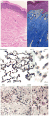
Figure 3
Tissue fibrosis in SSc. a | Skin biopsy from a patient with diffuse cutaneous SSc stained with hematoxylin and eosin (left) and Masson’s trichrome (right). Note the extensive dermal fibrosis, reduced cellularity and collagen deposition. b | Immunohistochemical EGR1 staining of lung tissue from a patient with SSc-associated end-stage lung fibrosis (upper panel) and a patient without lung fibrosis (lower panel). Note the dense pulmonary fibrosis, loss of normal alveolar architecture and strong upregulation of the profibrotic transcription factor EGR1 in the SSc specimen compared with normal tissue. Abbreviations: EGR1, early growth response protein 1; Sc, systemic sclerosis.
Similar articles
- Fibrosis in systemic sclerosis: emerging concepts and implications for targeted therapy.
Wei J, Bhattacharyya S, Tourtellotte WG, Varga J. Wei J, et al. Autoimmun Rev. 2011 Mar;10(5):267-75. doi: 10.1016/j.autrev.2010.09.015. Epub 2010 Sep 21. Autoimmun Rev. 2011. PMID: 20863909 Free PMC article. - Emerging roles of innate immune signaling and toll-like receptors in fibrosis and systemic sclerosis.
Bhattacharyya S, Varga J. Bhattacharyya S, et al. Curr Rheumatol Rep. 2015 Jan;17(1):474. doi: 10.1007/s11926-014-0474-z. Curr Rheumatol Rep. 2015. PMID: 25604573 Review. - Parvovirus B19 activates in vitro normal human dermal fibroblasts: a possible implication in skin fibrosis and systemic sclerosis.
Arvia R, Margheri F, Stincarelli MA, Laurenzana A, Fibbi G, Gallinella G, Ferri C, Del Rosso M, Zakrzewska K. Arvia R, et al. Rheumatology (Oxford). 2020 Nov 1;59(11):3526-3532. doi: 10.1093/rheumatology/keaa230. Rheumatology (Oxford). 2020. PMID: 32556240 Free PMC article. - Immune complexes containing scleroderma-specific autoantibodies induce a profibrotic and proinflammatory phenotype in skin fibroblasts.
Raschi E, Chighizola CB, Cesana L, Privitera D, Ingegnoli F, Mastaglio C, Meroni PL, Borghi MO. Raschi E, et al. Arthritis Res Ther. 2018 Aug 29;20(1):187. doi: 10.1186/s13075-018-1689-6. Arthritis Res Ther. 2018. PMID: 30157947 Free PMC article. - T cells in systemic sclerosis: a reappraisal.
O'Reilly S, Hügle T, van Laar JM. O'Reilly S, et al. Rheumatology (Oxford). 2012 Sep;51(9):1540-9. doi: 10.1093/rheumatology/kes090. Epub 2012 May 9. Rheumatology (Oxford). 2012. PMID: 22577083 Review.
Cited by
- Microangiopathy in Inflammatory Diseases-Strategies in Surgery of the Lower Extremity.
Biehl C, Biehl L, Tarner IH, Müller-Ladner U, Heiss C, Heinrich M. Biehl C, et al. Life (Basel). 2022 Jan 28;12(2):200. doi: 10.3390/life12020200. Life (Basel). 2022. PMID: 35207487 Free PMC article. - Serum adhesion molecule levels as prognostic markers in patients with early systemic sclerosis: a multicentre, prospective, observational study.
Hasegawa M, Asano Y, Endo H, Fujimoto M, Goto D, Ihn H, Inoue K, Ishikawa O, Kawaguchi Y, Kuwana M, Ogawa F, Takahashi H, Tanaka S, Sato S, Takehara K. Hasegawa M, et al. PLoS One. 2014 Feb 6;9(2):e88150. doi: 10.1371/journal.pone.0088150. eCollection 2014. PLoS One. 2014. PMID: 24516598 Free PMC article. - Stem cell-based therapy for fibrotic diseases: mechanisms and pathways.
Taherian M, Bayati P, Mojtabavi N. Taherian M, et al. Stem Cell Res Ther. 2024 Jun 18;15(1):170. doi: 10.1186/s13287-024-03782-5. Stem Cell Res Ther. 2024. PMID: 38886859 Free PMC article. Review. - Towards a Unified Approach in Autoimmune Fibrotic Signalling Pathways.
Sisto M, Lisi S. Sisto M, et al. Int J Mol Sci. 2023 May 21;24(10):9060. doi: 10.3390/ijms24109060. Int J Mol Sci. 2023. PMID: 37240405 Free PMC article. Review. - Clinical Treatment Options in Scleroderma: Recommendations and Comprehensive Review.
Zhao M, Wu J, Wu H, Sawalha AH, Lu Q. Zhao M, et al. Clin Rev Allergy Immunol. 2022 Apr;62(2):273-291. doi: 10.1007/s12016-020-08831-4. Epub 2021 Jan 15. Clin Rev Allergy Immunol. 2022. PMID: 33449302 Review.
References
- Rosenbloom J, Castro SV, Jimenez SA. Narrative review: fibrotic diseases: cellular and molecular mechanisms and novel therapies. Ann Intern Med. 2010;152:159–166. - PubMed
- Denton CP, Black CM, Abraham DJ. Mechanisms and consequences of fibrosis in systemic sclerosis. Nat Clin Pract Rheumatol. 2006;2:134–144. - PubMed
- Quillinan NP, Denton CP. Disease-modifying treatment in systemic sclerosis: current status. Curr Opin Rheumatol. 2009;21:636–641. - PubMed
Publication types
MeSH terms
LinkOut - more resources
Full Text Sources
Other Literature Sources
Medical
Miscellaneous


