Receptor for AGE (RAGE): signaling mechanisms in the pathogenesis of diabetes and its complications - PubMed (original) (raw)
Review
Receptor for AGE (RAGE): signaling mechanisms in the pathogenesis of diabetes and its complications
Ravichandran Ramasamy et al. Ann N Y Acad Sci. 2011 Dec.
Abstract
The receptor for advanced glycation endproducts (RAGE) was first described as a signal transduction receptor for advanced glycation endproducts (AGEs), the products of nonenzymatic glycation and oxidation of proteins and lipids that accumulate in diabetes and in inflammatory foci. The discovery that RAGE was a receptor for inflammatory S100/calgranulins and high mobility group box 1 (HMGB1) set the stage for linking RAGE to both the consequences and causes of types 1 and 2 diabetes. Recent discoveries regarding the structure of RAGE as well as novel intracellular binding partner interactions advance our understanding of the mechanisms by which RAGE evokes pathological consequences and underscore strategies by which antagonism of RAGE in the clinic may be realized. Finally, recent data tracking RAGE in the clinic suggest that levels of soluble RAGEs and polymorphisms in the gene encoding RAGE may hold promise for the identification of patients who are vulnerable to the complications of diabetes and/or are receptive to therapeutic interventions designed to prevent and reverse the damage inflicted by chronic hyperglycemia, irrespective of its etiology.
© 2011 New York Academy of Sciences.
Figures
Figure 1
PAI-1 (Serpine1), Tgf-β1, Tgf-β–induced, and α1-(IV) collagen mRNA transcripts and ROCK1 activity are lower in OVE26 RKO kidney cortex than in OVE26 kidney cortex at age seven months. Real-time PCR for PAI-1 (A), Tgf-β1 (B), Tgf-β1–induced (C), and α 1-(IV) collagen (D) gene products was performed, normalized to 18s transcript levels, and expressed as fold-change compared with the FVB or OVE26 group, as indicated in the figure (**P < 0.01, ***P < 0.005, ##P < 0.0005, ###P < 0.0001). n = at least 6 per group. (E) ROCK1 activity was measured as the amount of phosphorylated MYPT1 compared with total ROCK1 (*** P = 0.005). n = at least 3 per group. Reprinted with permission from Ref. 51.
Figure 2
Treatment with sRAGE reduces the expression of IL-1βand TNF-α in islets in NOD/scid mice subjected to adoptive transfer of diabetogenic splenocytes from NOD mice. The islets of NOD/scid mice that received splenocytes from diabetic NOD mice were studied for expression of IL-1β (A) and TNF-α (B) by immunohistochemistry. The area of cells staining with the anticytokine antibodies is indicated with arrows and was determined for each treatment group (control versus soluble RAGE). Representative sections from three separate recipients in each category are shown. The expression levels of both IL-1β and TNF-α were reduced by treatment with sRAGE. Reprinted with permission from Ref. 69.
Figure 3
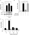
Hypoxia induces mDia-1 expression and mDia1 plays key roles in hypoxia-mediated upregulation of Egr-1. THP-1 cells and macrophages from mDia1 null mice were exposed to hypoxia (H; 0.5% of oxygen) or normoxia (N) for the indicated times. (A) Total protein was prepared from THP-1 cells, and immunoblotting with anti-mDia1 IgG was performed on 30 µg/lane of protein from THP-1 cells. Results of multiple experiments were quantified. (B and C) Total RNA was prepared from the indicated cells, and real-time PCR analysis of Egr-1 expression was performed. Data are represented as the relative expression of mRNA for Egr-1 normalized to 18 SirRNA. (B) RNA was prepared from THP-1 cells transfected with siRNA-mDia1 or scramble siRNA and then subjected to hypoxia for 15 min. (C) RNA was prepared from macrophages from wild-type or mDia1 (Drf1) null mice after exposure to hypoxia or normoxia for the indicated times. *P < 0.0001 and **P < 0.001 indicate statistical significance; #indicates no statistical significance. Note that Drf1 indicates mDia1. Adapted from Ref. 93.
Similar articles
- The RAGE axis: a fundamental mechanism signaling danger to the vulnerable vasculature.
Yan SF, Ramasamy R, Schmidt AM. Yan SF, et al. Circ Res. 2010 Mar 19;106(5):842-53. doi: 10.1161/CIRCRESAHA.109.212217. Circ Res. 2010. PMID: 20299674 Free PMC article. Review. - Unlocking the biology of RAGE in diabetic microvascular complications.
Manigrasso MB, Juranek J, Ramasamy R, Schmidt AM. Manigrasso MB, et al. Trends Endocrinol Metab. 2014 Jan;25(1):15-22. doi: 10.1016/j.tem.2013.08.002. Epub 2013 Sep 3. Trends Endocrinol Metab. 2014. PMID: 24011512 Free PMC article. Review. - Signal transduction in receptor for advanced glycation end products (RAGE): solution structure of C-terminal rage (ctRAGE) and its binding to mDia1.
Rai V, Maldonado AY, Burz DS, Reverdatto S, Yan SF, Schmidt AM, Shekhtman A. Rai V, et al. J Biol Chem. 2012 Feb 10;287(7):5133-44. doi: 10.1074/jbc.M111.277731. Epub 2011 Dec 21. J Biol Chem. 2012. PMID: 22194616 Free PMC article. - Glycation, inflammation, and RAGE: a scaffold for the macrovascular complications of diabetes and beyond.
Yan SF, Ramasamy R, Naka Y, Schmidt AM. Yan SF, et al. Circ Res. 2003 Dec 12;93(12):1159-69. doi: 10.1161/01.RES.0000103862.26506.3D. Circ Res. 2003. PMID: 14670831 Review. - Receptor for advanced glycation endproducts (RAGE) and the complications of diabetes.
Stern DM, Yan SD, Yan SF, Schmidt AM. Stern DM, et al. Ageing Res Rev. 2002 Feb;1(1):1-15. doi: 10.1016/s0047-6374(01)00366-9. Ageing Res Rev. 2002. PMID: 12039445 Review.
Cited by
- Age-Related Changes on CD40 Promotor Methylation and Immune Gene Expressions in Thymus of Chicken.
Li Y, Lei X, Lu H, Guo W, Wu S, Yin Z, Sun Q, Yang X. Li Y, et al. Front Immunol. 2018 Nov 21;9:2731. doi: 10.3389/fimmu.2018.02731. eCollection 2018. Front Immunol. 2018. PMID: 30519246 Free PMC article. - Receptor for advanced glycation end-products (RAGE) mediates phagocytosis in nonprofessional phagocytes.
Yang Y, Liu G, Li F, Carey LB, Sun C, Ling K, Tachikawa H, Fujita M, Gao XD, Nakanishi H. Yang Y, et al. Commun Biol. 2022 Aug 16;5(1):824. doi: 10.1038/s42003-022-03791-1. Commun Biol. 2022. PMID: 35974093 Free PMC article. - The RAGE/multiligand axis: a new actor in tumor biology.
Rojas A, Schneider I, Lindner C, Gonzalez I, Morales MA. Rojas A, et al. Biosci Rep. 2022 Jul 29;42(7):BSR20220395. doi: 10.1042/BSR20220395. Biosci Rep. 2022. PMID: 35727208 Free PMC article. Review. - Investigating the Molecular Mechanism of Quercetin Protecting against Podocyte Injury to Attenuate Diabetic Nephropathy through Network Pharmacology, MicroarrayData Analysis, and Molecular Docking.
Ma X, Hao C, Yu M, Zhang Z, Huang J, Yang W. Ma X, et al. Evid Based Complement Alternat Med. 2022 May 16;2022:7291434. doi: 10.1155/2022/7291434. eCollection 2022. Evid Based Complement Alternat Med. 2022. PMID: 35615688 Free PMC article. - Pro-inflammatory AGE-RAGE signaling is activated during arousal from hibernation in ground squirrel adipose.
Logan SM, Storey KB. Logan SM, et al. PeerJ. 2018 Jun 4;6:e4911. doi: 10.7717/peerj.4911. eCollection 2018. PeerJ. 2018. PMID: 29888131 Free PMC article.
References
- Forlenza GP, Paradise Black NM, McNamara EG, Sullivan SE. Ankyloglossia, exclusive breastfeeding, and failure to thrive. Pediatrics. 2010;125:e1500–e1504. - PubMed
- Peng H, Hagopian W. Environmental factors in the development of Type 1 diabetes. Rev. Endocr. Metab. Disord. 2006;7:149–162. - PubMed
- International Diabetes Federation IDF Diabetes Atlas. Epidemiology and Morbidity. International Diabetes Federation. Available from: http://www.idf.org.
- Sweat V, Bruzzese JM, Albert S, et al. The Banishing Obesity and Diabetes in Youth (BODY) Project: description and feasibility of a program to halt obesity-associated disease among urban high school students. J. Community Health. 2011 In press. - PubMed
Publication types
MeSH terms
Substances
LinkOut - more resources
Full Text Sources
Other Literature Sources
Medical


