DTI tractography and white matter fiber tract characteristics in euthymic bipolar I patients and healthy control subjects - PubMed (original) (raw)
DTI tractography and white matter fiber tract characteristics in euthymic bipolar I patients and healthy control subjects
Carinna M Torgerson et al. Brain Imaging Behav. 2013 Jun.
Abstract
With the introduction of diffusion tensor imaging (DTI), structural differences in white matter (WM) architecture between psychiatric populations and healthy controls can be systematically observed and measured. In particular, DTI-tractography can be used to assess WM characteristics over the entire extent of WM tracts and aggregated fiber bundles. Using 64-direction DTI scanning in 27 participants with bipolar disorder (BD) and 26 age-and-gender-matched healthy control subjects, we compared relative length, density, and fractional anisotrophy (FA) of WM tracts involved in emotion regulation or theorized to be important neural components in BD neuropathology. We interactively isolated 22 known white matter tracts using region-of-interest placement (TrackVis software program) and then computed relative tract length, density, and integrity. BD subjects demonstrated significantly shorter WM tracts in the genu, body and splenium of the corpus callosum compared to healthy controls. Additionally, bipolar subjects exhibited reduced fiber density in the genu and body of the corpus callosum, and in the inferior longitudinal fasciculus bilaterally. In the left uncinate fasciculus, however, BD subjects exhibited significantly greater fiber density than healthy controls. There were no significant differences between groups in WM tract FA for those tracts that began and ended in the brain. The significance of differences in tract length and fiber density in BD is discussed.
Figures
Figure 1
A) DTI tractography entails propagation of fibers along the path of least diffusivity from each voxel, creating a 3D map of all white matter connections. B) Programs like TrackVis enable an investigator to select voxels and view all fiber orientations that pass through the ROI. Coloration of fibers then allows the investigator to create ROIs (such as the pink sphere) which can turn off all voxels that pass through the original ROI but do not belong to the tract of interest. C) Each tract in this study was then assigned a different color. This panel depicts all of the tracts analyzed.
Figure 2
The first graph depicts the significant differences in fiber density. The values were obtained by dividing the number of fibers in the tract of interest by the total number of WM fibers in the subject’s DTI. BD subjects showed lower densities in all significant regions except the UF, in which we found significantly higher fiber density than in healthy controls. The second graph shows all of the significant differences in tract length (after normalizing lengths to head circumference of the subjects, yielding unitless results). BD subjects had shorter tracts in all significant regions. The third graph displays the significant FA differences
Figure 3
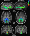
All fiber tracts are colored according to the value of their FA at each point; fibers with an FA close to 0 are shown as red, while increasing values shift toward blue, as elucidated by the colorbar in the upper right. Each row shows a comparison of the control and BD subjects in whom the tract of interest was closest to the median sample value for the group. The first row illustrates the greater fiber density in the ILF-R of controls. The second row depicts the greater length of splenium fibers in control subjects. Finally, the third row demonstrates the higher median FA in the CST-R2 of controls.
Similar articles
- A comparative diffusion tensor imaging study of corpus callosum subregion integrity in bipolar disorder and schizophrenia.
Li J, Kale Edmiston E, Chen K, Tang Y, Ouyang X, Jiang Y, Fan G, Ren L, Liu J, Zhou Y, Jiang W, Liu Z, Xu K, Wang F. Li J, et al. Psychiatry Res. 2014 Jan 30;221(1):58-62. doi: 10.1016/j.pscychresns.2013.10.007. Epub 2013 Nov 7. Psychiatry Res. 2014. PMID: 24300086 - Diffusion tensor imaging tractography study in bipolar disorder patients compared to first-degree relatives and healthy controls.
Mahapatra A, Khandelwal SK, Sharan P, Garg A, Mishra NK. Mahapatra A, et al. Psychiatry Clin Neurosci. 2017 Oct;71(10):706-715. doi: 10.1111/pcn.12530. Epub 2017 Jun 19. Psychiatry Clin Neurosci. 2017. PMID: 28419638 - A multicenter tractography study of deep white matter tracts in bipolar I disorder: psychotic features and interhemispheric disconnectivity.
Sarrazin S, Poupon C, Linke J, Wessa M, Phillips M, Delavest M, Versace A, Almeida J, Guevara P, Duclap D, Duchesnay E, Mangin JF, Le Dudal K, Daban C, Hamdani N, D'Albis MA, Leboyer M, Houenou J. Sarrazin S, et al. JAMA Psychiatry. 2014 Apr;71(4):388-96. doi: 10.1001/jamapsychiatry.2013.4513. JAMA Psychiatry. 2014. PMID: 24522197 - White matter modifications of corpus callosum in bipolar disorder: A DTI tractography review.
Videtta G, Squarcina L, Rossetti MG, Brambilla P, Delvecchio G, Bellani M. Videtta G, et al. J Affect Disord. 2023 Oct 1;338:220-227. doi: 10.1016/j.jad.2023.06.012. Epub 2023 Jun 9. J Affect Disord. 2023. PMID: 37301293 Review. - Neurobiological underpinnings of bipolar disorder focusing on findings of diffusion tensor imaging: a systematic review.
Duarte JA, de Araújo E Silva JQ, Goldani AA, Massuda R, Gama CS. Duarte JA, et al. Braz J Psychiatry. 2016 Mar 22;38(2):167-75. doi: 10.1590/1516-4446-2015-1793. Braz J Psychiatry. 2016. PMID: 27007148 Free PMC article. Review.
Cited by
- Comparison of tibial and femoral physeal diffusion tensor imaging in adolescents.
Santos L, Guariento A, Moustoufi-Moab S, Nguyen J, Tokaria R, Raya JM, Zurakowski D, Jambawalikar S, Jaramillo D. Santos L, et al. Pediatr Radiol. 2024 Nov 9. doi: 10.1007/s00247-024-06073-6. Online ahead of print. Pediatr Radiol. 2024. PMID: 39516384 - Alterations of Structural Network Efficiency in Early-Onset and Late-Onset Alzheimer's Disease.
Heo S, Yoon CW, Kim SY, Kim WR, Na DL, Noh Y. Heo S, et al. J Clin Neurol. 2024 May;20(3):265-275. doi: 10.3988/jcn.2023.0092. Epub 2024 Feb 5. J Clin Neurol. 2024. PMID: 38330417 Free PMC article. - Changes in the superior longitudinal fasciculus and anterior thalamic radiation in the left brain are associated with developmental dyscalculia.
Ayyıldız N, Beyer F, Üstün S, Kale EH, Mançe Çalışır Ö, Uran P, Öner Ö, Olkun S, Anwander A, Witte AV, Villringer A, Çiçek M. Ayyıldız N, et al. Front Hum Neurosci. 2023 Sep 28;17:1147352. doi: 10.3389/fnhum.2023.1147352. eCollection 2023. Front Hum Neurosci. 2023. PMID: 37868699 Free PMC article. - Diffusion tensor imaging of the physis: the ABC's.
Santos LA, Sullivan B, Kvist O, Jambawalikar S, Mostoufi-Moab S, Raya JM, Nguyen J, Marin D, Delgado J, Tokaria R, Nelson RR Jr, Kammen B, Jaramillo D. Santos LA, et al. Pediatr Radiol. 2023 Nov;53(12):2355-2368. doi: 10.1007/s00247-023-05753-z. Epub 2023 Sep 2. Pediatr Radiol. 2023. PMID: 37658251 Free PMC article. Review. - Genetic architecture of the white matter connectome of the human brain.
Sha Z, Schijven D, Fisher SE, Francks C. Sha Z, et al. Sci Adv. 2023 Feb 17;9(7):eadd2870. doi: 10.1126/sciadv.add2870. Epub 2023 Feb 17. Sci Adv. 2023. PMID: 36800424 Free PMC article.
References
- Adler CM, Holland SK, Schmithorst V, Wilke M, Weiss KL, Pan H, Strakowski SM. Abnormal frontal white matter tracts in bipolar disorder: a diffusion tensor imaging study. Bipolar Disord. 2004;6(3):197–203. - PubMed
- Altshuler LL. Bipolar disorder: are repeated episodes associated with neuroanatomic and cognitive changes? Biol Psychiatry. 1993;33(8–9):563–565. - PubMed
- Andreisek G, White LM, Kassner A, Tomlinson G, Sussman MS. Diffusion tensor imaging and fiber tractography of the median nerve at 1.5T: optimization of b value. Skeletal Radiol. 2009;38(1):51–59. - PubMed
- Ashtari M. Anatomy and functional role of the inferior longitudinal fasciculus: a search that has just begun. Dev Med Child Neurol. 2012;54(1):6–7. - PubMed
Publication types
MeSH terms
Substances
Grants and funding
- R21 MH086104/MH/NIMH NIH HHS/United States
- R21MH086104/MH/NIMH NIH HHS/United States
- R00 MH085944/MH/NIMH NIH HHS/United States
- R21 MH075944/MH/NIMH NIH HHS/United States
- K99 MH085944/MH/NIMH NIH HHS/United States
- R21MH085944/MH/NIMH NIH HHS/United States
LinkOut - more resources
Full Text Sources
Other Literature Sources
Medical


