Ginsenoside-Re ameliorates ischemia and reperfusion injury in the heart: a hemodynamics approach - PubMed (original) (raw)
Ginsenoside-Re ameliorates ischemia and reperfusion injury in the heart: a hemodynamics approach
Kyu Hee Lim et al. J Ginseng Res. 2013 Jul.
Abstract
Ginsenosides are divided into two groups based on the types of the panaxadiol group (e.g., ginsenoside-Rb1 and -Rc) and the panaxatriol group (e.g., ginsenoside-Rg1 and -Re). Among them, ginsenoside-Re (G-Re) is one of the compounds with the highest content in Panax ginseng and is responsible for pharmacological effects. However, it is not yet well reported if G-Re increases the hemodynamics functions on ischemia (30 min)/reperfusion (120 min) (I/R) induction. Therefore, in the present study, we investigated whether treatment of G-Re facilitated the recovery of hemodynamic parameters (heart rate, perfusion pressure, aortic flow, coronary flow, and cardiac output) and left ventricular developed pressure (±dp/dtmax). This research is designed to study the effects of G-Re by studying electrocardiographic changes such as QRS interval, QT interval and R-R interval, and inflammatory marker such as tissue necrosis factor-α (TNF-α) in heart tissue in I/R-induced heart. From the results, I/R induction gave a significant increase in QRS interval, QT interval and R-R interval, but showed decrease in all hemodynamic parameters. I/R induction resulted in increased TNF-α level. Treatment of G-Re at 30 and 100 μM doses before I/R induction significantly prevented the decrease in hemodynamic parameters, ameliorated the electrocardiographic abnormality, and inhibited TNF-α level. In this study, G-Re at 100 μM dose exerted more beneficial effects on cardiac function and preservation of myocardium in I/R injury than 30 μM. Collectively, these results indicate that G-Re has distinct cardioprotectective effects in I/R induced rat heart.
Keywords: Cardiac injury; Ginsenoside-Re; Hemodynamics; Myocardial preservation; Panax ginseng.
Figures
Fig. 1.. Experimental protocol. All experimental groups began with a 20 min perfusion period to allow for stabilization of the isolated hearts. Then, the hearts were divided into the normal control group (N/C), the ginsenoside-Re (G-Re) alone treated group (G-Re control), the ischemia for 30 min and reperfusion for 120 min group (ischemia/reperfusion [I/R] control), 30G-Re treated group, which received administration of 30 μM G-Re before ischemia induction (30G-Re+I/R), and 100G-Re treated group, which received administration of 100 μM G-Re before ischemia induction (100G-Re+I/R).
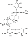
Fig. 2.. Chemical struture of ginsenoside-Re.
Fig. 3.. Relationships among perfusion pressure (A), aortic flow (B), coronary flow (C), and cardiac output changes (D) and each group on reperfusion for 120 min. Each bar represents the mean±SEM (_n_=9). N/C, normal control; G-Re, ginsenoside-Re; I/R, ischemia/reperfusion. *p<0.05, **p<0.01 compared with the I/R control group.
Fig. 4.. The changes in the maximal rate of change in left ventricular contraction (+dP/dtmax) (A) and the maximal rate of change in left ventricular relaxation (-dP/dtmin) (B). Values are expressed as mean±SD. N/C, normal control; G-Re, ginsenoside-Re; I/R, ischemia/reperfusion. *p<0.05, **p<0.01 compared with the I/R control group.
Fig. 5.. Effect of ginsenoside-Re (G-Re) on electrocardiographic pattern (A, normal control; B, G-Re control; C, ischemia/reperfusion [I/R] control; D, 30 μM G-Re+I/R; E, 100 μM G-Re+I/R) in isolated perfused heart.
Fig. 6.. Effect of ginsenoside-Re (G-Re) in QRS intervals on electrocardiographic pattern. Values are expressed as mean±SD for nine rats in each group. N/C, normal control; I/R, ischemia/reperfusion. *p<0.05, **p<0.01 compared with the I/R control group.
Fig. 7.. Effect of ginsenoside-Re (G-Re) on QT intervals against myocardial injury in isolated perfusion model. Values are expressed as mean±SD for nine rats in each group. N/C, normal control; I/R, ischemia/reperfusion. *p<0.05, **p<0.01 compared with the I/R control group.
Fig. 8.. Effect of ginsenoside-Re (G-Re) on R-R intervals against myocardial injury in isolated perfusion model. Values are expressed as mean±SD for nine rats in each group. N/C, normal control; I/R, ischemia/reperfusion. *p<0.05, **p<0.01 compared with the I/R control group.
Fig. 9.. Effect of ginsenoside-Re (G-Re) on cardiac tissue necrosis factor-α (TNF-α) levels in ischemia/reperfusion (I/R)-induced myocardial injury in isolated heart. Values are expressed as mean±SD for nine rats in each group. N/C, normal control. *p<0.05 and **p<0.01 as compared to I/R control group.
Similar articles
- The effects of ginseng total saponin, panaxadiol and panaxatriol on ischemia/reperfusion injury in isolated rat heart.
Kim TH, Lee SM. Kim TH, et al. Food Chem Toxicol. 2010 Jun;48(6):1516-20. doi: 10.1016/j.fct.2010.03.018. Epub 2010 Mar 29. Food Chem Toxicol. 2010. PMID: 20353807 - Neuroprotective Effects of Ginsenosides against Cerebral Ischemia.
Cheng Z, Zhang M, Ling C, Zhu Y, Ren H, Hong C, Qin J, Liu T, Wang J. Cheng Z, et al. Molecules. 2019 Mar 20;24(6):1102. doi: 10.3390/molecules24061102. Molecules. 2019. PMID: 30897756 Free PMC article. - Korean Red Ginseng Induced Cardioprotection against Myocardial Ischemia in Guinea Pig.
Lim KH, Kang CW, Choi JY, Kim JH. Lim KH, et al. Korean J Physiol Pharmacol. 2013 Aug;17(4):283-9. doi: 10.4196/kjpp.2013.17.4.283. Epub 2013 Jul 30. Korean J Physiol Pharmacol. 2013. PMID: 23946687 Free PMC article. - Protective Effects and Target Network Analysis of Ginsenoside Rg1 in Cerebral Ischemia and Reperfusion Injury: A Comprehensive Overview of Experimental Studies.
Xie W, Zhou P, Sun Y, Meng X, Dai Z, Sun G, Sun X. Xie W, et al. Cells. 2018 Dec 12;7(12):270. doi: 10.3390/cells7120270. Cells. 2018. PMID: 30545139 Free PMC article. Review. - Comparison of the pharmacological effects of Panax ginseng and Panax quinquefolium.
Chen CF, Chiou WF, Zhang JT. Chen CF, et al. Acta Pharmacol Sin. 2008 Sep;29(9):1103-8. doi: 10.1111/j.1745-7254.2008.00868.x. Acta Pharmacol Sin. 2008. PMID: 18718179 Review.
Cited by
- Ginsenoside Re promotes proliferation of murine bone marrow mesenchymal stem cells in vitro through estrogen-like action.
Luo L, Peng B, Xiong L, Wang B, Wang L. Luo L, et al. In Vitro Cell Dev Biol Anim. 2024 Oct;60(9):996-1008. doi: 10.1007/s11626-024-00969-1. Epub 2024 Sep 10. In Vitro Cell Dev Biol Anim. 2024. PMID: 39256290 - Inhibition of Cholesteryl Ester Transfer Protein Contributes to the Protection of Ginsenoside Re Against Isoproterenol-Induced Cardiac Hypertrophy.
Qiu Y, Xie M, Ding X, Zhang H, Li H, Wang H, Li T, Dong W, Jiang F, Tang X. Qiu Y, et al. Cureus. 2024 May 9;16(5):e59942. doi: 10.7759/cureus.59942. eCollection 2024 May. Cureus. 2024. PMID: 38854305 Free PMC article. - Reactive oxygen species-scavenging nanomaterials for the prevention and treatment of age-related diseases.
Dai Y, Guo Y, Tang W, Chen D, Xue L, Chen Y, Guo Y, Wei S, Wu M, Dai J, Wang S. Dai Y, et al. J Nanobiotechnology. 2024 May 15;22(1):252. doi: 10.1186/s12951-024-02501-9. J Nanobiotechnology. 2024. PMID: 38750509 Free PMC article. Review. - Ginsenoside Rb2 improves heart failure by down-regulating miR-216a-5p to promote autophagy and inhibit apoptosis and oxidative stress.
Peng Y, Liao B, Zhou Y, Zeng W. Peng Y, et al. J Appl Biomed. 2023 Dec;21(4):180-192. doi: 10.32725/jab.2023.024. Epub 2023 Dec 14. J Appl Biomed. 2023. PMID: 38112457
References
- Ostadal B. The past, the present and the future of experimental research on myocardial ischemia and protection. Pharmacol Rep. 2009;61:3–12. - PubMed
- Fleming PR. A short history of cardiology. Rodopi Publishers; Amsterdam: 1997. - PubMed
LinkOut - more resources
Full Text Sources
Other Literature Sources
Miscellaneous


