Preliminary review of Indian Eumenophorinae (Araneae: Theraphosidae) with description of a new genus and five new species from the Western Ghats - PubMed (original) (raw)
Preliminary review of Indian Eumenophorinae (Araneae: Theraphosidae) with description of a new genus and five new species from the Western Ghats
Zeeshan A Mirza et al. PLoS One. 2014.
Abstract
The theraphosid spider genera Heterophrictus Pocock, 1900 and Neoheterophrictus Siliwal & Raven, 2012 are rediagnosed in this paper and a new genus, Sahydroaraneus gen. nov. is described from Southern Western Ghats. Four new species (two each of Heterophrictus and Neoheterophrictus) and one of Sahydroaraneus gen. nov. are described from the Western Ghats. Plesiophrictus mahabaleshwari Tikader, 1977 is removed from the synonymy of Heterophrictus milleti Pocock, 1900 and is treated as a junior synonym of Heterophrictus blatteri (Gravely, 1935). Plesiophrictus bhori Gravely, 1915 is transferred to the genus Neoheterophrictus, Neoheterophrictus bhori (Gravely, 1915) new combination. The genus, Sahydroaraneus gen. nov., resembles tarantula belonging to the genus, Neoheterophrictus but differs with respect to structure of tibial apophysis and spermathecae. Detailed ultra-structure of setae type of the Indian Eumenophorinae is presented for the first time along with notes on their biogeography. Common elements among Africa, Madagascar and India like the Eumenophorinae and several other mygalomorph spiders advocate mygalomorphae as an important group for evolutionary investigation due to their inability for long distance dispersal rendering the members restrictive in distribution.
Conflict of interest statement
Competing Interests: The authors have declared that no competing interests exist.
Figures
Figure 1. Possible Indian odysseys: different models for the position of India approximately 65 million years ago.
a, The standard ‘biotic ferry’ model showing India isolated by large expanses of water. b, A limited ‘biotic (land) bridge’ model incorporating a narrow connection (Greater Somalia) with Africa. c, Another biotic bridge model assuming a different longitudinal position for India and showing connections with Madagascar, Africa and Asia (Hedges [42]).
Figure 2. Map depicting global distribution of genera of Eumenophorinae.
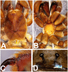
Figure 3. Heterophrictus millet.
A. Cephalothorax; b. Sternum, labium and maxillae; C. Chelicerae prolateral view; D. spermathecae.
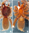
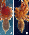
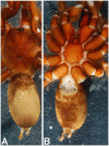
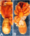
Figure 4. Heterophrictus raveni sp. nov. male holotype (ZSI/WRC/AR/418).
A. Cephalothorax and abdomen dorsal view; B. Sternum, labium, maxillae and abdomen ventral view.
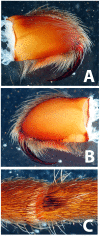
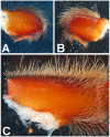
Figure 5. Heterophrictus raveni sp. nov. male holotype (ZSI/WRC/AR/418).
A. Chelicerae retrolateral view; B. Chelicerae prolateral view.
Figure 6. Heterophrictus raveni sp. nov. male holotype (ZSI/WRC/AR/418).
A. Cluster of spiniform setae on basal region of tibia I, retrolateral view; B. Cluster of spiniform setae tibia I dorsal view; C. Palp bulb dorsal view; D. Palp bulb prolateral view; E. Palp bulb retrolateral view.
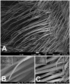
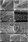
Figure 7. Scanning electron micrograph of Heterophrictus raveni sp. nov. male holotype tibia I (ZSI/WRC/AR/418).
A. Ultra-structure of spiniform setae and normal setae, dorsal view; B. Tip of spiniform setae; C. Surface texture of spiniform setae.
Figure 8. Heterophrictus raveni sp. nov. male holotype (ZSI/WRC/AR/418).
A. Palp bulb dorsal view; B. Palp bulb prolateral view; C. Palp bulb retrolateral view.
Figure 9
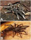
(A. & B.) Heterophrictus raveni sp. nov. female paratype (ZSI/WRC/AR/419) in life, photo by Zeeshan Mirza.
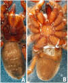
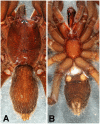
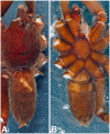
Figure 10. Heterophrictus raveni sp. nov. female paratype (ZSI/WRC/AR/419).
A. Cephalothorax and abdomen, dorsal view; B. Sternum, labium, maxilla and abdomen, ventral view.
Figure 11. Heterophrictus raveni sp. nov. female paratype (ZSI/WRC/AR/419).
A. Chelicerae retrolateral view; B. Chelicerae prolateral view.
Figure 12. Heterophrictus raveni sp. nov. female paratype (ZSI/WRC/AR/419).
A. Coxa of leg II prolateral view showing stridulatory setae; B. Spermathecae.
Figure 13
Scanning electron micrograph of Heterophrictus raveni sp. nov. female paratype (ZSI/WRC/AR/419), coxa II: A. Coxa of leg II prolateral view showing stridulatory setae; B. Basal half of horizontally aligned long pilose setae below coxal suture; C. Distal half of horizontally aligned long pilose setae; D. Ultra-structure of the surface texture of long pilose setae; D. Short pilose setae in posterior distal region of coxa of leg II; 31. Vertically aligned pyriform setae above coxal suture of leg II; E. Vertically aligned pyriform setae above coxal suture of leg II with curved tips; F. Vertically aligned pyriform setae above coxal suture of leg II basal region; G. Junction of coxal suture of leg II.
Figure 14. Scanning electron micrograph showing tarsal claws on leg IV of paratype female Heterophrictus raveni sp. nov. (ZSI/WRC/AR/419).
Figure 15
Heterophrictus aareyensis sp. nov. A. male holotype (ZSI/WRC/AR/420) in life, photo by Rajesh Sanap; B. female paratype BNHS Sp- 85 in life, photo by Zeeshan Mirza.
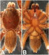
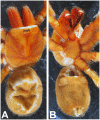
Figure 16. Heterophrictus aareyensis sp. nov. male holotype (ZSI/WRC/AR/420).
A. Cephalothorax and abdomen, dorsal view; B. Sternum, labium, maxillae, abdomen and chelicerae, ventral view.
Figure 17. Heterophrictus aareyensis sp. nov. male holotype (ZSI/WRC/AR/420).
A. Chelicerae retrolateral view; B. Chelicerae prolateral view; C. Cluster of spiniform setae tibia I.
Figure 18. Heterophrictus aareyensis sp. nov. male holotype (ZSI/WRC/AR/420).
A. palp bulb dorsal view; B. palp bulb prolateral view; C. palp bulb retrolateral view.
Figure 19. Heterophrictus aareyensis sp. nov. male holotype (ZSI/WRC/AR/420).
A. Palp bulb dorsal view; B. Palp bulb prolateral view; C. Palp bulb retrolateral view.
Figure 20. Heterophrictus aareyensis sp. nov. female paratype (BNHS SP-85).
A. Cephalothorax and abdomen, dorsal view; B. Sternum, labium, maxillae, abdomen and chelicerae, ventral view.
Figure 21. Heterophrictus aareyensis sp. nov. female paratype (BNHS SP-85).
A. Chelicerae prolateral view; B. chelicerae retrolateral view; C. eye; D. spermathecae.
Figure 22. Heterophrictus blatteri, male (BNHS SP-86).
A. Cephalothorax and abdomen, dorsal view; B. Sternum, labium, maxillae, abdomen and chelicerae, ventral view.
Figure 23. Heterophrictus blatteri, male (BNHS SP-86).
A. palp bulb prolateral view; B. palp bulb retrolateral view; C. spike setae on tibia I.
Figure 24. Heterophrictus blatteri, female (BMNH 16.5.2.15).
A. Chelicerae prodorsal view; A. coxa leg II, prolateral view; B. Spermathecae.
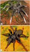
Figure 25. Neoheterophrictus smithi sp. nov. male holotype (ZSI/WRC/AR/421).
A. Neoheterophrictus smithi sp. nov male holotype in life; B. Neoheterophrictus smithi sp. nov female paratype in life, photos by Harshal Bhosale.
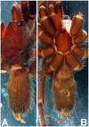
Figure 26. Neoheterophrictus smithi sp. nov. male holotype (ZSI/WRC/AR/421).
A. Cephalothorax and abdomen, dorsal view; B. Sternum, labium, maxillae, abdomen and chelicerae, ventral view.
Figure 27. Neoheterophrictus smithi sp. nov. male holotype (ZSI/WRC/AR/421).
A. Chelicerae retrolateral view; B. Chelicerae prolateral view; C. Retrolateral view of maxilla showing stridulatory setae aligned in a dorso-ventral series.
Figure 28. Neoheterophrictus smithi sp. nov. male holotype (ZSI/WRC/AR/421).
A. Palp bulb dorsal view; B. Palp bulb prolateral view; C. palp bulb retrolateral view.
Figure 29. Neoheterophrictus smithi sp. nov. male holotype (ZSI/WRC/AR/421).
A. Palp bulb dorsal view; B. Palp bulb retrolateral view; C. Palp bulb prolateral view.
Figure 30. Neoheterophrictus smithi sp. nov. male holotype (ZSI/WRC/AR/421).
A. Tibial apophysis retrolateral view; B. Tibial apophysis prolateral view; C. Tibial apophysis ventral view.
Figure 31. Neoheterophrictus smithi sp. nov. male holotype (ZSI/WRC/AR/421).
A. Tibial apophysis retrolateral view; B. Tibial apophysis prolateral view; C. Tibial apophysis ventral view.
Figure 32. Neoheterophrictus smithi sp. nov. female (ZSI/WRC/AR/422).
A. Cephalothorax and abdomen, dorsal view; B. Sternum, labium, maxilla, chelicerae and abdomen ventral view.
Figure 33. Neoheterophrictus smithi sp. nov. female (ZSI/WRC/AR/422).
A. Chelicerae retrolateral view; B. Chelicerae prolateral view.
Figure 34. Scanning electron micrograph of Neoheterophrictus smithi sp. nov. female (ZSI/WRC/AR/422).
A. Cheliceral prolateral broader showing rastellum inter-mixed with normal setae; B. Prolateral cheliceral border showing stout spines.
Figure 35. Scanning electron micrograph of Neoheterophrictus smithi sp. nov. female (ZSI/WRC/AR/422).
A. Ventral view of tarsus showing dividing spike setae; B. Base of spike setae; C. Base of spike setae; D. Spike setae inter-mixed with scopulae setae.
Figure 36. Neoheterophrictus amboli sp. nov. male holotype (ZSI/WRC/AR/423) in life.
Photo by Aditya Malgaonkar.
Figure 37. Neoheterophrictus amboli sp. nov. male holotype (ZSI/WRC/AR/423).
A. Cephalothorax and abdomen, dorsal view; B. Sternum, labium, maxillae, abdomen and chelicerae, ventral view.
Figure 38. Neoheterophrictus amboli sp. nov. male holotype (ZSI/WRC/AR/423).
A. Chelicerae prolateral view; B. Chelicerae retrolateral view; C. Maxilla retrolateral view showing stridulatory setae intermixed with normal setae between palp-I on the retrolateral basal region.
Figure 39. Neoheterophrictus amboli sp. nov. male holotype (ZSI/WRC/AR/423) palp bulb.
A. dorsal view; B. Prolateral view; C. Rretrolateral view.
Figure 40. Neoheterophrictus amboli sp. nov. male holotype (ZSI/WRC/AR/423) palp bulb.
A. dorsal view; B. Prolateral view; C. Retrolateral view.
Figure 41. Neoheterophrictus amboli sp. nov. male holotype (ZSI/WRC/AR/423) tibial apophysis.
A. retrolateral view; B. prolateral view; C. Ventral view.
Figure 42. Neoheterophrictus amboli sp. nov. male holotype (ZSI/WRC/AR/423) tibial apophysis.
A. Retrolateral view; B. Prolateral view; C. Ventral view.
Figure 43. Neoheterophrictus bhori female (Type BMNH 16.5.2.16).
A. Cephalothorax and abdomen, dorsal view; B. Sternum, labium, maxillae, abdomen and chelicerae, ventral view.
Figure 44. Neoheterophrictus bhori female (Type BMNH 16.5.2.16).
A. Chelicerae pro-dorsal view B. coxa leg III, prolateral view; C. spermathecae.
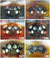
Figure 45. Eyes in Heterophrictus and Neoheterophrictus.
A. Heterophrictus raveni sp. nov. holotype male; B. Heterophrictus raveni sp. nov. paratype female; C. Heterophrictus aareyensis sp. nov. holotype male; D. Neoheterophrictus amboli sp. nov. holotype male; E. Neoheterophrictus smithi sp. nov. holotype male; F. Neoheterophrictus smithi sp. nov. female.
Figure 46. Sahydroaraneus hirsti sp. nov. male holotype BMNH 16.5.2.12.
A. eye, B. coxa leg I prolateral view, C. chelicerae prolateral view, D. coxa leg I retrolateral view.
Figure 47. Sahydroaraneus hirsti sp. nov. male holotype BMNH 16.5.2.12.
Palp bulb, A. retrolateral view; B. prolateral view; C. dorsal view.
Figure 48. Sahydroaraneus hirsti sp. nov. male holotype BMNH 16.5.2.12.
Tibial apophysis, A. retrolateral view; B. prolateral view; C. dorsal view.
Figure 49. Sahydroaraneus hirsti sp. nov. male holotype BMNH 16.5.2.12.
Leg scopulae, A. leg III, ventral view; B. leg IV, ventral view.
Figure 50. Sahydroaraneus raja female type BMNH 16.5.2.17.
A. Cephalothorax and abdomen, dorsal view; B. Sternum, labium, maxillae, abdomen and chelicerae, ventral view.
Figure 51. Sahydroaraneus raja female type BMNH 16.5.2.17.
A. chelicerae pro-dorsal view, coxa leg I, proalteral view; C. absence of spermathecae indicating an immature specimen.
Figure 52. Sahydroaraneus collinus female type BMNH 19.16.29. A. eye; B. spermathecae.
Figure 53. Map showing location of Western Ghats in India and biogeographic zones within Western Ghats based on floral composition .
Figure 54. Map showing distribution of Eumenophorinae in India.
(A) Heterophrictus, (B) Neoheterophrictus, (C) Sahydroaraneus gen. nov.
Similar articles
- Aguapanela, a new tarantula genus from the Colombian Andes (Araneae, Theraphosidae).
Perafán C, Cifuentes Y, Estrada-Gomez S. Perafán C, et al. Zootaxa. 2015 Oct 27;4033(4):529-42. doi: 10.11646/zootaxa.4033.4.4. Zootaxa. 2015. PMID: 26624422 - A revision of ant-mimicking spiders of the family Corinnidae (Araneae) in the Western Pacific.
Raven RJ. Raven RJ. Zootaxa. 2015 May 20;3958:1-258. doi: 10.11646/zootaxa.3958.1.1. Zootaxa. 2015. PMID: 26249225 - A review of the genus Sphingius Thorell, 1890 from India (Araneae: Liocranidae).
Sankaran PM, Caleb JTD, Sebastian PA. Sankaran PM, et al. Zootaxa. 2020 Dec 23;4896(4):zootaxa.4896.4.3. doi: 10.11646/zootaxa.4896.4.3. Zootaxa. 2020. PMID: 33756846 Review. - A review of the spider genus Iberina (Araneae, Hahniidae).
Rika V. Rika V. Zootaxa. 2022 May 6;5133(4):555-566. doi: 10.11646/zootaxa.5133.4.6. Zootaxa. 2022. PMID: 36101083 Review.
Cited by
- Discovery of a deeply divergent new lineage of vine snake (Colubridae: Ahaetuliinae: Proahaetulla gen. nov.) from the southern Western Ghats of Peninsular India with a revised key for Ahaetuliinae.
Mallik AK, Achyuthan NS, Ganesh SR, Pal SP, Vijayakumar SP, Shanker K. Mallik AK, et al. PLoS One. 2019 Jul 17;14(7):e0218851. doi: 10.1371/journal.pone.0218851. eCollection 2019. PLoS One. 2019. PMID: 31314800 Free PMC article.
References
- Dippenaar-Schoeman AS (2002) Baboon and Trapdoor Spiders of Southern Africa: An Introduction Manual. Plant Protection Research Institute Handbook No. 13, Agricultural Research Council, Pretoria, 128pp.
- Platnick NI (2013) The World Spider Catalog, version 14.0. American Museum of Natural History. Available: http://research.amnh.org/iz/spiders/catalog. DOI:10.5531/db.iz.0001. - DOI
- Siliwal M (2009) Revalidating the taxonomic position of the Indian Ischnocolus spp. (Araneae:Theraphosidae). J Threat Tax 10: 533–534.
- Siliwal M, Molur S (2009) Redescription, distribution and status of the Karwar Large Burrowing Spider Thrigmopoeus truculentus Pocock, 1899 (Araneae: Theraphosidae) a Western Ghats endemic ground mygalomorph. J Threat Tax 1(6): 331–339.
- Siliwal M, Gupta N, Raven R (2012) A new genus of the family Theraphosidae (Araneae: Mygalomorphae) with description of three new species from the Western Ghats of Karnataka, India. J Threat Tax 4(14): 3233–3254.
Publication types
MeSH terms
Grants and funding
The study was partly funded by The Newby Trust Limited, London through a travel grant to visit the Natural History Museum to ZAM. The funders had no role in study design, data collection and analysis, decision to publish, or preparation of the manuscript. No additional external funding was received for this study.
LinkOut - more resources
Full Text Sources
Other Literature Sources


