Torvosaurus gurneyi n. sp., the largest terrestrial predator from Europe, and a proposed terminology of the maxilla anatomy in nonavian theropods - PubMed (original) (raw)
Torvosaurus gurneyi n. sp., the largest terrestrial predator from Europe, and a proposed terminology of the maxilla anatomy in nonavian theropods
Christophe Hendrickx et al. PLoS One. 2014.
Abstract
The Lourinhã Formation (Kimmeridgian-Tithonian) of Central West Portugal is well known for its diversified dinosaur fauna similar to that of the Morrison Formation of North America; both areas share dinosaur taxa including the top predator Torvosaurus, reported in Portugal. The material assigned to the Portuguese T. tanneri, consisting of a right maxilla and an incomplete caudal centrum, was briefly described in the literature and a thorough description of these bones is here given for the first time. A comparison with material referred to Torvosaurus tanneri allows us to highlight some important differences justifying the creation of a distinct Eastern species. Torvosaurus gurneyi n. sp. displays two autapomorphies among Megalosauroidea, a maxilla possessing fewer than eleven teeth and an interdental wall nearly coincidental with the lateral wall of the maxillary body. In addition, it differs from T. tanneri by a reduced number of maxillary teeth, the absence of interdental plates terminating ventrally by broad V-shaped points and falling short relative to the lateral maxillary wall, and the absence of a protuberant ridge on the anterior part of the medial shelf, posterior to the anteromedial process. T. gurneyi is the largest theropod from the Lourinhã Formation of Portugal and the largest land predator discovered in Europe hitherto. This taxon supports the mechanism of vicariance that occurred in the Iberian Meseta during the Late Jurassic when the proto-Atlantic was already well formed. A fragment of maxilla from the Lourinhã Formation referred to Torvosaurus sp. is ascribed to this new species, and several other bones, including a femur, a tibia and embryonic material all from the Kimmeridgian-Tithonian of Portugal, are tentatively assigned to T. gurneyi. A standard terminology and notation of the theropod maxilla is also proposed and a record of the Torvosaurus material from Portugal is given.
Conflict of interest statement
Competing Interests: The authors have declared that no competing interests exist.
Figures
Figure 1. Proposed terminology and annotation of the nonavian theropod maxilla.
Right maxilla of Allosaurus fragilis (USNM 8335) in A, lateral; B, anterior; C, medial and D, posterior views, with details of E, promaxillary recess and maxillary antrum in medial view; and F, ascending ramus and dorsal margin of vestibular bulla in dorsal view. Abbreviations: ammf, anteromedial maxillary fenestra; amp, anteromedial process; anr, anterior ramus; aor, antorbital ridge; asr, ascending ramus; idw, interdental wall; ifs, interfenestral strut; juc, jugal contact; lac, lacrimal contact; laof, lateral antorbital fossa; law, lateral wall; maf, maxillary alveolar foramina; man, maxillary antrum; maof, medial antorbital fossa; mbo, maxillary body; mcf, maxillary circumfenestra foramina; mes, medial shelf; mew, medial wall; mfe, maxillary fenestra; mfo, maxillary fossa; mmf, medial maxillary foramina; mx1, first maxillary tooth; nac, nasal contact; nuf, nutrient foramina; nug, nutrient groove; pac, palatine contact; pmc, premaxillary contact; pmmf, posteromedial maxillary fenestra; pmr, promaxillary recess; pne, pneumatic excavation; poas, postantral strut; pras, preantral strut; snf, subnarial foramen; suas, suprantral strut; veb, vestibular bulla. Scale bars = 5 cm.
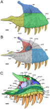
Figure 2. Proposed terminology and annotation of the nonavian theropod maxilla.
Left maxillae of Tyrannosaurus rex in A–B, lateral view (CMNH 9380, reversed); and C, medial view (BHI 3033; modified from [32]). Abbreviations: ammf, anteromedial maxillary fenestra; amp, anteromedial process; anb, anterior body; aofe, antorbital fenestra; asr, ascending ramus; ear, epiantral recess; idg, interdental gap; idp, interdental plate; ifs, interfenestral strut; juc, jugal contact; jur, jugal ramus; lac, lacrimal contact; laof, lateral antorbital fossa; law, lateral wall; maf, maxillary alveolar foramina; man, maxillary antrum; mbo, maxillary body; mcf, maxillary circumfenestra foramina; mes, medial shelf; mew, medial wall; mfe, maxillary fenestra; mx9, ninth maxillary tooth; nac, nasal contact; nuf, nutrient foramina; nug, nutrient groove; pab, preantorbital body; pac, palatine contact; pmc, premaxillary contact; pmf, promaxillary fenestra; pmmf, posteromedial maxillary fenestra; pmr, promaxillary recess; pne, pneumatic excavation; poas, postantral strut; pras, preantral strut; prms, promaxillary strut; snf, subnarial foramen. Scale bars = 5 cm.
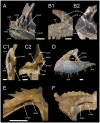
Figure 3. Proposed terminology and annotation of the nonavian theropod maxilla.
A, Right maxilla of Allosaurus fragilis (AMNH 600) in posteromedial view; B, lateral antorbital fossae of Ceratosaurus in lateral view; B1, right maxilla of Ceratosaurus magnicornis (MWC 1) and; B2, left maxilla of Ceratosaurus dentisulcatus (UMNH VP 5278; courtesy of Roger Benson); C, left maxilla of Tyrannosaurus rex (CMNH 9380) in posterodorsal (C1) and dorsal (C2) views; D, left maxilla of Tarbosaurus baatar (ZPAL MgD-I/4; courtesy of Stephen Brusatte) in lateral view; E, right maxilla of Duriavenator hesperis (BMNH R332) in dorsomedial view; and F, left maxilla of Piatnitzkysaurus floresi (PVL 4073) in dorsomedial view (courtesy of Martin Ezcurra). Abbreviations: amf, accessory maxillary fenestra; ammf, anteromedial maxillary fenestra; ampr anteromedial pneumatic recess; iar, interalveolar recess; mal, maxillary alveoli; mes, medial shelf; mfe, maxillary fenestra; mfo, maxillary fossa; pmf, promaxillary fenestra; pmmf, posteromedial maxillary fenestra; pmr, promaxillary recess; pne, pneumatic recess; poas, postantral strut; pras, preantral strut; ptmf, postmaxillary fenestra; ptms, postmaxillary strut; trb, tooth root bulge; vmpr, ventromedial pneumatic recess. Scale bars = 5 cm.
Figure 4. Reconstruction of Torvosaurus gurneyi in lateral view.
A, Skeletal reconstruction of Torvosaurus gurneyi in lateral view illustrating, in red, the elements present in the holotype specimen (ML 1100) and, in blue, the elements tentatively assigned to this species (artwork by Scott Hartman, used with permission and modified; drawing of man by Carol Abraczinskas, University of Chicago, used with permission). B, Skull reconstruction of Torvosaurus gurneyi in lateral view illustrating the incomplete left maxilla(ML 1100) of the holotype specimen (artwork by Simão Mateus, used with permission and modified). Scale bars = 1 m (A) and 10 cm (B).
Figure 5. Maxilla of Torvosaurus gurneyi (ML 1100) and comparison with T. tanneri.
Incomplete left maxilla of the holotype specimen of Torvosaurus gurneyi (ML 1100) in A, lateral; B, medial; C, ventral; D, dorsal; E, anterior; F, posterior views with details of G, Anterodorsal margin of jugal ramus in dorsomedial view; and H, Posterior part of jugal ramus in dorsal view. I–J, Anterior part of interdental wall of I, T. gurneyi; and J, T. tanneri (BYUVP 9122) in medial view. K–L, Anteromedial process of K, T. gurneyi; and L, T. tanneri (BYUVP 9122) in medial views. Scale bars = 10 cm (A–H), 5 cm (G–L).
Figure 6. Maxilla of Torvosaurus gurneyi (ML 1100) and comparison with T. tanneri.
Interpretive line drawing of the left maxilla of the holotype specimen of Torvosaurus gurneyi (ML 1100) in A, lateral; B, medial; C, ventral; D, dorsal; E, anterior; F, posterior views with details of G, anterodorsal margin of jugal ramus in dorsomedial view; and H, posterior part of jugal ramus in dorsal view. I–J, Interpretive line drawing of the anterior part of interdental wall of I, T. gurneyi; and J, T. tanneri (BYUVP 9122) in medial view. K–L, Interpretive line drawing of the anteromedial process of K, T. gurneyi; and L, T. tanneri (BYUVP 9122) in medial views. Hatched areas represents missing parts, light grey tone indicates reconstructed part, and dark grey tone corresponds to the pneumatopores, foramina, and alveoli, with alveoli 9 and 10 being reconstructed. Abbreviations: adc, anterodorsal crest; adr, anterodorsal ridge of the anteromedial process; afo, anterior foramina; al, alveolus; amg, anteromedial groove of the anteromedial process; amp, anteromedial process; amr, anteromedial ridge; anr, anterior ramus; aor, antorbital ridge; asr, ascending ramus; avg, anteroventral groove of the anteromedial process; avr, anteroventral ridge on the anteromedial process; dmg, dorsomedial groove; idw, interdental wall; juc, jugal contact; lac, lacrimal contact; laof, lateral antorbital fossa; law, lateral wall; maf, maxillary alveolar foramina; mcf, maxillary circumfenestra foramina; mes, medial shelf; mew, medial wall; mfo, maxillary fossa; mx, maxillary teeth; nac, nasal contact; nuf, nutrient foramina; nug, nutrient groove; nvo, neurovascular opening; pmc, premaxillary contact; snf, subnarial foramen. Scale bars = 10 cm (A–H), 5 cm (G–L).
Figure 7. Dentition of Torvosaurus gurneyi (ML 1100).
A, C, E–H, Second maxillary tooth; and B, D, third non-erupted maxillary tooth of the holotype specimen of Torvosaurus gurneyi in A–B, labial; C–D, lingual; E, mesial; F, distal; G, basal; and H, apical views. I–J, Distal; and K–M, mesial denticles of the second maxillary tooth in lateral view. M, Distal serrations showing the interdenticular sulci; and N, enamel texture of the third non-erupted tooth in labial view. Abbreviations: cd, cervix dentis; dca, distal carina; del, dentine layer; ent, enamel texture; ids, interdenticular sulci; idsp, interdenticular space; mca, mesial carina; lic, lingual concavity for the erupting tooth; puc, pulp cavity; ro, root; uet, unerupted tooth; und, transversal undulation. Scale bars = 5 cm (A–F), 3 cm (G–H), 3 mm (I, K, M–N), 1 mm (J, L).
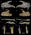
Figure 8. Caudal vertebra of Torvosaurus gurneyi (ML 1100).
A–D, Posterior part of an anterior caudal centrum of the holotype specimen of Torvosaurus gurneyi (ML 1100) in A, anterior; B, posterior; C, right lateral; D, left lateral; E, dorsal; and F, ventral views. Abbreviations: nc, neural canal; st, striation. Scale bar = 5 cm.
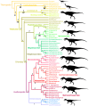
Figure 9. Cladogram of basal Theropoda and phylogenetic position of Torvosaurus gurneyi.
Strict consensus cladogram from 71 most parsimonious trees after pruning Magnosaurus, Poekilopleuron, Streptospondylus and Xuanhanosaurus from the full set of most parsimonious trees. Initial analysis used New Technology Search using TNT v.1.1 of a data matrix comprising 353 characters for two outgroup (Eoraptor and Herrerasaurus) and 60 nonavian theropod taxa. Tree length = 1022 steps; CI = 0.414, RI = 0.685. Bremer support values are in regular and bootstrap values are in bold. Dinosaur silhouettes by Scott Hartman (all but Metriacanthosauridae; used with permission) and Gregory S. Paul (Metriacanthosauridae; used with permission).
Figure 10. Comparison of the maxillae of Torvosaurus gurneyi and Torvosaurus tanneri.
Left maxillae of the holotype specimen of Torvosaurus gurneyi (ML 1100) in A, lateral; B, medial; E, ventral; F, dorsal; I, anterior; and K, posterior views. Left maxillae of a specimen referred to Torvosaurus tanneri (BYUVP 9122) in C, lateral; D, medial; G, ventral; H, dorsal; J, anterior; and L, posterior views. Abbreviations: adc, anterodorsal crest; adr, anterodorsal ridge of the anteromedial process; afo, anterior foramina; al1, first alveolus; al8, eighth alveolus; al10, tenth alveolus; amp, anteromedial process; aor, antorbital ridge; avg, anteroventral groove of the anteromedial process; avr, anteroventral ridge on the anteromedial process; idw, interdental wall; ldr, laterodorsal ridge within the anterior corner of the lateral antorbital fossa; mfo, maxillary fossa; nuf, nutrient foramina; nug, nutrient groove; nvo, neurovascular opening. Scale bars = 5 cm.
Figure 11. Femur and tibia of Megalosauridae from the Late Jurassic of Portugal.
Distal portion of a left femur (ML 632) of a megalosaurid tentatively referred to Torvosaurus gurneyi in A, anterior; B, lateral; C, posterior; D, medial; E, proximal; and F, distal views. Incomplete left tibia (ML 430) of Torvosaurus sp. (and tentatively referred to Torvosaurus gurneyi), with reconstruction of missing part of cnemial crest, in A, anterior; B, lateral; C, posterior; D, medial; E, proximal; and F, distal views. Abbreviations: asc, contact with astragalus; ccr, cnemial crest ridge; cnc, basal part of cnemial crest; ctf, crista tibiofibularis; dep, anterodistal depression; dir, distal ridges; exg, extensor groove; ffl, fibular flange; flg, flexor groove; lc, lateral condyle; mc, medial condyle; mdc, medio-distal crest; sab, supracetabular buttress. Scale bars = 10 cm.
Similar articles
- Filling the gaps of dinosaur eggshell phylogeny: Late Jurassic Theropod clutch with embryos from Portugal.
Araújo R, Castanhinha R, Martins RM, Mateus O, Hendrickx C, Beckmann F, Schell N, Alves LC. Araújo R, et al. Sci Rep. 2013;3:1924. doi: 10.1038/srep01924. Sci Rep. 2013. PMID: 23722524 Free PMC article. - New evidence of shared dinosaur across Upper Jurassic Proto-North Atlantic: Stegosaurus from Portugal.
Escaso F, Ortega F, Dantas P, Malafaia E, Pimentel NL, Pereda-Suberbiola X, Sanz JL, Kullberg JC, Kullberg MC, Barriga F. Escaso F, et al. Naturwissenschaften. 2007 May;94(5):367-74. doi: 10.1007/s00114-006-0209-8. Epub 2006 Dec 23. Naturwissenschaften. 2007. PMID: 17187254 - The theropod furcula.
Nesbitt SJ, Turner AH, Spaulding M, Conrad JL, Norell MA. Nesbitt SJ, et al. J Morphol. 2009 Jul;270(7):856-79. doi: 10.1002/jmor.10724. J Morphol. 2009. PMID: 19206153 Review. - Evolution of the respiratory system in nonavian theropods: evidence from rib and vertebral morphology.
Schachner ER, Lyson TR, Dodson P. Schachner ER, et al. Anat Rec (Hoboken). 2009 Sep;292(9):1501-13. doi: 10.1002/ar.20989. Anat Rec (Hoboken). 2009. PMID: 19711481 Review.
Cited by
- Bite and tooth marks on sauropod dinosaurs from the Morrison Formation.
Lei R, Tschopp E, Hendrickx C, Wedel MJ, Norell M, Hone DWE. Lei R, et al. PeerJ. 2023 Nov 14;11:e16327. doi: 10.7717/peerj.16327. eCollection 2023. PeerJ. 2023. PMID: 38025762 Free PMC article. - A Morrison stem gekkotan reveals gecko evolution and Jurassic biogeography.
Meyer D, Brownstein CD, Jenkins KM, Gauthier JA. Meyer D, et al. Proc Biol Sci. 2023 Nov 29;290(2011):20232284. doi: 10.1098/rspb.2023.2284. Epub 2023 Nov 29. Proc Biol Sci. 2023. PMID: 38018104 Free PMC article. - Evolutionary process toward avian-like cephalic thermoregulation system in Theropoda elucidated based on nasal structures.
Tada S, Tsuihiji T, Matsumoto R, Hanai T, Iwami Y, Tomita N, Sato H, Tsogtbaatar K. Tada S, et al. R Soc Open Sci. 2023 Apr 12;10(4):220997. doi: 10.1098/rsos.220997. eCollection 2023 Apr. R Soc Open Sci. 2023. PMID: 37063996 Free PMC article. - A European giant: a large spinosaurid (Dinosauria: Theropoda) from the Vectis Formation (Wealden Group, Early Cretaceous), UK.
Barker CT, Lockwood JAF, Naish D, Brown S, Hart A, Tulloch E, Gostling NJ. Barker CT, et al. PeerJ. 2022 Jun 9;10:e13543. doi: 10.7717/peerj.13543. eCollection 2022. PeerJ. 2022. PMID: 35702254 Free PMC article. - A new theropod dinosaur from the early cretaceous (Barremian) of Cabo Espichel, Portugal: Implications for spinosaurid evolution.
Mateus O, Estraviz-López D. Mateus O, et al. PLoS One. 2022 Feb 16;17(2):e0262614. doi: 10.1371/journal.pone.0262614. eCollection 2022. PLoS One. 2022. PMID: 35171930 Free PMC article.
References
- Rauhut OWM (2000) The dinosaur fauna from the Guimarota mine. In: Martin T, Krebs B, editors. Guimarota - A Jurassic ecosystem. München. pp. 75–82.
- Antunes MT, Mateus O (2003) Dinosaurs of Portugal. Comptes Rendus Palevol 2: 77–95.
- Mateus O (2006) Late Jurassic dinosaurs from the Morrison Formation (USA), the Lourinhã and Alcobaça Formations (Portugal), and the Tendaguru beds (Tanzania): a comparison. New Mexico Museum of Natural History and Science Bulletin 36: 223–232.
- Mateus O, Walen A, Antunes MT (2006) The large theropod fauna of the Lourinhã Formation (Portugal) and its similarity to the Morrison Formation, with a description of a new species of Allosaurus . New Mexico Museum of Natural History and Science Bulletin 36: 123–129.
- Mateus O, Antunes MT (2000) Ceratosaurus sp. (Dinosauria: Theropoda) in the Late Jurassic of Portugal. Abstracts, 31st International Geological Congress.
Publication types
MeSH terms
Grants and funding
This research was supported by the Fundação para a Ciência e a Tecnologia (FCT) scholarship SFRH/BD/62979/2009 (Ministério da Ciência, Tecnologia e Ensino superior, Portugal) and the Dinoeggs Project (PTDC/BIA-EVF/113222/2009) financed by FCT/MEC (PIDDAC). The funders had no role in study design, data collection and analysis, decision to publish, or preparation of the manuscript.
LinkOut - more resources
Full Text Sources
Other Literature Sources


