Cellular scent of influenza virus infection - PubMed (original) (raw)
Cellular scent of influenza virus infection
Alexander A Aksenov et al. Chembiochem. 2014.
Abstract
Volatile organic compounds (VOCs) emanating from humans have the potential to revolutionize non-invasive diagnostics. Yet, little is known about how these compounds are generated by complex biological systems, and even less is known about how these compounds are reflective of a particular physiological state. In this proof-of-concept study, we examined VOCs produced directly at the cellular level from B lymphoblastoid cells upon infection with three live influenza virus subtypes: H9N2 (avian), H6N2 (avian), and H1N1 (human). Using a single cell line helped to alleviate some of the complexity and variability when studying VOC production by an entire organism, and it allowed us to discern marked differences in VOC production upon infection of the cells. The patterns of VOCs produced in response to infection were unique for each virus subtype, while several other non-specific VOCs were produced after infections with all three strains. Also, there was a specific time course of VOC release post infection. Among emitted VOCs, production of esters and other oxygenated compounds was particularly notable, and these may be attributed to increased oxidative stress resulting from infection. Elucidating VOC signatures that result from the host cells response to infection may yield an avenue for non-invasive diagnostics and therapy of influenza and other viral infections.
Keywords: breath analysis; esters; gas chromatography; influenza; mass spectrometry; volatile organic compounds.
© 2014 WILEY-VCH Verlag GmbH & Co. KGaA, Weinheim.
Figures
Figure 1
GC/MS analysis of volatile organic compounds produced by C1R cells infected with H1N1 at MOI 10 and incubated for 24 h. A representative chromatogram from 12 replicates is shown. Inset: detail illustrates the high information content in the experimental data.
Figure 2
Overlay of GC chromatograms differentiates between uninfected C1R cells and those infected as indicated and incubated for 48 h. Peak C1 (Tables 1, 2, S1, and S2) was identified as 2-methoxy-ethanol. Representative chromatograms of 12 replicates are shown. The p value p ≤ 0.05 was used throughout.
Figure 3
Overlay of GC chromatograms shows abundance differences at different incubation times. C1R cells were infected and incubated as indicated. Peak C12 (Tables 1, 2, S1, and S2) was identified as 3,7-dimethyloctan-3-ol, and is evident after 48 h incubation with H9N2 and H6N2 strains, but essentially not present under all other conditions. Representative chromatograms of 12 replicates are shown. Appearance of peak C12 was consistent with observed morphological changes in cells only after 48 h incubation. The p value _p_≤ 0.05 was used throughout.
Figure 4
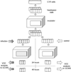
Work flow: C1R cells were placed into 12 vials and incubated. Vials were infected with influenza virus (n = 9 total, gray; n = 3 H9N2, n = 3 H6N2, and n = 3 H1N1, MOI 10) or untreated (controls; n = 3, white). After re-suspension in medium and further incubation, vials were removed at either 24 h (n = 9 gray and n = 3 white) or 48 h (n = 9 gray and n = 3 white). All vials underwent the same VOC sampling. All experiments were repeated four times. The H1N1 experiment at MOI 1 was conducted independently (n = 12 at 24-hours, n = 12 at 48-hours). Totals: virus-infected 96; controls 24; VOC chromatograms 120.
Similar articles
- Volatile scents of influenza A and S. pyogenes (co-)infected cells.
Traxler S, Barkowsky G, Saß R, Klemenz AC, Patenge N, Kreikemeyer B, Schubert JK, Miekisch W. Traxler S, et al. Sci Rep. 2019 Dec 11;9(1):18894. doi: 10.1038/s41598-019-55334-0. Sci Rep. 2019. PMID: 31827195 Free PMC article. - Identifying viral infections through analysis of head space volatile organic compounds.
Sanmark E, Marjanen P, Virtanen J, Aaltonen K, Tauriainen S, Österlund P, Mäkelä M, Saari S, Roine A, Rönkkö T, Vartiainen VA. Sanmark E, et al. J Breath Res. 2024 Oct 30;19(1). doi: 10.1088/1752-7163/ad89f0. J Breath Res. 2024. PMID: 39437816 - Volatile organic compounds generated by cultures of bacteria and viruses associated with respiratory infections.
Abd El Qader A, Lieberman D, Shemer Avni Y, Svobodin N, Lazarovitch T, Sagi O, Zeiri Y. Abd El Qader A, et al. Biomed Chromatogr. 2015 Dec;29(12):1783-90. doi: 10.1002/bmc.3494. Epub 2015 May 27. Biomed Chromatogr. 2015. PMID: 26033043 - Volatile organic compounds at swine facilities: a critical review.
Ni JQ, Robarge WP, Xiao C, Heber AJ. Ni JQ, et al. Chemosphere. 2012 Oct;89(7):769-88. doi: 10.1016/j.chemosphere.2012.04.061. Epub 2012 Jun 7. Chemosphere. 2012. PMID: 22682363 Review. - Evidence of endogenous volatile organic compounds as biomarkers of diseases in alveolar breath.
Sarbach C, Stevens P, Whiting J, Puget P, Humbert M, Cohen-Kaminsky S, Postaire E. Sarbach C, et al. Ann Pharm Fr. 2013 Jul;71(4):203-15. doi: 10.1016/j.pharma.2013.05.002. Epub 2013 Jun 17. Ann Pharm Fr. 2013. PMID: 23835018 Review.
Cited by
- Point-of-care breath sample analysis by semiconductor-based E-Nose technology discriminates non-infected subjects from SARS-CoV-2 pneumonia patients: a multi-analyst experiment.
Woehrle T, Pfeiffer F, Mandl MM, Sobtzick W, Heitzer J, Krstova A, Kamm L, Feuerecker M, Moser D, Klein M, Aulinger B, Dolch M, Boulesteix AL, Lanz D, Choukér A. Woehrle T, et al. MedComm (2020). 2024 Oct 24;5(11):e726. doi: 10.1002/mco2.726. eCollection 2024 Nov. MedComm (2020). 2024. PMID: 39465142 Free PMC article. - High and low pathogenicity avian influenza virus discrimination and prediction based on volatile organic compounds signature by SIFT-MS: a proof-of-concept study.
Filaire F, Sécula A, Bessière P, Pagès-Homs M, Guérin JL, Violleau F, Till U. Filaire F, et al. Sci Rep. 2024 Jul 24;14(1):17051. doi: 10.1038/s41598-024-67219-y. Sci Rep. 2024. PMID: 39048690 Free PMC article. - A comprehensive meta-analysis and systematic review of breath analysis in detection of COVID-19 through Volatile organic compounds.
Long GA, Xu Q, Sunkara J, Woodbury R, Brown K, Huang JJ, Xie Z, Chen X, Fu XA, Huang J. Long GA, et al. Diagn Microbiol Infect Dis. 2024 Jul;109(3):116309. doi: 10.1016/j.diagmicrobio.2024.116309. Epub 2024 Apr 27. Diagn Microbiol Infect Dis. 2024. PMID: 38692202 Review. - Tuning phosphorene and MoS2 2D materials for detecting volatile organic compounds associated with respiratory diseases.
Allosh A, Pantis-Simut CA, Filipoiu N, Preda AT, Necula G, Ghitiu I, Anghel DV, Dulea MA, Nemnes GA. Allosh A, et al. RSC Adv. 2024 Jan 8;14(3):1803-1812. doi: 10.1039/d3ra07685g. eCollection 2024 Jan 3. RSC Adv. 2024. PMID: 38192312 Free PMC article. - A feasibility study on exhaled breath analysis using UV spectroscopy to detect COVID-19.
Sutaria SR, Morris JD, Xie Z, Cooke EA, Silvers SM, Long GA, Balcom D, Marimuthu S, Parrish LW, Aliesky H, Arnold FW, Huang J, Fu XA, Nantz MH. Sutaria SR, et al. J Breath Res. 2023 Nov 2;18(1):016004. doi: 10.1088/1752-7163/ad0646. J Breath Res. 2023. PMID: 37875100 Free PMC article.
References
- Adams S, Sandrock C. Med. Princ. Pract. 2010;19:421–432. - PubMed
- Cardona CJ, Xing Z, Sandrock CE, Davis CE. Comp. Immunol. Microbiol. Infect. Dis. 2009;32:255–273. - PubMed
- Uyeki TM. N. Engl. J. Med. 2010;362:2221–2223. - PubMed
Publication types
MeSH terms
Substances
Grants and funding
- T32 ES007059/ES/NIEHS NIH HHS/United States
- 8KL2TR000134-07K12/TR/NCATS NIH HHS/United States
- T35 ES007301/ES/NIEHS NIH HHS/United States
- UL1 TR000002/TR/NCATS NIH HHS/United States
- T32-ES007059/ES/NIEHS NIH HHS/United States
- T32-HL007013/HL/NHLBI NIH HHS/United States
- T32 HL007013/HL/NHLBI NIH HHS/United States
- KL2 TR000134/TR/NCATS NIH HHS/United States
LinkOut - more resources
Full Text Sources
Other Literature Sources
Medical


