Hippo coactivator YAP1 upregulates SOX9 and endows esophageal cancer cells with stem-like properties - PubMed (original) (raw)
. 2014 Aug 1;74(15):4170-82.
doi: 10.1158/0008-5472.CAN-13-3569. Epub 2014 Jun 6.
Jaffer A Ajani 2, Soichiro Honjo 2, Dipen M Maru 3, Qiongrong Chen 2, Ailing W Scott 2, Todd R Heallen 4, Lianchun Xiao 5, Wayne L Hofstetter 6, Brian Weston 7, Jeffrey H Lee 7, Roopma Wadhwa 2, Kazuki Sudo 2, John R Stroehlein 7, James F Martin 4, Mien-Chie Hung 8, Randy L Johnson 9
Affiliations
- PMID: 24906622
- PMCID: PMC4136429
- DOI: 10.1158/0008-5472.CAN-13-3569
Hippo coactivator YAP1 upregulates SOX9 and endows esophageal cancer cells with stem-like properties
Shumei Song et al. Cancer Res. 2014.
Abstract
Cancer stem cells (CSC) are purported to initiate and maintain tumor growth. Deregulation of normal stem cell signaling may lead to the generation of CSCs; however, the molecular determinants of this process remain poorly understood. Here we show that the transcriptional coactivator YAP1 is a major determinant of CSC properties in nontransformed cells and in esophageal cancer cells by direct upregulation of SOX9. YAP1 regulates the transcription of SOX9 through a conserved TEAD binding site in the SOX9 promoter. Expression of exogenous YAP1 in vitro or inhibition of its upstream negative regulators in vivo results in elevated SOX9 expression accompanied by the acquisition of CSC properties. Conversely, shRNA-mediated knockdown of YAP1 or SOX9 in transformed cells attenuates CSC phenotypes in vitro and tumorigenicity in vivo. The small-molecule inhibitor of YAP1, verteporfin, significantly blocks CSC properties in cells with high YAP1 and a high proportion of ALDH1(+). Our findings identify YAP1-driven SOX9 expression as a critical event in the acquisition of CSC properties, suggesting that YAP1 inhibition may offer an effective means of therapeutically targeting the CSC population.
©2014 American Association for Cancer Research.
Conflict of interest statement
Disclosure of Potential Conflicts of Interest
No potential conflicts of interest were disclosed.
Figures
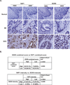
Figure 1. Nuclear YAP1 and SOX9 Expression are Positively Correlated in human Esophageal Adenocarcinoma Tissues
A. EAC tissue microarray slides were immunohistochemically stained using SOX9 and YAP1 antibodies as described in Materials & Methods. Representative YAP1 and SOX9 staining are shown in normal, BE and EAC. B. The upper table demonstrates the correlation of YAP1 combined (percentage and intensity) score vs SOX9 combined scores in EAC tissues; the lower table demonstrates the correlation of nuclear staining intensity between YAP1 and SOX9.
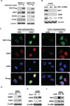
Figure 2. YAP1 Enhances SOX9 Expression in both Normal and Transformed Cells
A. SKGT-4 and KATO-TN cells were transduced with lentiviral PIN20YAP1 plasmid containing inducible YAP1 cDNA. YAP1 expression was induced by doxycycline at 1µg/ml. Immunoblotting using antibodies against Flag, YAP1, SOX9 and CTGF were performed. B. Immunoblotting of YAP1, SOX9 and CTGF was performed in JHESO cells with two independent YAP1 shRNAs. C. Immunofluorescent staining of flag-YAP and SOX9 in SKGT-4 (PIN20YAP1) and KATO-TN (PIN20YAP1) EC cells with or without doxycycline induction. D. SOX-9 and YAP1 were detected by immunoblotting in MEFs cells from Lats1/2 mutant mice and compared with that from wt mice. E. SOX9 and YAP1 were examined by immunoblotting in B299 cells with or without deletion of Sav1. F. 293T cells were transfected with either mutant YAPS127A or wt YAP1 expression vectors followed by immunoblotting to detect YAP1 and SOX9.
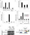
Figure 3. YAP1 Induces SOX9 Transcription and requires an intact TEAD binding site
A. SOX9 luciferase promoter activity was determined by transient transfection of SOX9 luciferase promoter reporter in SKGT-4, YES-6 and KATO-TN EC cells with or without induced YAP1. B. SOX9 promoter activity was detected in JHESO cells with or without YAP1 knockdown. C. SOX9 promoter activity was detected in 293T cells after co-transfection of YAP1, NICD, or the combination of YAP1 and Tead2. D. SOX9 promoter activity was determined in 293T cells after co-transfection of wild-type (wt) or mutant SOX9 promoter luciferase with either YAP1 or YAP1 in combination with Tead2. E. ChIP assay was performed using YAP1 and normal IgG pull down of chromatin from SKGT-4 with (DOX+) or without (DOX-) YAP1 induction using primers that amplify SOX9 promoter containing the TEAD binding site and primers that amplify a control promoter region that does not contain the TEAD binding site (Control site). F. SOX9 promoter primers designed for ChIP assays in the SOX9 promoter containing a TEAD binding site.
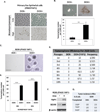
Figure 4. YAP1 Endows Sphere and Tumor Forming Capacity to Untransformed Mouse Cells
A. Representative microscopic images of primary esophageal epithelial cells (Eso) transduced with YAP1S127 cDNA (PIN20YAP1) with or without doxycycline induction at low (less than 5) or high passage (over 10). B. Representative sphere image and number in Eso cells (PIN20YAP1) with or without doxycycline induction. C. Representative images of spheres in B299 cells with YAP1 induction by doxycycline at 1µg/ml (DOX+) and without YAP1 induction (DOX-). D. Representative bar graph demonstrating the sphere numbers in the 4th and 6th generation of B299 cells with or without YAP1 induction. Data are represented as mean and SD from three experiments.***p<0.0001. E. Quantification of sphere numbers per 2500 B299 cells (PIN20YAP1) at one through ten passages with (DOX+) or without (DOX-) YAP1 induction. F. Immunoblotting analysis for YAP1, SOX9 expression in B299 cells with (DOX+) or without (DOX-) YAP1 induction. G. Tumorigenecity of B299 cells with or without YAP1 induction by subcutaneous injection into nude mice. 5 mice/group.
Figure 5. YAP1 Confers CSCs Properties in EC Cells
A. ALDH1 labeling and ALDH1+/CD44+ double labeling was performed in JHESO and KATO-TN cells as described in Materials & Methods. B. Labeling index for ALDH+ cells or double labeling for CD44+ and ALDH1+ cells in JHESO and KATO-TN cells. C. Immunoblotting for YAP1 and SOX9 was performed in JHESO and KATO-TN cells. D. Immunofluorescent staining of ALDH1 and OCT4 in JHESO and KATO-TN cells. The images on the right are the overlay of ALDH1, OCT4 and Dapi. E. Representative images of tumorspheres (top) and quantification of tumorsphere numbers (low) in KATO-TN cells with (DOX+) or without (DOX-) YAP1 induction. F. Representative images of tumorspheres (top) and quantification of tumorsphere numbers (low) in JHESO cells with YAP1 shRNA knockdown (Sh2 and Sh3) and Control. **p<0.01
Figure 6. Pharmacological Inhibition of YAP1 Suppress CSCs Properties in Vitro and Tumorigenecity in Vivo
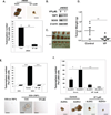
A. Images (upper) and quantification (lower) of tumorsphere numbers in JHESO cells treated with VP at 1µM and control cells. B. Immunoblotting was performed to detect YAP1 and SOX9 in JHESO cells treated with VP at 1µM. C. JHESO cells (1.5×10⁶) were injected subcutaneously in nude mice, 5 mice/group. Representative tumors after 5 weeks are shown. D. Each point represents mean tumor weight and SD from five mice. E. Quantification (upper) and images (low) of tumorspheres in B299 cells with YAP1 induction and treated with VP at 1 µM. F. Representative images (lower) and quantification of tumorspheres (upper) in ALDH1+ or ALDH1− cells sorted from JHESO EC cells. Data are represented as mean and SD from three experiments.***p<0.0001,**p<0.01, *p<0.05.
Figure 7. shRNA-Mediated Knockdown YAP1 or SOX9 inhibits CSC properties in vitro and tumorigenecity in vivo
A&B. Representative images of tumorspheres (A) and quantification of tumorsphere numbers (B) in SKGT-4 (PIN20YAP1) cells with (DOX+) or without (DOX-) YAP1 induction and further knockdown of YAP1 or SOX9. C,D&E. SKGT-4 (PIN20YAP1) cells with (DOX+) or without (DOX-) YAP1 induction and shYAP1 or shSOX9 were inoculated into nude mice (n=5 per group). Representative tumors after 6 weeks are shown (C).Tumor volume (D) and weight (E) were calculated as described in Materials & Methods. F. Immunohistochemistry for YAP1, SOX9 and Ki67 was performed in mouse tumor tissues derived from xenograft nude mice from Figure 7C. G. Proposed model by which YAP1 endows CSCs properties by up-regulating SOX9 in EC cells.
Similar articles
- Targeting Hippo coactivator YAP1 through BET bromodomain inhibition in esophageal adenocarcinoma.
Song S, Li Y, Xu Y, Ma L, Pool Pizzi M, Jin J, Scott AW, Huo L, Wang Y, Lee JH, Bhutani MS, Weston B, Shanbhag ND, Johnson RL, Ajani JA. Song S, et al. Mol Oncol. 2020 Jun;14(6):1410-1426. doi: 10.1002/1878-0261.12667. Epub 2020 Apr 7. Mol Oncol. 2020. PMID: 32175692 Free PMC article. - The Hippo Coactivator YAP1 Mediates EGFR Overexpression and Confers Chemoresistance in Esophageal Cancer.
Song S, Honjo S, Jin J, Chang SS, Scott AW, Chen Q, Kalhor N, Correa AM, Hofstetter WL, Albarracin CT, Wu TT, Johnson RL, Hung MC, Ajani JA. Song S, et al. Clin Cancer Res. 2015 Jun 1;21(11):2580-90. doi: 10.1158/1078-0432.CCR-14-2191. Epub 2015 Mar 4. Clin Cancer Res. 2015. PMID: 25739674 Free PMC article. - A Novel YAP1 Inhibitor Targets CSC-Enriched Radiation-Resistant Cells and Exerts Strong Antitumor Activity in Esophageal Adenocarcinoma.
Song S, Xie M, Scott AW, Jin J, Ma L, Dong X, Skinner HD, Johnson RL, Ding S, Ajani JA. Song S, et al. Mol Cancer Ther. 2018 Feb;17(2):443-454. doi: 10.1158/1535-7163.MCT-17-0560. Epub 2017 Nov 22. Mol Cancer Ther. 2018. PMID: 29167315 Free PMC article. - Role of Yes-associated protein 1 in gliomas: pathologic and therapeutic aspects.
Liu YC, Wang YZ. Liu YC, et al. Tumour Biol. 2015 Apr;36(4):2223-7. doi: 10.1007/s13277-015-3297-2. Epub 2015 Mar 7. Tumour Biol. 2015. PMID: 25750037 Review. - The Hippo Pathway as Drug Targets in Cancer Therapy and Regenerative Medicine.
Nagashima S, Bao Y, Hata Y. Nagashima S, et al. Curr Drug Targets. 2017;18(4):447-454. doi: 10.2174/1389450117666160112115641. Curr Drug Targets. 2017. PMID: 26758663 Review.
Cited by
- Unveiling promising targets in gastric cancer therapy: A comprehensive review.
Li W, Wei J, Cheng M, Liu M. Li W, et al. Mol Ther Oncol. 2024 Aug 5;32(3):200857. doi: 10.1016/j.omton.2024.200857. eCollection 2024 Sep 19. Mol Ther Oncol. 2024. PMID: 39280587 Free PMC article. Review. - Context-Dependent Distinct Roles of SOX9 in Combined Hepatocellular Carcinoma-Cholangiocarcinoma.
Park Y, Hu S, Kim M, Oertel M, Singhi A, Monga SP, Liu S, Ko S. Park Y, et al. Cells. 2024 Aug 29;13(17):1451. doi: 10.3390/cells13171451. Cells. 2024. PMID: 39273023 Free PMC article. - Drug Delivery Opportunities in Esophageal Cancer: Current Treatments and Future Prospects.
Sabatelle RC, Colson YL, Sachdeva U, Grinstaff MW. Sabatelle RC, et al. Mol Pharm. 2024 Jul 1;21(7):3103-3120. doi: 10.1021/acs.molpharmaceut.4c00246. Epub 2024 Jun 18. Mol Pharm. 2024. PMID: 38888089 Free PMC article. Review. - Modelling esophageal adenocarcinoma and Barrett's esophagus with patient-derived organoids.
Milne JV, Mustafa EH, Clemons NJ. Milne JV, et al. Front Mol Biosci. 2024 Apr 24;11:1382070. doi: 10.3389/fmolb.2024.1382070. eCollection 2024. Front Mol Biosci. 2024. PMID: 38721276 Free PMC article. Review. - SRY-Box transcription factor 9 triggers YAP nuclear entry via direct interaction in tumors.
Qian H, Ding CH, Liu F, Chen SJ, Huang CK, Xiao MC, Hong XL, Wang MC, Yan FZ, Ding K, Cui YL, Zheng BN, Ding J, Luo C, Zhang X, Xie WF. Qian H, et al. Signal Transduct Target Ther. 2024 Apr 24;9(1):96. doi: 10.1038/s41392-024-01805-4. Signal Transduct Target Ther. 2024. PMID: 38653754 Free PMC article.
References
Publication types
MeSH terms
Substances
Grants and funding
- P30 CA016672/CA/NCI NIH HHS/United States
- CA129906/CA/NCI NIH HHS/United States
- R01 HL118761/HL/NHLBI NIH HHS/United States
- R21 CA129906/CA/NCI NIH HHS/United States
- R01 DE023177/DE/NIDCR NIH HHS/United States
- R01 CA172741/CA/NCI NIH HHS/United States
- R01 CA138671/CA/NCI NIH HHS/United States
LinkOut - more resources
Full Text Sources
Other Literature Sources
Medical
Research Materials
Miscellaneous
