Endogenous biotin-binding proteins: an overlooked factor causing false positives in streptavidin-based protein detection - PubMed (original) (raw)
Endogenous biotin-binding proteins: an overlooked factor causing false positives in streptavidin-based protein detection
Hanne L P Tytgat et al. Microb Biotechnol. 2015 Jan.
Abstract
Biotinylation is widely used in DNA, RNA and protein probing assays as this molecule has generally no impact on the biological activity of its substrate. During the streptavidin-based detection of glycoproteins in Lactobacillus rhamnosus GG with biotinylated lectin probes, a strong positive band of approximately 125 kDa was observed, present in different cellular fractions. This potential glycoprotein reacted heavily with concanavalin A (ConA), a lectin that specifically binds glucose and mannose residues. Surprisingly, this protein of 125 kDa could not be purified using a ConA affinity column. Edman degradation of the protein, isolated via cation and anion exchange chromatography, lead to the identification of the band as pyruvate carboxylase, an enzyme of 125 kDa that binds biotin as a cofactor. Detection using only the streptavidin conjugate resulted in more false positive signals of proteins, also in extracellular fractions, indicating biotin-associated proteins. Indeed, biotin is a known cofactor of numerous carboxylases. The potential occurence of false positive bands with biotinylated protein probes should thus be considered when using streptavidin-based detection, e.g. by developing a blot using only the streptavidin conjugate. To circumvent these false positives, alternative approaches like detection based on digoxigenin labelling can also be used.
© 2014 The Authors. Microbial Biotechnology published by John Wiley & Sons Ltd and Society for Applied Microbiology.
Figures
Fig 1
Detection of glycoproteins on Western blot using biotinylated lectins and streptavidin results in false positive hits.A. Biotinylated lectin blots – The exoproteome of wild type L . rhamnosus GG and its Δ_dltD_::TcR mutant were subjected to SDS-PAGE on NuPAGE® Novex® 12% Bis-Tris gels (Life Technologies) and subsequently blotted to PVDF membranes (Life Technologies). The prestained Kaleidoscope™ ladder (Bio-Rad) was added as a molecular weight marker. The Western blots were developed using biotinylated probes, in this case lectins. In the left panel, the Western blot was probed with biotinylated ConA, which specifically binds glucose and terminal mannose. Incubation with streptavidin conjugated to alkaline phosphatase (Roche) enabled the visual detection of positive bands using NBT and BCIP. In the right panel, the blot was developed using the same principle, but initial probing was performed using a mix of biotinylated lectins: ConA (Glc, Man), GNA (Man), HHA (Man), WGA (GlcNAc), DSL (GlcNAc), UDA (GlcNAc), Nictaba (GlcNAc), RSA (Gal, GalNAc) and PNA (Gal, GalNAc). In both Western blots and for both strains, a peculiarly strong band appeared at approximately 125 kDa.B. Using a streptavidin conjugate to sample the proteome for false positive hits – The blotted exoproteome of L . rhamnosus GG and the Δ_dltD_::TcR mutant were probed directly with a streptavidin conjugate. This resulted in the appearance of several bands, among which the strong 125 kDa band. Based on this result, we suggest that the band is not caused by a glycoprotein, but is a false positive.C. PAS glycostain does not react with the 125 kDa protein – An SDS-PAGE gel of the proteome of L . rhamnosus GG was post-stained with Periodic Acid Schiff base stain (PAS, Pro-Q® Emerald 488 stain, Life Technologies), a method to specifically stain glycosylated proteins in a gel. At 125 kDa, no band could be detected, which further supports our hypothesis that the 125 kDa signal perceived on the lectin blots is the result of a false positive hit.
Fig 2
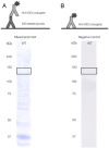
The digoxigenin–anti-digoxigenin detection as an alternative to avoid false positive hits caused by proteins binding endogenous biotin.A. DIG-labelled lectin blots – The wild type exoproteome of L . rhamnosus GG was Western blotted and developed using a mix of DIG-labelled lectins: ConA (Glc, Man), GNA (Man), HHA (Man), WGA (GlcNAc), DSL (GlcNAc), UDA (GlcNAc), Nictaba (GlcNAc), RSA (Gal, GalNAc) and PNA (Gal, GalNAc). These lectins were labelled using digoxigenin-3-O-methyl-ε-aminocaproix acid-N-hydroxysuccinimide ester (Roche). Anti-DIG Fab antibody fragments (Roche) were used to detect proteins that reacted positively with the lectin probes. Here, we clearly see that the false positive band at 125 kDa is absent.B. Negative control with anti-DIG – Direct application of the anti-DIG Fab antibody fragments (Roche) to the Western blotted proteome of L . rhamnosus GG results in a blot on which no bands can be perceived. This confirms that the DIG–anti-DIG detection method is a good alternative for the biotin–streptavidin system, without causing false positive hits.
Similar articles
- Detection of endogenous biotin-containing proteins in bone and cartilage cells with streptavidin systems.
Praul CA, Brubaker KD, Leach RM, Gay CV. Praul CA, et al. Biochem Biophys Res Commun. 1998 Jun 18;247(2):312-4. doi: 10.1006/bbrc.1998.8757. Biochem Biophys Res Commun. 1998. PMID: 9642122 - Pre-hybridisation: an efficient way of suppressing endogenous biotin-binding activity inherent to biotin-streptavidin detection system.
Ahmed R, Spikings E, Zhou S, Thompsett A, Zhang T. Ahmed R, et al. J Immunol Methods. 2014 Apr;406:143-7. doi: 10.1016/j.jim.2014.03.010. Epub 2014 Mar 19. J Immunol Methods. 2014. PMID: 24657589 - Stable, high-affinity streptavidin monomer for protein labeling and monovalent biotin detection.
Lim KH, Huang H, Pralle A, Park S. Lim KH, et al. Biotechnol Bioeng. 2013 Jan;110(1):57-67. doi: 10.1002/bit.24605. Epub 2012 Aug 8. Biotechnol Bioeng. 2013. PMID: 22806584 - [(Biotin-Ala-Ser-Lys-Lys-Pro-Lys-Arg-Asn-Ile-Lys-Ala)4-dendrimer]-streptavidin-[biotin-gadolinium 1,4,7-tris(carboxymethyl)-10-(2’-hydroxypropyl)-1,4,7,10-tetraazacyclododecane-loaded apoferritin].
Zhang H. Zhang H. 2007 Dec 27 [updated 2008 Feb 4]. In: Molecular Imaging and Contrast Agent Database (MICAD) [Internet]. Bethesda (MD): National Center for Biotechnology Information (US); 2004–2013. 2007 Dec 27 [updated 2008 Feb 4]. In: Molecular Imaging and Contrast Agent Database (MICAD) [Internet]. Bethesda (MD): National Center for Biotechnology Information (US); 2004–2013. PMID: 20641469 Free Books & Documents. Review. - Streptavidin-biotin technology: improvements and innovations in chemical and biological applications.
Dundas CM, Demonte D, Park S. Dundas CM, et al. Appl Microbiol Biotechnol. 2013 Nov;97(21):9343-53. doi: 10.1007/s00253-013-5232-z. Epub 2013 Sep 22. Appl Microbiol Biotechnol. 2013. PMID: 24057405 Review.
Cited by
- Supramolecular latching system based on ultrastable synthetic binding pairs as versatile tools for protein imaging.
Kim KL, Sung G, Sim J, Murray J, Li M, Lee A, Shrinidhi A, Park KM, Kim K. Kim KL, et al. Nat Commun. 2018 Apr 27;9(1):1712. doi: 10.1038/s41467-018-04161-4. Nat Commun. 2018. PMID: 29703887 Free PMC article. - Endogenous TOM20 Proximity Labeling: A Swiss-Knife for the Study of Mitochondrial Proteins in Human Cells.
Meurant S, Mauclet L, Dieu M, Arnould T, Eyckerman S, Renard P. Meurant S, et al. Int J Mol Sci. 2023 May 31;24(11):9604. doi: 10.3390/ijms24119604. Int J Mol Sci. 2023. PMID: 37298552 Free PMC article. - Formulations for Bacteriophage Therapy and the Potential Uses of Immobilization.
Rosner D, Clark J. Rosner D, et al. Pharmaceuticals (Basel). 2021 Apr 13;14(4):359. doi: 10.3390/ph14040359. Pharmaceuticals (Basel). 2021. PMID: 33924739 Free PMC article. Review. - Prodrug-conjugated tumor-seeking commensals for targeted cancer therapy.
Shen H, Zhang C, Li S, Liang Y, Lee LT, Aggarwal N, Wun KS, Liu J, Nadarajan SP, Weng C, Ling H, Tay JK, Wang Y, Yao SQ, Hwang IY, Lee YS, Chang MW. Shen H, et al. Nat Commun. 2024 May 21;15(1):4343. doi: 10.1038/s41467-024-48661-y. Nat Commun. 2024. PMID: 38773197 Free PMC article. - Probiotic Gut Microbiota Isolate Interacts with Dendritic Cells via Glycosylated Heterotrimeric Pili.
Tytgat HL, van Teijlingen NH, Sullan RM, Douillard FP, Rasinkangas P, Messing M, Reunanen J, Satokari R, Vanderleyden J, Dufrêne YF, Geijtenbeek TB, de Vos WM, Lebeer S. Tytgat HL, et al. PLoS One. 2016 Mar 17;11(3):e0151824. doi: 10.1371/journal.pone.0151824. eCollection 2016. PLoS One. 2016. PMID: 26985831 Free PMC article.
References
- Chaiet L. Wolf FJ. The properties of streptavidin, a biotin-binding protein produced by streptomycetes. Arch Biochem Biophys. 1964;106:1–5. - PubMed
- Chapman-Smith A. Cronan JE., Jr Molecular biology of biotin attachment to proteins. J Nutr. 1999;129(2S Suppl):477S–484S. - PubMed
- Chevalier J, Yi J, Michel O. Tang XM. Biotin and digoxigenin as labels for light and electron microscopy in situ hybridization probes: where do we stand? J Histochem Cytochem. 1997;45:481–491. - PubMed
- Coyne MJ, Reinap B, Lee MM. Comstock LE. Human symbionts use a host-like pathway for surface fucosylation. Science. 2005;307:1778–1781. - PubMed
Publication types
MeSH terms
Substances
LinkOut - more resources
Full Text Sources
Other Literature Sources

