PINK1 and Parkin – mitochondrial interplay between phosphorylation and ubiquitylation in Parkinson's disease - PubMed (original) (raw)
Review
. 2015 Jan;282(2):215-23.
doi: 10.1111/febs.13127. Epub 2014 Nov 20.
Affiliations
- PMID: 25345844
- PMCID: PMC4368378
- DOI: 10.1111/febs.13127
Review
PINK1 and Parkin – mitochondrial interplay between phosphorylation and ubiquitylation in Parkinson's disease
Agne Kazlauskaite et al. FEBS J. 2015 Jan.
Abstract
The discovery of mutations in genes encoding protein kinase PTEN-induced kinase 1 (PINK1) and E3 ubiquitin ligase Parkin in familial Parkinson's disease and their association with mitochondria provides compelling evidence that mitochondrial dysfunction is a major contributor to neurodegeneration in Parkinson's disease. In recent years, tremendous progress has been made in the understanding of how PINK1 and Parkin enzymes are regulated and how they influence downstream mitochondrial signalling processes. We provide a critical overview of the key advances in the field and also discuss the outstanding questions, including novel ways in which this knowledge could be exploited to develop therapies against Parkinson's disease.
Keywords: PINK1; Parkinson's disease; kinase; mitochondria; parkin; ubiquitin.
© 2014 The Authors. FEBS Journal published by John Wiley & Sons Ltd on behalf of FEBS.
Figures
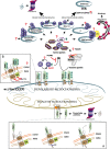
Figure 1
PINK1-Parkin signalling. Under healthy conditions, PINK1 is imported to the mitochondria via a multisubunit complex, including TOM40 on the outer mitochondrial membrane (OMM) and TIM23 in the inner mitochondrial membrane (IMM). Upon entry, PINK1 is sequentially cleaved by proteases MPP and PARL between residues 103 and 104. The resulting C-terminal fragment is rapidly degraded via the N-end rule pathway (inset A). Upon mitochondrial depolarization (e.g. induced by uncouplers), mitochondrial import and cleavage by PARL is inhibited, resulting in PINK1 stabilization and accumulation at the OMM, which in turn leads to PINK1 autophosphorylation and activation (inset B). Parkin exists in an inactive state mediated by multiple autoinhibitory surface interactions. Upon activation, PINK1 phosphorylates Parkin at Ser65 within its Ubl domain and ubiquitin at Ser65. Phosphorylation of Parkin Ubl Ser65 and binding of ubiquitin Ser65 confers maximal activation of Parkin E3 ubiquitin ligase activity. Multiple Parkin substrates have been identified, implicating its role in distinct mitochondrial signalling processes, although only a few substrates have been characterized in detail. Parkin-dependent ubiquitylated OMM substrates interact with ubiquitin binding domain-containing proteins (UBD) e.g. p62 that stimulate recruitment of autophagy machinery to induce mitophagy. Targeting of mitochondrial GTPases Miro1 and Mfn1/2 may influence mitochondrial transport and dynamics, respectively. The negative regulators of this pathway remain largely unknown, although recent work has suggested roles for USP15 and USP30 as deubiquitylases.
Similar articles
- The endoplasmic reticulum/mitochondria interface: a subcellular platform for the orchestration of the functions of the PINK1-Parkin pathway?
Erpapazoglou Z, Corti O. Erpapazoglou Z, et al. Biochem Soc Trans. 2015 Apr;43(2):297-301. doi: 10.1042/BST20150008. Biochem Soc Trans. 2015. PMID: 25849933 Review. - PTEN-induced kinase 1 (PINK1) and Parkin: Unlocking a mitochondrial quality control pathway linked to Parkinson's disease.
Agarwal S, Muqit MMK. Agarwal S, et al. Curr Opin Neurobiol. 2022 Feb;72:111-119. doi: 10.1016/j.conb.2021.09.005. Epub 2021 Oct 27. Curr Opin Neurobiol. 2022. PMID: 34717133 Review. - PINK1, Parkin, and Mitochondrial Quality Control: What can we Learn about Parkinson's Disease Pathobiology?
Truban D, Hou X, Caulfield TR, Fiesel FC, Springer W. Truban D, et al. J Parkinsons Dis. 2017;7(1):13-29. doi: 10.3233/JPD-160989. J Parkinsons Dis. 2017. PMID: 27911343 Free PMC article. Review. - PINK1/PARKIN signalling in neurodegeneration and neuroinflammation.
Quinn PMJ, Moreira PI, Ambrósio AF, Alves CH. Quinn PMJ, et al. Acta Neuropathol Commun. 2020 Nov 9;8(1):189. doi: 10.1186/s40478-020-01062-w. Acta Neuropathol Commun. 2020. PMID: 33168089 Free PMC article. Review. - Pink1, Parkin, DJ-1 and mitochondrial dysfunction in Parkinson's disease.
Dodson MW, Guo M. Dodson MW, et al. Curr Opin Neurobiol. 2007 Jun;17(3):331-7. doi: 10.1016/j.conb.2007.04.010. Epub 2007 May 11. Curr Opin Neurobiol. 2007. PMID: 17499497 Review.
Cited by
- Ataxia-telangiectasia mutated interacts with Parkin and induces mitophagy independent of kinase activity. Evidence from mantle cell lymphoma.
Sarkar A, Stellrecht CM, Vangapandu HV, Ayres M, Kaipparettu BA, Park JH, Balakrishnan K, Burks JK, Pandita TK, Hittelman WN, Neelapu SS, Gandhi V. Sarkar A, et al. Haematologica. 2021 Feb 1;106(2):495-512. doi: 10.3324/haematol.2019.234385. Haematologica. 2021. PMID: 32029507 Free PMC article. - Aminochrome Induces Irreversible Mitochondrial Dysfunction by Inducing Autophagy Dysfunction in Parkinson's Disease.
Segura-Aguilar J, Huenchuguala S. Segura-Aguilar J, et al. Front Neurosci. 2018 Mar 13;12:106. doi: 10.3389/fnins.2018.00106. eCollection 2018. Front Neurosci. 2018. PMID: 29593482 Free PMC article. No abstract available. - Cardiomyocyte Death and Genome-Edited Stem Cell Therapy for Ischemic Heart Disease.
Cho HM, Cho JY. Cho HM, et al. Stem Cell Rev Rep. 2021 Aug;17(4):1264-1279. doi: 10.1007/s12015-020-10096-5. Epub 2021 Jan 25. Stem Cell Rev Rep. 2021. PMID: 33492627 Free PMC article. Review. - Deciphering the dual role and prognostic potential of PINK1 across cancer types.
Dai K, Radin DP, Leonardi D. Dai K, et al. Neural Regen Res. 2021 Apr;16(4):659-665. doi: 10.4103/1673-5374.295314. Neural Regen Res. 2021. PMID: 33063717 Free PMC article. - Generation and Release of Mitochondrial-Derived Vesicles in Health, Aging and Disease.
Picca A, Guerra F, Calvani R, Coelho-Junior HJ, Bossola M, Landi F, Bernabei R, Bucci C, Marzetti E. Picca A, et al. J Clin Med. 2020 May 12;9(5):1440. doi: 10.3390/jcm9051440. J Clin Med. 2020. PMID: 32408624 Free PMC article. Review.
References
- Schulz JB. Beal MF. Mitochondrial dysfunction in movement disorders. Curr Opin Neurol. 1994;7:333–339. &. - PubMed
- Langston JW, Ballard P, Tetrud JW. Irwin I. Chronic Parkinsonism in humans due to a product of meperidine-analog synthesis. Science. 1983;219:979–980. &. - PubMed
- Nicklas WJ, Vyas I. Heikkila RE. Inhibition of NADH-linked oxidation in brain mitochondria by 1-methyl-4-phenyl-pyridine, a metabolite of the neurotoxin, 1-methyl-4-phenyl-1,2,5,6-tetrahydropyridine. Life Sci. 1985;36:2503–2508. &. - PubMed
- Mizuno Y, Ohta S, Tanaka M, Takamiya S, Suzuki K, Sato T, Oya H, Ozawa T. Kagawa Y. Deficiencies in complex I subunits of the respiratory chain in Parkinson's disease. Biochem Biophys Res Commun. 1989;163:1450–1455. &. - PubMed
- Schapira AH, Cooper JM, Dexter D, Jenner P, Clark JB. Marsden CD. Mitochondrial complex I deficiency in Parkinson's disease. Lancet. 1989;1:1269. &. - PubMed
Publication types
MeSH terms
Substances
Grants and funding
- 101022/WT_/Wellcome Trust/United Kingdom
- G-1506/PUK_/Parkinson's UK/United Kingdom
- MRC_/Medical Research Council/United Kingdom
- 101022/Z/13/Z/WT_/Wellcome Trust/United Kingdom
LinkOut - more resources
Full Text Sources
Other Literature Sources
Medical
Research Materials
