The vigorous immune microenvironment of microsatellite instable colon cancer is balanced by multiple counter-inhibitory checkpoints - PubMed (original) (raw)
doi: 10.1158/2159-8290.CD-14-0863. Epub 2014 Oct 30.
Michael Cruise 2, Ada Tam 3, Elizabeth C Wicks 4, Elizabeth M Hechenbleikner 4, Janis M Taube 2, Richard L Blosser 3, Hongni Fan 1, Hao Wang 5, Brandon S Luber 5, Ming Zhang 6, Nickolas Papadopoulos 6, Kenneth W Kinzler 6, Bert Vogelstein 6, Cynthia L Sears 7, Robert A Anders 2, Drew M Pardoll 8, Franck Housseau 9
Affiliations
- PMID: 25358689
- PMCID: PMC4293246
- DOI: 10.1158/2159-8290.CD-14-0863
The vigorous immune microenvironment of microsatellite instable colon cancer is balanced by multiple counter-inhibitory checkpoints
Nicolas J Llosa et al. Cancer Discov. 2015 Jan.
Abstract
We examined the immune microenvironment of primary colorectal cancer using immunohistochemistry, laser capture microdissection/qRT-PCR, flow cytometry, and functional analysis of tumor-infiltrating lymphocytes. A subset of colorectal cancer displayed high infiltration with activated CD8(+) cytotoxic T lymphocyte (CTL) as well as activated Th1 cells characterized by IFNγ production and the Th1 transcription factor TBET. Parallel analysis of tumor genotypes revealed that virtually all of the tumors with this active Th1/CTL microenvironment had defects in mismatch repair, as evidenced by microsatellite instability (MSI). Counterbalancing this active Th1/CTL microenvironment, MSI tumors selectively demonstrated highly upregulated expression of multiple immune checkpoints, including five-PD-1, PD-L1, CTLA-4, LAG-3, and IDO-currently being targeted clinically with inhibitors. These findings link tumor genotype with the immune microenvironment, and explain why MSI tumors are not naturally eliminated despite a hostile Th1/CTL microenvironment. They further suggest that blockade of specific checkpoints may be selectively efficacious in the MSI subset of colorectal cancer.
Significance: The findings reported in this article are the first to demonstrate a link between a genetically defined subtype of cancer and its corresponding expression of immune checkpoints in the tumor microenvironment. The mismatch repair-defective subset of colorectal cancer selectively upregulates at least five checkpoint molecules that are targets of inhibitors currently being clinically tested.
©2014 American Association for Cancer Research.
Figures
Figure 1. Geographic distribution in situ of MSI and MSS CRC-infiltrating lymphocytes
FFPE tissue sections were characterized by IHC for CD4+, CD8+ and Foxp3+ cell infiltration. Three distinct histological areas designated as tumor infiltrating lymphocytes (TIL), tumor stroma and invasive front (where tumor invaded normal lamina propria) were histologically identified and separately analyzed. Invasive front (A) and TIL/Stroma (B) areas of a representative MSS (bottom panel) and MSI (top panel) are shown (20X magnification). Dashed lines in (A) delineate the invasive front with the tumor tissue on the top side and the normal tissue on the bottom side. Red stars and blue arrows in (B) indicate the tumor stroma and tumor epithelium-infiltrating immune cells, respectively. Scale bars, 100μm. C, cell density was scored in 14 MSS (blue) and 9 MSI (red) specimens by determining the average number of stained cells in 5 distinct hpf (0.0028mm2/hpf). The graphs display the mean for each group and * indicates statistically significant differences between MSS and MSI (p<0.05 using Mann-Whitney U test).
Figure 2. Th1 and CTL based immune signature and elevated checkpoint expression in MSI CRC
RNA was extracted from tissue samples laser-microdissected representing TIL in tumor nests (A), stroma surrounding tumor (B) and invasive front (C) areas of MSS (blue squares) and MSI (red circles) CRC specimens. Immune-related gene expression profiles were assessed using Taqman-based qRT-PCR for selected genes. Sets of genes were defined by functional relevance (Th1/Tc1, CTL, Th17, Treg, pro-inflammation, immune checkpoints and metabolism). The Y axis represents an arbitrary unit of expression 2−ΔCt, ΔCt representing cycle threshold (Ct) of the gene of interest normalized by Ct of ubiquitous genes (GUSB, GAPDH). The graphs display the geometric means. Their differential representation between MSS and MSI specimens was analyzed using adjusted Wilcoxon Mann Whitney test as described in Methods section. *, Wilcoxon p-value < 0.05. D, gene group comparison in TIL, tumor stroma and invasive front areas between MSS and MSI specimens.Permutation Test Results based on the maximum Wilcoxon Mann Whitney test statistic within the gene groups Th1/Tc1, CTL, Th17 and immune checkpoints. * indicates statistically significant differences between MSS and MSI (p<0.05).
Figure 3. PD-1 and LAG-3 expression in MSI and MSS CRC specimens
IHC analysis of PD-1 and LAG-3 expression in invasive front (A) and TIL/Stroma (B) areas was performed on FFPE tissue sections of a representative set of MSI (top panel) and MSS (bottom panel) CRC specimens. Magnification, X20; Scale bars, 100μM; Red stars and blue arrows in (B) indicate the tumor stroma and tumor epithelium infiltrating immune cells, respectively.
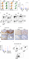
Fig. 4. MSI CRC are characterized by IFN-γ-producing PD1hi TIL and PDL-1+ tumor-infiltrating myeloid cells
A, Freshly dissociated MSS and MSI colon tumors (T) as well as patient-matched normal tissue (N) were assessed by MFC for the expression of PD-1 on infiltrating CD4+ and CD8+ T cells. PD-1 expression in tumor was normalized to the normal tissue ran simultaneously and both histograms were aligned to delineate in tumor samples the PD1hi cells when compared to normal tissue. B, proportion of PD-1hi CD4+ and CD8+ cells among CD3+ lymphocytes infiltrating MSS (blue squares) and MSI (red circles) specimens. In each group the mean is indicated and * represents statistically significant differences between MSS and MSI (*p<0.05, ****p<0.0001; non-parametric Mann-Whitney U test). C, representative intracellular staining for IFN-γ production by in vitro PMA/ionomycin activated T cells (3hrs). The dot plots show the co-expression of PD-1 and IFN-γ in CD4+ T cells and CD8+ T cells in a representative set of MSS (Left) and MSI (Right) CRC (Top) and patient-matched normal (Bottom) specimens. The gates delineate PD1hi and PD1lo cells. D, co-localization of CD163 and PD-L1 expression in invasive front (left panel) and TIL/Stroma (right panel) areas of a representative set of MSS (lower panels) and MSI (upper panel) CRC specimens were assessed by IHC; x20 magnification. Scale bars, 100μM. Red stars indicate the tumor stroma. E, PD-L1 expression scores in 7 MSS (blue) and 7 MSI (red) CRC specimens (average of 5 hpf per sample). F, MFC analysis of PD-L1 expression on MSI CRC-infiltrating myeloid cells. Dot plots represent the expression of myeloid associated markers on CD11b+HLA-DR−/low cells. Infiltrating myeloid cells were characterized as CD15−CD14+CD33+CD11c+ cells. PD-L1 expression (dark gray) is overlaid with corresponding isotype control (light gray).
Comment in
- The microsatellite instable subset of colorectal cancer is a particularly good candidate for checkpoint blockade immunotherapy.
Xiao Y, Freeman GJ. Xiao Y, et al. Cancer Discov. 2015 Jan;5(1):16-8. doi: 10.1158/2159-8290.CD-14-1397. Cancer Discov. 2015. PMID: 25583798 Free PMC article.
Similar articles
- The microsatellite instable subset of colorectal cancer is a particularly good candidate for checkpoint blockade immunotherapy.
Xiao Y, Freeman GJ. Xiao Y, et al. Cancer Discov. 2015 Jan;5(1):16-8. doi: 10.1158/2159-8290.CD-14-1397. Cancer Discov. 2015. PMID: 25583798 Free PMC article. - New insights into the inflamed tumor immune microenvironment of gastric cancer with lymphoid stroma: from morphology and digital analysis to gene expression.
Gullo I, Oliveira P, Athelogou M, Gonçalves G, Pinto ML, Carvalho J, Valente A, Pinheiro H, Andrade S, Almeida GM, Huss R, Das K, Tan P, Machado JC, Oliveira C, Carneiro F. Gullo I, et al. Gastric Cancer. 2019 Jan;22(1):77-90. doi: 10.1007/s10120-018-0836-8. Epub 2018 May 19. Gastric Cancer. 2019. PMID: 29779068 - Cytolytic activity correlates with the mutational burden and deregulated expression of immune checkpoints in colorectal cancer.
Zaravinos A, Roufas C, Nagara M, de Lucas Moreno B, Oblovatskaya M, Efstathiades C, Dimopoulos C, Ayiomamitis GD. Zaravinos A, et al. J Exp Clin Cancer Res. 2019 Aug 20;38(1):364. doi: 10.1186/s13046-019-1372-z. J Exp Clin Cancer Res. 2019. PMID: 31429779 Free PMC article. - Why is immunotherapy effective (or not) in patients with MSI/MMRD tumors?
Nebot-Bral L, Coutzac C, Kannouche PL, Chaput N. Nebot-Bral L, et al. Bull Cancer. 2019 Feb;106(2):105-113. doi: 10.1016/j.bulcan.2018.08.007. Epub 2018 Oct 17. Bull Cancer. 2019. PMID: 30342749 Review. - Tumor-Infiltrating Lymphocytes in Colorectal Cancer: The Fundamental Indication and Application on Immunotherapy.
Bai Z, Zhou Y, Ye Z, Xiong J, Lan H, Wang F. Bai Z, et al. Front Immunol. 2022 Jan 14;12:808964. doi: 10.3389/fimmu.2021.808964. eCollection 2021. Front Immunol. 2022. PMID: 35095898 Free PMC article. Review.
Cited by
- Systemic Inflammatory Markers of Resectable Colorectal Cancer Patients with Different Mismatch Repair Gene Status.
Li J, Zhang Y, Xu Q, Wang G, Jiang L, Wei Q, Luo C, Chen L, Ying J. Li J, et al. Cancer Manag Res. 2021 Mar 30;13:2925-2935. doi: 10.2147/CMAR.S298885. eCollection 2021. Cancer Manag Res. 2021. PMID: 33833576 Free PMC article. - Role of DNA repair defects in predicting immunotherapy response.
Zhang J, Shih DJH, Lin SY. Zhang J, et al. Biomark Res. 2020 Jun 29;8:23. doi: 10.1186/s40364-020-00202-7. eCollection 2020. Biomark Res. 2020. PMID: 32612833 Free PMC article. Review. - Targeting MEK/COX-2 axis improve immunotherapy efficacy in dMMR colorectal cancer with PIK3CA overexpression.
Peng K, Liu Y, Liu S, Wang Z, Zhang H, He W, Jin Y, Wang L, Xia X, Xia L. Peng K, et al. Cell Oncol (Dordr). 2024 Jun;47(3):1043-1058. doi: 10.1007/s13402-024-00916-y. Epub 2024 Feb 5. Cell Oncol (Dordr). 2024. PMID: 38315285 - Molecular cartography uncovers evolutionary and microenvironmental dynamics in sporadic colorectal tumors.
Heiser CN, Simmons AJ, Revetta F, McKinley ET, Ramirez-Solano MA, Wang J, Kaur H, Shao J, Ayers GD, Wang Y, Glass SE, Tasneem N, Chen Z, Qin Y, Kim W, Rolong A, Chen B, Vega PN, Drewes JL, Markham NO, Saleh N, Nikolos F, Vandekar S, Jones AL, Washington MK, Roland JT, Chan KS, Schürpf T, Sears CL, Liu Q, Shrubsole MJ, Coffey RJ, Lau KS. Heiser CN, et al. Cell. 2023 Dec 7;186(25):5620-5637.e16. doi: 10.1016/j.cell.2023.11.006. Cell. 2023. PMID: 38065082 Free PMC article. - Immunotherapy for Early Stage Colorectal Cancer: A Glance into the Future.
Cohen R, Shi Q, André T. Cohen R, et al. Cancers (Basel). 2020 Jul 21;12(7):1990. doi: 10.3390/cancers12071990. Cancers (Basel). 2020. PMID: 32708216 Free PMC article. Review.
References
- Fridman WH, Pages F, Sautes-Fridman C, Galon J. The immune contexture in human tumours: impact on clinical outcome. Nat Rev Cancer. 2012;12:298–306. - PubMed
- Galon J, Costes A, Sanchez-Cabo F, Kirilovsky A, Mlecnik B, Lagorce-Pages C, et al. Type, density, and location of immune cells within human colorectal tumors predict clinical outcome. Science. 2006;313:1960–4. - PubMed
- Tosolini M, Kirilovsky A, Mlecnik B, Fredriksen T, Mauger S, Bindea G, et al. Clinical impact of different classes of infiltrating T cytotoxic and helper cells (Th1, th2, treg, th17) in patients with colorectal cancer. Cancer Res. 2011;71:1263–71. - PubMed
- Shankaran V, Ikeda H, Bruce AT, White JM, Swanson PE, Old LJ, et al. IFNgamma and lymphocytes prevent primary tumour development and shape tumour immunogenicity. Nature. 2001;410:1107–11. - PubMed
Publication types
MeSH terms
Grants and funding
- 5T32CA126607-05/CA/NCI NIH HHS/United States
- P50 CA062924/CA/NCI NIH HHS/United States
- P30 CA006973/CA/NCI NIH HHS/United States
- P30 DK089502/DK/NIDDK NIH HHS/United States
- R01 CA151393/CA/NCI NIH HHS/United States
- T32 CA126607/CA/NCI NIH HHS/United States
- K08 DK087856/DK/NIDDK NIH HHS/United States
LinkOut - more resources
Full Text Sources
Other Literature Sources
Research Materials


