A noncanonical Frizzled2 pathway regulates epithelial-mesenchymal transition and metastasis - PubMed (original) (raw)
A noncanonical Frizzled2 pathway regulates epithelial-mesenchymal transition and metastasis
Taranjit S Gujral et al. Cell. 2014.
Abstract
Wnt signaling plays a critical role in embryonic development, and genetic aberrations in this network have been broadly implicated in colorectal cancer. We find that the Wnt receptor Frizzled2 (Fzd2) and its ligands Wnt5a/b are elevated in metastatic liver, lung, colon, and breast cancer cell lines and in high-grade tumors and that their expression correlates with markers of epithelial-mesenchymal transition (EMT). Pharmacologic and genetic perturbations reveal that Fzd2 drives EMT and cell migration through a previously unrecognized, noncanonical pathway that includes Fyn and Stat3. A gene signature regulated by this pathway predicts metastasis and overall survival in patients. We have developed an antibody to Fzd2 that reduces cell migration and invasion and inhibits tumor growth and metastasis in xenografts. We propose that targeting this pathway could provide benefit for patients with tumors expressing high levels of Fzd2 and Wnt5a/b.
Figures
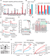
Figure 1. Fzd2 and its cognate ligands Wnt5a/b are overexpressed in late stage cancers and their expression correlates with mesenchymal markers
A. Heatmaps showing correlation of Fzd2 and its ligands Wnt5a/b with mesenchymal markers in 59 breast, 62 colon, 28 liver and 186 lung cancer cell lines. B. Bar graph showing Fzd2 mRNA expression is significantly increased in late stages (Stage III and IV) of primary liver and lung cancers compared with normal tissue (P<0.05). C. Fzd2 regulates cell migration. Top, Relative wound density (RWD) of Fzd2-shRNA or control-shRNA (sh-Ctl) expressing FOCUS and BT549 mesenchymal cells. Bottom, RWD of Fzd2-expressing or control-vector expressing Huh7 and Dld1 epithelial cells. RWD is a measure of the spatial cell density in the wound area relative to the spatial cell density outside of the wound area at every time point. D. Fzd2 signaling regulates EMT program. Representative images showing expression of Fzd2 in Huh7 cells decreased levels of epithelial markers, E-cadherin and Occludin and increased levels of mesenchymal markers, Foxc1 and Vimentin. Blue-nucleus stain. E. Volume plot of 75 EMT genes measured by qPCR in FOCUS cells expressing Ctl-shRNA or Fzd2-shRNA (left) or Huh7 cells expressing vector only or Fzd2 expression vector (right). A set of genes which were significantly downregulated (p<0.05) upon knockdown of Fzd2 are shown in red while significantly upregulated (p<0.05) genes are shown in green. See also Figure S1, S2
Figure 2. Stat3 is a key mediator of Fzd2-mediated downstream signaling, EMT program and cellular migration
A. Comparison of 45 different signal transduction pathways in FOCUS cells transfected with Fzd2 or control shRNA using a 45-transcription factor reporter array. Signaling pathways which showed significant change in Fzd2 knockdown samples are indicated. Neg and Pos denotes negative and positive luciferase controls. B. Bar graph showing increase in transcription activity of Stat3 upon Wnt5a stimulation in Fzd2-expressing Huh7 cells. C. Bar graph showing decrease in phosphorylation of Stat3, Erk1 and Mek1 upon Fzd2 knockdown in FOCUS cells. The relative phosphorlation of Akt (Ser473) is unchanged in Fzd2-shRNA expressing cells. D. Wnt5a stimulation increases phosphorylation of Stat3, Erk and Mek in a Fzd2-dependent manner. E. Treatment with Stat3 inhibitor reduces FOCUS cell migration. Dose response curves showing EC50 (50% reduction in cell migration compared with DMSO control) in FOCUS, and SNU449 liver cancer cell lines treated with Stat3 or Mek inhibitors. F. Western blots showing Fzd2 and Stat3 associate in a co-immunoprecipitation assay. Lysates immunoblotted with anti-Stat3, and anti-Fzd2 are also shown. G. Perturbing Stat3 expression reverses EMT in FOCUS cells. Bar graph showing expression of epithelial and mesenchymal marker genes in FOCUS cells with knockdown of Stat3. H. Stat3 activity regulates cell migration. Knocking down expression of Stat3 decreases Fzd2-mediated cell migration in FOCUS cells (left) while expression of constitutively active Stat3 (Stat3C) increased migration of Dld1 epithelial cells (middle). Treatment with Stattic (Stat3 inhibitor) decreased migration of Fzd2 over-expressing Huh7 cells (right) in a dose dependent manner. See also Figures S3, S4, S5 and Table S1
Figure 3. Fyn kinase is critical regulator of Fzd2-mediated Stat3 activity
A. Identification of informative kinases in Fzd2-mediated cell migration using Kinome Regularization. Plot show LOOCV error using elastic net regularization fit. The error bars represent cross-validation error plus 1 SD. The kinases identified at absolute minima (blue dashed line) were termed the most informative kinases. B. Evolution of regression coefficients. Plot showing regression coefficients for Fyn kinase against value of elasticnet penalty α. Nonzero regression coefficients for kinases picked at α >0.5 (gray region) are considered significantly informative. C. Src family kinase inhibitor reduces Fzd2-mediated cellular migration. Relative wound density of cancer cells treated with varying concentration of Dasatinib was monitored for 96 h. Dose-response curves of Dastatinib treatment in seven cancer cell lines and respective EC50 are shown. D. Knockdown of Fzd2 expression reduces phosphorylation of Src Family Kinases in FOCUS cells while overexpression of Fzd2 increases Src phosphorylation in Huh7 cells. E. Fyn kinase phosphorylates Stat3. Western blots showing phosphorylation of Stat3 upon knockdown of Fyn in FOCUS cells. F. Wnt5-Fzd2-dependent Stat3 transcription activity can be rescued by overexpression of active Src in Fzd2 or Fyn knockdown cells. G. Overexpression of active Src Family Kinase (SrcY527F) in Huh7 cells increased transcriptional activity of Stat3. See also Figures S6
Figure 4. Fyn regulates Fzd2-mediated EMT program and cellular migration
A. Perturbing Fzd2-dependent Fyn activity reverses EMT. Plots showing mRNA expression of selected EMT genes measured by quantitative PCR in FOCUS cells expressing shRNA against Fyn or (B) Huh7 cells expressing active Src (Src Y527F). C. Representative images showing expression of active Src in Huh7 cells decreased levels of epithelial markers, E-cadherin and Occludin and increased levels of mesenchymal markers, Foxc1, Slug and Vimentin. Blue-nucleus stain. Scale bar; 100 pixels. D. Heat map showing affect of Fyn inhibitor (Dasatinib) on expression of EMT-associated genes. E, F. Fyn-shRNA showed significant decrease in Fzd2-mediated cell migration in FOCUS cells while expression of SrcY527F increases cell migration in Huh7 cells. Treatment with Stattic (Stat3 inhibitor) decreased migration of Huh7 cells expressing SrcY527F to the wild-type Huh7 levels. See also Figure S6
Figure 5. Fzd2 is tyrosine phosphorylated and directly associate with Fyn-SH2 domain
A. Schematics showing domain structures of Fyn-kinase and Fzd2 proteins. Fyn contains an SH3 domain, SH2 domain and a kinase domain. Fzd2 is a seven transmembrane domain containing protein. Tyrosine residues in the first cytosolic loop (Y275) and in the C-terminal tail (Y552) are highlighted. Dvl binding sequence in the C-terminal domain of Fzd2 is also shown. B. Fzd2 is tyrosine phosphorylated in three HCC cell lines. Western blots showing tyrosine phosphorylation of Fzd2 detected by immunoblotting with anti-phosphotyrosine antibody (pY100) in immunoprecipitates. Total protein levels of Fzd2 and β-actin in whole cell lysates are also shown. C. Fzd2-pY552 binds directly to the SH2 domain of Fyn. A peptide array consisting of phosphorylated tyrosine 275 (pY275), pY552, non phosphorylated tyrosine 275 (Y275), Y552 and a peptide containing proline rich region from the first cytoplasmic loop of Fzd2 (P276) were incubated with purified SH2 domain of Fyn, SH3 domain of Fyn, purified Stat3 of GST control proteins. Protein-peptide interaction was measured by probing arrays with anti-GST antibody. A plot of relative fluorescence intensity measured on the array is shown. D. Western blots showing GST-pull down of Fzd2 and Stat3 using purified SH2 domain of Fyn. The pull downs were subjected to western blotting and immunoblotted with anti-Fzd2, Stat3 and GST antibodies. Total protein levels of Fzd2 and β-actin in whole cell lysates are also shown. E. SH2 domain of Fyn is critical for Wnt5/Fzd2-mediated Stat3 transcription activity. Stat3 transcriptional activity was measured in FOCUS cells transfected with indicated constructs. F. A bar graph showing Fzd2 tyrosine phosphorylation in FOCUS cells treated with DMSO, Dasatinib (1µM) or Staurosporine (100nM) for 30 minutes. Fzd2 phosphorylation was detected by immunoblotting with anti-phosphotyrosine antibody (pY100) in Fzd2 immunoprecipitates. Data are the mean of at least two independent samples and error bars indicate SEM. See also Figures S6
Figure 6. Fzd2 knockdown or treatment with an anti-Fzd2 antibody reduces tumor growth, and metastasis in mouse xenograft
A. Knockdown of Fzd2 expression reduces tumor growth in nude mice. FOCUS cells were injected s.c. into athymic mice and the ability of cells to form tumor outgrowths was monitored in the presence (red shade) or absence (green shade) of siRNA against Fzd2. B. Treatment with two different clones of anti-Fzd2 antibodies reduced tumor growth in nude mice in a dose-dependent manner. We subcutaneously injected FOCUS-Luc cells into athymic mice. When the outgrowths were approximately 200 mm3, mice were divided at random into three groups (vehicle control, mAb-Fzd2 10 mg/kg, mAb-Fzd2 30 mg/kg) for clone1 while into two groups (vehicle control and mAb-Fzd2 30 mg/kg) for clone 2 treatment. The treated group received mAb-Fzd2 injection twice a week for two weeks, while the control group received s.c injection of vehicle. C. Ex-vivo detection of metastasis after subcutaneous injection of FOCUS-luc or Huh7-Luc cells in nude mice. Liver and lungs were dissected from mice treated with 30 mg/kg antibody clone 1 or 28 mg/kg antibody clone 2 as well as the vehicle-treated control group to examine metastasis. Liver and lungs were dissected from mice injected with Huh7 cells expressing Fzd2 or vector only controls. D. Overexpression of Fzd2 expression in Huh7 cells does not affect tumor growth in nude mice. Huh7 cells transfected with either empty vector or vector encoding Fzd2 gene were injected s.c. into athymic mice and the ability of cells to form tumor outgrowths was monitored. E. A Fzd2-gene signature (55 genes), 3-gene signature (Fzd2, E-cadherin and MMP9) and Fzd2-only correctly predicted metastasis in 46 cases of HCC. AUC represent area under the curve. F. Kaplan-Meier survival curves for 46 HCC patients. The statistical p value was generated by the Cox-Mantel log-rank test. See also Figure S7 and Table S2
Figure 7. A schematic of novel noncanonical Fzd2 pathway
Wnt5-Fzd2-Fyn-Stat3 axis contributes to EMT program, cellular migration and tumor metastasis. Dashed line indicated provisional nature of this pathway.
Similar articles
- Promotion of epithelial-mesenchymal transition by Frizzled2 is involved in the metastasis of endometrial cancer.
Bian Y, Chang X, Liao Y, Wang J, Li Y, Wang K, Wan X. Bian Y, et al. Oncol Rep. 2016 Aug;36(2):803-10. doi: 10.3892/or.2016.4885. Epub 2016 Jun 17. Oncol Rep. 2016. PMID: 27373314 - Abi1 loss drives prostate tumorigenesis through activation of EMT and non-canonical WNT signaling.
Nath D, Li X, Mondragon C, Post D, Chen M, White JR, Hryniewicz-Jankowska A, Caza T, Kuznetsov VA, Hehnly H, Jamaspishvili T, Berman DM, Zhang F, Kung SHY, Fazli L, Gleave ME, Bratslavsky G, Pandolfi PP, Kotula L. Nath D, et al. Cell Commun Signal. 2019 Sep 18;17(1):120. doi: 10.1186/s12964-019-0410-y. Cell Commun Signal. 2019. PMID: 31530281 Free PMC article. - Frizzled2 signaling regulates growth of high-risk neuroblastomas by interfering with β-catenin-dependent and β-catenin-independent signaling pathways.
Zins K, Schäfer R, Paulus P, Dobler S, Fakhari N, Sioud M, Aharinejad S, Abraham D. Zins K, et al. Oncotarget. 2016 Jul 19;7(29):46187-46202. doi: 10.18632/oncotarget.10070. Oncotarget. 2016. PMID: 27323822 Free PMC article. - Wnt/PCP Signaling Contribution to Carcinoma Collective Cell Migration and Metastasis.
VanderVorst K, Dreyer CA, Konopelski SE, Lee H, Ho HH, Carraway KL 3rd. VanderVorst K, et al. Cancer Res. 2019 Apr 15;79(8):1719-1729. doi: 10.1158/0008-5472.CAN-18-2757. Epub 2019 Apr 5. Cancer Res. 2019. PMID: 30952630 Free PMC article. Review.
Cited by
- Depletion of VPS35 attenuates metastasis of hepatocellular carcinoma by restraining the Wnt/PCP signaling pathway.
Liu Y, Deng H, Liang L, Zhang G, Xia J, Ding K, Tang N, Wang K. Liu Y, et al. Genes Dis. 2020 Jul 25;8(2):232-240. doi: 10.1016/j.gendis.2020.07.009. eCollection 2021 Mar. Genes Dis. 2020. PMID: 33997170 Free PMC article. - FYN: emerging biological roles and potential therapeutic targets in cancer.
Peng S, Fu Y. Peng S, et al. J Transl Med. 2023 Feb 5;21(1):84. doi: 10.1186/s12967-023-03930-0. J Transl Med. 2023. PMID: 36740671 Free PMC article. Review. - Proteomic and Transcriptomic Profiling Reveals Mitochondrial Oxidative Phosphorylation as Therapeutic Vulnerability in Androgen Receptor Pathway Active Prostate Tumors.
Xue C, Corey E, Gujral TS. Xue C, et al. Cancers (Basel). 2022 Mar 29;14(7):1739. doi: 10.3390/cancers14071739. Cancers (Basel). 2022. PMID: 35406510 Free PMC article. - Dual inhibition of CDK4 and FYN leads to selective cell death in KRAS-mutant colorectal cancer.
Wang Y, Lin R, Ling H, Ke Y, Zeng Y, Xiong Y, Zhou Q, Zhou F, Zhou Y. Wang Y, et al. Signal Transduct Target Ther. 2019 Nov 29;4:52. doi: 10.1038/s41392-019-0088-z. eCollection 2019. Signal Transduct Target Ther. 2019. PMID: 31815009 Free PMC article. No abstract available. - Bioengineered BERA-Wnt5a siRNA Targeting Wnt5a/FZD2 Signaling Suppresses Advanced Prostate Cancer Tumor Growth and Enhances Enzalutamide Treatment.
Ning S, Liu C, Lou W, Yang JC, Lombard AP, D'Abronzo LS, Batra N, Yu AM, Leslie AR, Sharifi M, Evans CP, Gao AC. Ning S, et al. Mol Cancer Ther. 2022 Oct 7;21(10):1594-1607. doi: 10.1158/1535-7163.MCT-22-0216. Mol Cancer Ther. 2022. PMID: 35930737 Free PMC article.
References
- Deka J, Wiedemann N, Anderle P, Murphy-Seiler F, Bultinck J, Eyckerman S, Stehle J-C, André S, Vilain N, Zilian O. Bcl9/Bcl9l are critical for Wnt-mediated regulation of stem cell traits in colon epithelium and adenocarcinomas. Cancer research. 2010;70:6619–6628. - PubMed
- Dissanayake SK, Wade M, Johnson CE, O'Connell MP, Leotlela PD, French AD, Shah KV, Hewitt KJ, Rosenthal DT, Indig FE. The Wnt5A/protein kinase C pathway mediates motility in melanoma cells via the inhibition of metastasis suppressors and initiation of an epithelial to mesenchymal transition. Journal of Biological Chemistry. 2007;282:17259–17271. - PMC - PubMed
Publication types
MeSH terms
Substances
Grants and funding
- R01 GM072872/GM/NIGMS NIH HHS/United States
- P50 GM68762/GM/NIGMS NIH HHS/United States
- U54 HG006097/HG/NHGRI NIH HHS/United States
- R01 HD073104/HD/NICHD NIH HHS/United States
- R01 GM103785/GM/NIGMS NIH HHS/United States
- P50 GM068762/GM/NIGMS NIH HHS/United States
LinkOut - more resources
Full Text Sources
Other Literature Sources
Molecular Biology Databases
Research Materials
Miscellaneous


