miR30a inhibits LOX expression and anaplastic thyroid cancer progression - PubMed (original) (raw)
. 2015 Jan 15;75(2):367-77.
doi: 10.1158/0008-5472.CAN-14-2304. Epub 2014 Dec 8.
Naris Nilubol 1, Lisa Zhang 1, Sudheer Kumar Gara 1, Samira M Sadowski 1, Amit Mehta 2, Mei He 1, Sean Davis 3, Jennifer Dreiling 4, John A Copland 5, Robert C Smallridge 6, Martha M Quezado 4, Electron Kebebew 7
Affiliations
- PMID: 25488748
- PMCID: PMC6986377
- DOI: 10.1158/0008-5472.CAN-14-2304
miR30a inhibits LOX expression and anaplastic thyroid cancer progression
Myriem Boufraqech et al. Cancer Res. 2015.
Abstract
Anaplastic thyroid cancer (ATC) is one of the most lethal human malignancies, but its genetic drivers remain little understood. In this study, we report losses in expression of the miRNA miR30a, which is downregulated in ATC compared with differentiated thyroid cancer and normal tissue. miR30a downregulation was associated with advanced differentiated thyroid cancer and higher mortality. Mechanistically, we found miR30a decreased cellular invasion and migration, epithelial-mesenchymal transition marker levels, lysyl oxidase (LOX) expression, and metastatic capacity. LOX was identified as a direct target of miR30a that was overexpressed in ATC and associated with advanced differentiated thyroid cancer and higher mortality rate. Consistent with its role in other cancers, we found that LOX inhibited cell proliferation, cellular invasion, and migration and metastasis in vitro and in vivo. Together, our findings establish a critical functional role for miR30a downregulation in mediating LOX upregulation and thyroid cancer progression, with implications for LOX targeting as a rational therapeutic strategy in ATC.
©2014 American Association for Cancer Research.
Conflict of interest statement
Disclosure of Potential Conflicts of Interest
No potential conflicts of interest were disclosed.
Figures
Figure 1.
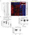
miR30a expression in thyroid tissue. A, heatmap of differentially expressed miRNA in thyroid cancer samples. Supervised hierarchical clustering of miRNA expression in 20 tissue samples: 6 N, 6 PTC, and 8 ATC. Red, overexpression; green, underexpression. N, normal. B, miR30a expression is downregulated in the aggressive thyroid cancer PDTC:17N, 21 PTC, 10 ATC, and 6 PDTC samples were analyzed. *, P < 0.05; ***, P< 0.001; ****, P < 0.0001. C, miR30a expression by thyroid cancer risk group as defined by the MACIS prognostic scoring system. Data from NCI’s TCGA program (accessed at
explorer.cancerregulome.org
) showed significantly lower expression of miR30a in high-risk thyroid cancer, as defined by the MACIS risk group. *, P < 0.05; ns, not significant. D, miR30a expression is lower in patients who died from conventional PTC. *, P < 0.05.
Figure 2.
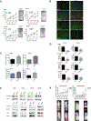
Effect of miR30a on cellular proliferation, migration and invasion, apoptosis, and metastasis. A, miR30a inhibits cellular proliferation. The effect of miR30a overexpression was determined in four ATC cell lines, 8505C, THJ-11T, THJ-16T, and SW1736, using a CyQUANT Cell Proliferation Assay and a clonogenic assay after transfection with 25 nmol/L of miR30a or miR-C. miR30a transfection significantly inhibited cell proliferation and colonies formation compared with miR-C. *, P < 0.05; **, P < 0.01; ***, P < 0.001. B, representative images of ATC cells obtained using fluorescent microscopy. Viable cells exhibit green fluorescence (calcein-AM), whereas membrane-permeable, nonviable cells exhibit red fluorescence (ethidium homodimer-1). Cells are shown 72 hours after transfection. C, ectopic expression of miR30a increased caspase-3/7 activity. Data shown are from 72 hours after transfection. **, P < 0.01; ***, P < 0.001. D, miR30a inhibits cellular migration and invasion. The Transwell assay showed that overexpression of miR30a markedly decreased invasion and migration in four ATC cell lines. These results are representative of at least three independent experiments. E, ectopic expression of miR30a reduces N-cadherin, vimentin, and CD44 expression compared with miR-C. F, effect of miR30a on tumor growth and metastasis in vivo. Nude mice were injected subcutaneously with 8505C-Luc cells expressing either miR-C or miR30a. Luciferase activity (mean±SD) was measured over time in flank xenograft (left) and metastatic (right) models. There was no significant difference in growth in the flank xenograft, but there was a significant difference in the number and volume of metastatic lesions in mice that had tail-vein injections of 8505C-Luc cells. Luciferase activity images of lung metastases in mice that received tail-vein injection of cells overexpressing miR30a (3 mice) or miR-C (3 mice). RFU, relative fluorescence unit. *, P < 0.05; **, P < 0.01; ***, P < 0.001.
Figure 3.
Identification of LOX as a target gene of miR30a. A, miR30a regulates LOX expression. Western blot analysis shows reduced expression of LOX 72 hours after miR30a transfection. B, miR30a directly targets the 3’UTR of LOX mRNA. A luciferase vector was designed to include the region of the candidate target sequence for miR30a. Bar graph shows luciferase activity of the 3’UTR-LOX luciferase vector after cotransfection with miR-C or miR30a in THJ-16Tcells. *, P < 0.05.
Figure 4.
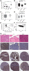
Analysis of LOX expression in thyroid cancer samples. A, LOX expression, assayed by RT-PCR, is upregulated in our ATC cohort. B, validation of upregulated LOX expression in aggressive thyroid cancer in the thyroid cancer dataset from TCGA. C, LOX expression is higher in patients who die from thyroid cancer. D, high LOX expression in tumors is associated with local invasion. E and F, correlation between miR30a and LOX expression in our cohort (E) and the dataset from TCGA (F). G, representative LOX protein expression, as detected by immunohistochemistry, in TC-PTC, cPTC, and N thyroid tissue. LOX expression is restricted to the tumor tissue, and its levels are higher in TC-PTC than in cPTC. N, normal. H, representative immunohistochemical staining of LOX protein in six ATCsamples. Magnification, ×6 and ×40. *, P < 0.05; **, P < 0.01; ***, P < 0.001; ****, P < 0.0001.
Figure 5.
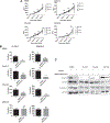
Effect of LOX activity inhibition on thyroid cancer biology in vitro and in vivo. A, inhibition of LOX activity does not affect cellular proliferation. BAPN (100, 250, and 500 μmol/L) was used with a CyQUANT assay to evaluate the effect of LOX inhibition on cellular proliferation. B, LOX activity inhibition reduces cellular migration and invasion in ATC cell lines. Transwell migration and invasion assays were performed after 24 hours of treatment with and without 100 μmol/L of BAPN. C, LOX inhibition reduces only vimentin expression in ATC cells. The cell lines were treated with and without 100 μmol/L of BAPN over 48 hours. RFU, relative fluorescence units.
Figure 6.
Effect of LOX knockdown on cell invasion, migration, apoptosis, and metastasis. A, LOX knockdown, using 28 nmol/L of siRNA, assayed by Western blot analysis, RT-PCR, and immunofluorescent staining. B, LOX knockdown reduced cell proliferation and colony formation, as determined by a CyQUANT cell proliferation assay and a clonogenic assay. C, LOX knockdown resulted in increased apoptosis. The apoptosis marker, caspase-3/7 activity, was analyzed 72 hours after siRNA transfection. D, LOX knockdown decreases cellular migration and invasion. These results are representative of at least three independent experiments. E, immunoblot showing the effect of siLOX on the EMT markers N-cadherin and vimentin 72 hours after transfection. F, LOX knockdown reduces metastasis. Representative ex vivo images and hematoxylin and eosin (H&E)-stained tissue of LOX knockdown samples (left side) and metastasis in mice as measured by luciferase activity (right side) of the 8505c-Luc cells that were injected into the tail veins. The relative luminescence values (RLU) of the three siControl (siC) mice and the three siLOX mice are presented here; *, P < 0.05. G, effect of miR30a on LOX and EMT marker expression in our thyroid cancer metastasis mouse model. Lung metastases show lower LOX, vimentin, and N-cadherin expression when miR30a is overexpressed. Representative immunohistochemical images for LOX, vimentin, and N-cadherin in lung metastases of mice injected with 8505C-Luc cells transfected with miR30a or miR-C. Magnification, ×20.
Similar articles
- Targeting ubiquitin-specific protease 22 suppresses growth and metastasis of anaplastic thyroid carcinoma.
Zhao HD, Tang HL, Liu NN, Zhao YL, Liu QQ, Zhu XS, Jia LT, Gao CF, Yang AG, Li JT. Zhao HD, et al. Oncotarget. 2016 May 24;7(21):31191-203. doi: 10.18632/oncotarget.9098. Oncotarget. 2016. PMID: 27145278 Free PMC article. - MiR-125b inhibits anaplastic thyroid cancer cell migration and invasion by targeting PIK3CD.
Bu Q, You F, Pan G, Yuan Q, Cui T, Hao L, Zhang J. Bu Q, et al. Biomed Pharmacother. 2017 Apr;88:443-448. doi: 10.1016/j.biopha.2016.11.090. Epub 2017 Jan 22. Biomed Pharmacother. 2017. PMID: 28122310 - Lysyl Oxidase (LOX) Transcriptionally Regulates SNAI2 Expression and TIMP4 Secretion in Human Cancers.
Boufraqech M, Zhang L, Nilubol N, Sadowski SM, Kotian S, Quezado M, Kebebew E. Boufraqech M, et al. Clin Cancer Res. 2016 Sep 1;22(17):4491-504. doi: 10.1158/1078-0432.CCR-15-2461. Epub 2016 Mar 30. Clin Cancer Res. 2016. PMID: 27029493 Free PMC article. - Expression of MicroRNAs in Thyroid Carcinoma.
Zhu G, Xie L, Miller D. Zhu G, et al. Methods Mol Biol. 2017;1617:261-280. doi: 10.1007/978-1-4939-7046-9_19. Methods Mol Biol. 2017. PMID: 28540691 Review. - Genomic Landscape of poorly Differentiated and Anaplastic Thyroid Carcinoma.
Xu B, Ghossein R. Xu B, et al. Endocr Pathol. 2016 Sep;27(3):205-12. doi: 10.1007/s12022-016-9445-4. Endocr Pathol. 2016. PMID: 27372303 Review.
Cited by
- The Thyroid Tumor Microenvironment: Potential Targets for Therapeutic Intervention and Prognostication.
MacDonald L, Jenkins J, Purvis G, Lee J, Franco AT. MacDonald L, et al. Horm Cancer. 2020 Oct;11(5-6):205-217. doi: 10.1007/s12672-020-00390-6. Epub 2020 Jun 17. Horm Cancer. 2020. PMID: 32548798 Free PMC article. Review. - Noncoding RNAs in Thyroid-Follicular-Cell-Derived Carcinomas.
De Martino M, Esposito F, Capone M, Pallante P, Fusco A. De Martino M, et al. Cancers (Basel). 2022 Jun 23;14(13):3079. doi: 10.3390/cancers14133079. Cancers (Basel). 2022. PMID: 35804851 Free PMC article. Review. - Lysyl Oxidase (LOX) Family Proteins: Key Players in Breast Cancer Occurrence and Progression.
Li J, Wang X, Liu R, Zi J, Li Y, Li Z, Xiong W. Li J, et al. J Cancer. 2024 Aug 13;15(16):5230-5243. doi: 10.7150/jca.98688. eCollection 2024. J Cancer. 2024. PMID: 39247609 Free PMC article. Review. - New insights into the mechanisms of the extracellular matrix and its therapeutic potential in anaplastic thyroid carcinoma.
Xia J, Shi Y, Chen X. Xia J, et al. Sci Rep. 2024 Sep 9;14(1):20977. doi: 10.1038/s41598-024-72020-y. Sci Rep. 2024. PMID: 39251678 Free PMC article. Review. - circMTO1 sponges microRNA-219a-5p to enhance gallbladder cancer progression via the TGF-β/Smad and EGFR pathways.
Wang P, Zhou C, Li D, Zhang D, Wei L, Deng Y. Wang P, et al. Oncol Lett. 2021 Jul;22(1):563. doi: 10.3892/ol.2021.12824. Epub 2021 May 27. Oncol Lett. 2021. PMID: 34113391 Free PMC article.
References
- Smallridge RC, Ain KB, Asa SL, Bible KC, Brierley JD, Burman KD, et al. American Thyroid Association guidelines for management of patients with anaplastic thyroid cancer. Thyroid 2012;22:1104–39. - PubMed
- Kebebew E, Greenspan FS, Clark OH, Woeber KA, McMillan A. Anaplastic thyroid carcinoma. Treatment outcome and prognostic factors. Cancer 2005;103:1330–5. - PubMed
- McIver B, Hayl D, Giuffrida DF, Dvorak CE, Grant CS, Thompson GB, et al. Anaplastic thyroid carcinoma: a 50-year experience at a single institution. Surgery 2001;130:1028–34. - PubMed
- De Crevoisier R, Baudin E, Bachelot A, Leboulleux S, Travagli JP, Caillou B, et al. Combined treatment of anaplastic thyroid carcinoma with surgery, chemotherapy, and hyperfractionated accelerated external radiotherapy. Int J Radiat Oncol Biol Phys 2004;60:1137–43. - PubMed
Publication types
MeSH terms
Substances
LinkOut - more resources
Full Text Sources
Other Literature Sources
Medical
Research Materials


