Pathway activation profiling reveals new insights into age-related macular degeneration and provides avenues for therapeutic interventions - PubMed (original) (raw)
Pathway activation profiling reveals new insights into age-related macular degeneration and provides avenues for therapeutic interventions
Evgeny Makarev et al. Aging (Albany NY). 2014 Dec.
Abstract
Age-related macular degeneration (AMD) is a major cause of blindness in older people and is caused by loss of the central region of the retinal pigment epithelium (RPE). Conventional methods of gene expression analysis have yielded important insights into AMD pathogenesis, but the precise molecular pathway alterations are still poorly understood. Therefore we developed a new software program, "AMD Medicine", and discovered differential pathway activation profiles in samples of human RPE/choroid from AMD patients and controls. We identified 29 pathways in RPE-choroid AMD phenotypes: 27 pathways were activated in AMD compared to controls, and 2 pathways were activated in controls compared to AMD. In AMD, we identified a graded activation of pathways related to wound response, complement cascade, and cell survival. Also, there was downregulation of two pathways responsible for apoptosis. Furthermore, significant activation of pro-mitotic pathways is consistent with dedifferentiation and cell proliferation events, which occur early in the pathogenesis of AMD. Significantly, we discovered new global pathway activation signatures of AMD involved in the cell-based inflammatory response: IL-2, STAT3, and ERK. The ultimate aim of our research is to achieve a better understanding of signaling pathways involved in AMD pathology, which will eventually lead to better treatments.
Conflict of interest statement
Conflict of interest statement
The authors of this manuscript declare no conflict of interest.
Figures
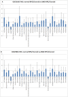
Figure 1. Pathway activation strength (PAS) for selected pathways
PAS values have been calculated according to OncoFinder algorithm. PAS presented on this figure passed the following filters PAS<−1.5 and PAS>1.5 in both datasets. Blue bars represent PAS average for each pathway, and error bars represents standard deviation A. PAS derived from GSE50195 dataset. B. PAS derived from GSE50195 that cell-based inflammatory responses within the RPE-choroid are a core feature of AMD. However, cellular sources and targets of pro-inflammatory secreted factors are still need to be determined along with the regulatory mechanism of the chemokine network.
Figure 2. Heatmap of differentially activated pathways shown in Figure 1A
Complement factor H genetic background (rs1061170 SNP) for PAS values derived from GSE50195 dataset shown for high-risk YH/HH and low-risk YY genotype. Blue shading indicates pathway downregulation; red shading indicates pathway upregulation. Samples with names ending in CTRL indicates control samples; samples with names ending in ARM indicates AMD samples.
Figure 3. Comparison of GSE29801 derived PAS distribution and GSE50195 derived PAS distribution
Box plots of GSE29801 (right) derived PAS and GSE50195 (left) derived PAS for each pathway. All PAS values for each pathway from two independent data sets are comparable; moreover box plots for GSE50195 derived PAS lay inside of box plots for GSE50195 derived PAS. Box plot whiskers represent min and max values for each pathways.
Figure 4. An example of how multiple pathways are activated and down-regulated during AMD
This figure also serves as a working hypothesis for the pathogenesis of AMD. Proposed steps and interactions are as follows: A. Environmental Stress in the form of aging, obesity, inflammation, or diet causes B. senescence and loss of proliferation of the retinal pigment epithelial cells leading to C. activation of the MAPK, ERK, p38 and AKT pathways in the cytoplasmic components of the cells. This cellular senescence also has several consequences, primary of which are D. upregulation of the SASP, interleukin, and inflammatory cytokine networks, and E. downregulation of the caspase cascade and mitochondrial apoptosis. These pathways also interact; for example the upregulation of the SASP, interleukin, and inflammatory cytokine pathways causes downregulation of the caspase and mitochondrial apoptosis pathways. Green arrows represent upregulated pathways, red arrows represent downregulated pathways, and blue dotted arrows represent connected pathways.
Similar articles
- Altered gene expression in dry age-related macular degeneration suggests early loss of choroidal endothelial cells.
Whitmore SS, Braun TA, Skeie JM, Haas CM, Sohn EH, Stone EM, Scheetz TE, Mullins RF. Whitmore SS, et al. Mol Vis. 2013 Nov 16;19:2274-97. eCollection 2013. Mol Vis. 2013. PMID: 24265543 Free PMC article. - Comparison of Mouse and Human Retinal Pigment Epithelium Gene Expression Profiles: Potential Implications for Age-Related Macular Degeneration.
Bennis A, Gorgels TG, Ten Brink JB, van der Spek PJ, Bossers K, Heine VM, Bergen AA. Bennis A, et al. PLoS One. 2015 Oct 30;10(10):e0141597. doi: 10.1371/journal.pone.0141597. eCollection 2015. PLoS One. 2015. PMID: 26517551 Free PMC article. - Dry age-related macular degeneration like pathology in aged 5XFAD mice: Ultrastructure and microarray analysis.
Park SW, Im S, Jun HO, Lee K, Park YJ, Kim JH, Park WJ, Lee YH, Kim JH. Park SW, et al. Oncotarget. 2017 Jun 20;8(25):40006-40018. doi: 10.18632/oncotarget.16967. Oncotarget. 2017. PMID: 28467791 Free PMC article. - Regulatory role of HIF-1alpha in the pathogenesis of age-related macular degeneration (AMD).
Arjamaa O, Nikinmaa M, Salminen A, Kaarniranta K. Arjamaa O, et al. Ageing Res Rev. 2009 Oct;8(4):349-58. doi: 10.1016/j.arr.2009.06.002. Epub 2009 Jul 7. Ageing Res Rev. 2009. PMID: 19589398 Review. - Stem cell based therapies for age-related macular degeneration: The promises and the challenges.
Nazari H, Zhang L, Zhu D, Chader GJ, Falabella P, Stefanini F, Rowland T, Clegg DO, Kashani AH, Hinton DR, Humayun MS. Nazari H, et al. Prog Retin Eye Res. 2015 Sep;48:1-39. doi: 10.1016/j.preteyeres.2015.06.004. Epub 2015 Jun 23. Prog Retin Eye Res. 2015. PMID: 26113213 Review.
Cited by
- Gene expression levels of the insulin-like growth factor family in patients with AMD before and after ranibizumab intravitreal injections.
Strzalka-Mrozik B, Kimsa-Furdzik M, Kabiesz A, Michalska-Malecka K, Nita M, Mazurek U. Strzalka-Mrozik B, et al. Clin Interv Aging. 2017 Sep 5;12:1401-1408. doi: 10.2147/CIA.S135030. eCollection 2017. Clin Interv Aging. 2017. PMID: 28919726 Free PMC article. - Radioprotectors.org: an open database of known and predicted radioprotectors.
Aliper AM, Bozdaganyan ME, Sarkisova VA, Veviorsky AP, Ozerov IV, Orekhov PS, Korzinkin MB, Moskalev A, Zhavoronkov A, Osipov AN. Aliper AM, et al. Aging (Albany NY). 2020 Aug 15;12(15):15741-15755. doi: 10.18632/aging.103815. Epub 2020 Aug 15. Aging (Albany NY). 2020. PMID: 32805729 Free PMC article. - Interleukin-2 induces extracellular matrix synthesis and TGF-β2 expression in retinal pigment epithelial cells.
Jing R, Qi T, Wen C, Yue J, Wang G, Pei C, Ma B. Jing R, et al. Dev Growth Differ. 2019 Sep;61(7-8):410-418. doi: 10.1111/dgd.12630. Epub 2019 Oct 13. Dev Growth Differ. 2019. PMID: 31608440 Free PMC article. - Topical administration of Esculetin as a potential therapy for experimental dry eye syndrome.
Jiang D, Liu X, Hu J. Jiang D, et al. Eye (Lond). 2017 Dec;31(12):1724-1732. doi: 10.1038/eye.2017.117. Epub 2017 Jun 23. Eye (Lond). 2017. PMID: 28643798 Free PMC article. - The effect of systemic levels of TNF-alpha and complement pathway activity on outcomes of VEGF inhibition in neovascular AMD.
Khan AH, Pierce CO, De Salvo G, Griffiths H, Nelson M, Cree AJ, Menon G, Lotery AJ. Khan AH, et al. Eye (Lond). 2022 Nov;36(11):2192-2199. doi: 10.1038/s41433-021-01824-3. Epub 2021 Nov 8. Eye (Lond). 2022. PMID: 34750590 Free PMC article.
References
- Bourne RR, Stevens GA, White RA, Smith JL, Flaxman SR, Price H, Jonas JB, Keeffe J, Leasher J, Naidoo K, Pesudovs K, Resnikoff S, Taylor HR. Causes of vision loss worldwide, 1990-2010: a systematic analysis. The Lancet Global health. 2013;1:e339–349. - PubMed
- Wong WL, Su X, Li X, Cheung CM, Klein R, Cheng CY, Wong TY. Global prevalence of age-related macular degeneration and disease burden projection for 2020 and 2040: a systematic review and meta-analysis. The Lancet Global health. 2014;2:e106–116. - PubMed
MeSH terms
Substances
LinkOut - more resources
Full Text Sources
Other Literature Sources
Medical
Miscellaneous


