NOS1AP Functionally Associates with YAP To Regulate Hippo Signaling - PubMed (original) (raw)
NOS1AP Functionally Associates with YAP To Regulate Hippo Signaling
Leanne Clattenburg et al. Mol Cell Biol. 2015 Jul.
Abstract
Deregulation of cellular polarity proteins and their associated complexes leads to changes in cell migration and proliferation. The nitric oxide synthase 1 adaptor protein (NOS1AP) associates with the tumor suppressor protein Scribble to control cell migration and oncogenic transformation. However, how NOS1AP is linked to the cell signaling events that curb oncogenic progression has remained elusive. Here we identify several novel NOS1AP isoforms, NOS1APd, NOS1APe, and NOS1APf, with distinct cellular localizations. We show that isoforms with a membrane-interacting phosphotyrosine binding (PTB) domain can associate with Scribble and recognize acidic phospholipids. In a screen to identify novel binding proteins, we have discovered a complex consisting of NOS1AP and the transcriptional coactivator YAP linking NOS1AP to the Hippo signaling pathway. Silencing of NOS1AP reduces the phosphorylation of YAP and of the upstream kinase Lats1. Conversely, expression of NOS1AP promotes YAP and Lats1 phosphorylation, which correlates with reduced TEAD activity and restricted cell proliferation. Together, these data implicate a role for NOS1AP in the regulation of core Hippo signaling and are consistent with the idea that NOS1AP functions as a tumor suppressor.
Copyright © 2015, American Society for Microbiology. All Rights Reserved.
Figures
FIG 1
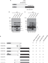
Identification of multiple NOS1AP isoforms. (A) Schematic of NOS1APa and NOS1APc showing the regions of each protein that were used to generate the isoform-specific antibodies used in this study. (B and C) NOS1AP isoforms were precipitated (I.P.) from rat brain lysate with the indicated antibodies. Bound complexes were analyzed by immunoblotting using the antibodies indicated at the bottom. All NOS1AP antibodies tested precipitated and detected bands at 95 kDa (arrows) and at 70 kDa (arrowheads). Two faster-migrating bands at ∼40 kDa and 25 kDa were detected in both panels B and C when lysates were precipitated with pep-NOS1APC antibody (arrows with one or two asterisks). WB, Western blotting. (D, left) Schematic of NOS1AP isoforms (NOS1APb to -h) detected from the cerebellar cDNA screen and amino acids representing splice sites in NOS1APa. (Right) Summary of the antibodies used in this study and their relative affinities for the different NOS1AP isoforms. Shaded boxes in NOS1APb, -g, and -h represent the unique 5′ exon identified for NOS1APb. The small insert in the NOS1APe PTB domain represents the schematic where the LLLLQ insert is found.
FIG 2
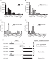
qPCR analysis of different NOS1AP transcripts. (A) mRNA copy numbers of NOS1AP isoforms that contain the PTB domain from different tissues. (B) mRNA copy numbers of NOS1AP isoforms that contain the C-terminal region of exon 10 of rat NOS1APa. (C) Copy numbers of NOS1AP isoforms that contain the unique C-terminal region found in NOS1APc, -d, -e, and -f. (D) Copy numbers of NOS1AP constructs that contain the unique 5′ region of NOS1APf. (E, left) Schematic of the regions (shaded boxes with symbols) in each isoform (*, NOS1APa; &, NOS1APa/b; #, NOS1APc, -d, -e, and -f; ‡, NOS1APf) that were analyzed by quantitative PCR in panels A to D. (Right) Tissues with the highest mRNA expression levels for the given NOS1AP isoforms. Abbreviations: Crtx, cortex; Cereb, cerebellum; Hippo, hippocampus; OB, olfactory bulb; Striat, striatum; BS, brain stem; MB, midbrain; SC, spinal cord; SM, skeletal muscle.
FIG 3
NOS1AP isoforms with different subcellular localizations. (A) Spinning-disk confocal images of MCF7 cells stably expressing YFP, YFP-NOS1APa, NOS1APc, NOS1APd, NOS1APe, and NOS1APf. Note a faint enrichment of YFP-NOS1APc at cell-cell contacts (arrows) and low levels of nuclear localization in YFP, YFP-NOS1APc, NOS1APd, and NOS1APf (arrowheads). Bar = 20 μm. (B) NMR structure model of the Drosophila NUMB PTB domain (42). The arrows point to the insert in NOS1APe positioned in the predicted surface-exposed loop in the protein. (C) A membrane covered with different phospholipids was probed with purified GST, GST-PTB, or GST-PTBinsert (the NOS1APe PTB domain containing the LLLLQ insert). LPA, lysophosphatidic acid; LPC, lysophosphatidylcholine; PE, phosphatidylethanolamine; PS, phosphatidylserine; PtdIns, phosphatidylinositol.
FIG 4
NOS1AP isoforms containing the PTB domain interact with Scribble. (A) Endogenous NOS1AP isoforms were precipitated (I.P.) from rat brain lysate with the indicated antibodies. Bound complexes were analyzed by immunoblotting using anti-Scribble, anti-nNOS, and pep-NOS1APc antibodies, as indicated at the bottom. (B and C) cDNA constructs encoding the indicated proteins were transfected into HEK293T cells. (B) Bound complexes were analyzed with anti-myc and anti-GFP antibodies. (C) Whole-cell lysates with input levels.
FIG 5
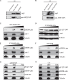
NOS1AP binds to and modulates the phosphorylation of YAP. (A) Rat brain lysate was precipitated with pan-NOS1AP or Scribble antibodies. Coprecipitated complexes were analyzed by immunoblotting with YAP antibodies (top) and pan-NOS1AP (bottom). (B) HEK293T cell lysate was precipitated with pep-NOS1APc or GST-NOS1APc antibodies. The resulting precipitated complexes were probed with YAP (top) or pep-NOS1APc (bottom) antibodies. (C to F) HEK293T cells were transfected with increasing amounts of a plasmid encoding YFP-NOS1APa (C and D), YFP-NOS1APc (E), or the YFP-PTB domain alone (F). The lysates were analyzed for endogenous Lats1 and YAP and with phospho-Lats1 (detecting S909) and phospho-YAP (detecting S127) antibodies. (G and H) IEC-18 cells transfected with NOS1AP-targeting siRNA (siNOS1AP) (G) or control siRNA (Csi) (H) were lysed at the indicated time points. Equal amounts of cell lysate were prepared for Western blotting and analyzed with the indicated antibodies.
FIG 6
NOS1APa reduces YAP nuclear accumulation. MCF7 cells stably expressing YFP or YFP-NOS1APa were fixed and costained with anti-GFP and anti-YAP antibodies. Hoechst stains cell nuclei. Bar = 20 μm.
FIG 7
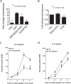
The PTB domain is sufficient to reduce YAP-mediated TEAD activity. (A and B) HEK293T cells transfected with TEAD-luciferase together with the plasmids indicated and lysates were prepared for luciferase measurements after 24 h. Data show luciferase activity normalized against β-galactosidase activity. Histograms show means ± SEM from 3 independent experiments. Differences were considered significant by Student's t test at a P value of <0.05. (C and D) MCF7 cells stably expressing YFP-NOS1APa incorporate less radiolabeled thymidine over a 72-h period without (C) or with (D) 5% serum than do MCF7 cells stably expressing YFP. Differences were considered significant by Student's t test at a P value of <0.05 (asterisk).
Similar articles
- The Hippo component YAP localizes in the nucleus of human papilloma virus positive oropharyngeal squamous cell carcinoma.
Alzahrani F, Clattenburg L, Muruganandan S, Bullock M, MacIsaac K, Wigerius M, Williams BA, Graham ME, Rigby MH, Trites JR, Taylor SM, Sinal CJ, Fawcett JP, Hart RD. Alzahrani F, et al. J Otolaryngol Head Neck Surg. 2017 Feb 22;46(1):15. doi: 10.1186/s40463-017-0187-1. J Otolaryngol Head Neck Surg. 2017. PMID: 28222762 Free PMC article. - NOS1AP associates with Scribble and regulates dendritic spine development.
Richier L, Williton K, Clattenburg L, Colwill K, O'Brien M, Tsang C, Kolar A, Zinck N, Metalnikov P, Trimble WS, Krueger SR, Pawson T, Fawcett JP. Richier L, et al. J Neurosci. 2010 Mar 31;30(13):4796-805. doi: 10.1523/JNEUROSCI.3726-09.2010. J Neurosci. 2010. PMID: 20357130 Free PMC article. - Dynamic alterations in Hippo signaling pathway and YAP activation during liver regeneration.
Grijalva JL, Huizenga M, Mueller K, Rodriguez S, Brazzo J, Camargo F, Sadri-Vakili G, Vakili K. Grijalva JL, et al. Am J Physiol Gastrointest Liver Physiol. 2014 Jul 15;307(2):G196-204. doi: 10.1152/ajpgi.00077.2014. Epub 2014 May 29. Am J Physiol Gastrointest Liver Physiol. 2014. PMID: 24875096 - Research progress in NOS1AP in neurological and psychiatric diseases.
Wang J, Jin L, Zhu Y, Zhou X, Yu R, Gao S. Wang J, et al. Brain Res Bull. 2016 Jul;125:99-105. doi: 10.1016/j.brainresbull.2016.05.014. Epub 2016 May 26. Brain Res Bull. 2016. PMID: 27237129 Review. - YAP and TAZ Take Center Stage in Cancer.
Zhang K, Qi HX, Hu ZM, Chang YN, Shi ZM, Han XH, Han YW, Zhang RX, Zhang Z, Chen T, Hong W. Zhang K, et al. Biochemistry. 2015 Nov 3;54(43):6555-66. doi: 10.1021/acs.biochem.5b01014. Epub 2015 Oct 20. Biochemistry. 2015. PMID: 26465056 Review.
Cited by
- CRISPR-cas9 screening identified lethal genes enriched in Hippo kinase pathway and of predictive significance in primary low-grade glioma.
Mijiti M, Maimaiti A, Chen X, Tuersun M, Dilixiati M, Dilixiati Y, Zhu G, Wu H, Li Y, Turhon M, Abulaiti A, Maimaitiaili N, Yiming N, Kasimu M, Wang Y. Mijiti M, et al. Mol Med. 2023 May 14;29(1):64. doi: 10.1186/s10020-023-00652-3. Mol Med. 2023. PMID: 37183261 Free PMC article. - Effects of Long Form of CAPON Overexpression on Glioma Cell Proliferation are Dependent on AKT/mTOR/P53 Signaling.
Liang D, Song Y, Fan G, Ji D, Zhang T, Nie E, Liu X, Liang J, Yu R, Gao S. Liang D, et al. Int J Med Sci. 2019 Apr 25;16(4):614-622. doi: 10.7150/ijms.31579. eCollection 2019. Int J Med Sci. 2019. PMID: 31171914 Free PMC article. - The Physiological Function of nNOS-Associated CAPON Proteins and the Roles of CAPON in Diseases.
Xie W, Xing N, Qu J, Liu D, Pang Q. Xie W, et al. Int J Mol Sci. 2023 Oct 31;24(21):15808. doi: 10.3390/ijms242115808. Int J Mol Sci. 2023. PMID: 37958792 Free PMC article. Review. - Hepatic nitric oxide synthase 1 adaptor protein regulates glucose homeostasis and hepatic insulin sensitivity in obese mice depending on its PDZ binding domain.
Mu K, Sun Y, Zhao Y, Zhao T, Li Q, Zhang M, Li H, Zhang R, Hu C, Wang C, Jia W. Mu K, et al. EBioMedicine. 2019 Sep;47:352-364. doi: 10.1016/j.ebiom.2019.08.033. Epub 2019 Aug 28. EBioMedicine. 2019. PMID: 31473185 Free PMC article. - The Hippo component YAP localizes in the nucleus of human papilloma virus positive oropharyngeal squamous cell carcinoma.
Alzahrani F, Clattenburg L, Muruganandan S, Bullock M, MacIsaac K, Wigerius M, Williams BA, Graham ME, Rigby MH, Trites JR, Taylor SM, Sinal CJ, Fawcett JP, Hart RD. Alzahrani F, et al. J Otolaryngol Head Neck Surg. 2017 Feb 22;46(1):15. doi: 10.1186/s40463-017-0187-1. J Otolaryngol Head Neck Surg. 2017. PMID: 28222762 Free PMC article.
References
Publication types
MeSH terms
Substances
LinkOut - more resources
Full Text Sources
Molecular Biology Databases
Research Materials


