Experimental Inoculation of Egyptian Rousette Bats (Rousettus aegyptiacus) with Viruses of the Ebolavirus and Marburgvirus Genera - PubMed (original) (raw)
Experimental Inoculation of Egyptian Rousette Bats (Rousettus aegyptiacus) with Viruses of the Ebolavirus and Marburgvirus Genera
Megan E B Jones et al. Viruses. 2015.
Abstract
The Egyptian rousette bat (Rousettus aegyptiacus) is a natural reservoir for marburgviruses and a consistent source of virus spillover to humans. Cumulative evidence suggests various bat species may also transmit ebolaviruses. We investigated the susceptibility of Egyptian rousettes to each of the five known ebolaviruses (Sudan, Ebola, Bundibugyo, Taï Forest, and Reston), and compared findings with Marburg virus. In a pilot study, groups of four juvenile bats were inoculated with one of the ebolaviruses or Marburg virus. In ebolavirus groups, viral RNA tissue distribution was limited, and no bat became viremic. Sudan viral RNA was slightly more widespread, spurring a second, 15-day Sudan virus serial euthanasia study. Low levels of Sudan viral RNA disseminated to multiple tissues at early time points, but there was no viremia or shedding. In contrast, Marburg virus RNA was widely disseminated, with viremia, oral and rectal shedding, and antigen in spleen and liver. This is the first experimental infection study comparing tissue tropism, viral shedding, and clinical and pathologic effects of six different filoviruses in the Egyptian rousette, a known marburgvirus reservoir. Our results suggest Egyptian rousettes are unlikely sources for ebolaviruses in nature, and support a possible single filovirus-single reservoir host relationship.
Keywords: Ebolavirus; Egyptian rousette bat; Marburgvirus; Rousettus aegyptiacus; experimental infection study; reservoir host.
Figures
Figure 1
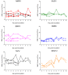
Blood chemistry measurements for bats inoculated with six different filoviruses in the pilot study and euthanized at 5 and 10 days post inoculation (DPI). Mock = mock-inoculated controls, MARV = Marburg virus, SUDV = Sudan virus, EBOV = Ebola virus, BDBV = Bundibugyo virus, TAFV = Taï Forest virus, and RESTV = Reston virus. ALT = alanine aminotransferase, AST = aspartate aminotransferase, ALP = alkaline phosphatase, ALB = albumin, BUN = blood urea nitrogen.
Figure 2
Complete white blood cell (WBC) counts for bats inoculated with six different filoviruses in the pilot study and euthanized at 5 and 10 days post inoculation (DPI). Mock = mock-inoculated controls, MARV = Marburg virus, SUDV = Sudan virus, EBOV = Ebola virus, BDBV = Bundibugyo virus, TAFV = Taï Forest virus, and RESTV = Reston virus.
Figure 3
Viral loads, as determined by Q-RT-PCR and expressed as 50% tissue culture infective dose (TCID50) equivalents per mL, in four bats inoculated with Marburg virus in the Pilot Study and euthanized at 5 (n = 2) and 10 (n = 2) DPI. (A) Marburg viral RNA in blood is evidence of viremia in all four Marburg virus-inoculated bats; (B) Marburg viral RNA in oral (filled bars) and rectal (open bars) swabs.
Figure 4
Photomicrographs of tissues from Egyptian rousette bats experimentally inoculated with filoviruses. (A) Liver, MARV-inoculated bat, day 5 post-inoculation (pilot study). A focus of histiocytic infiltrate and rare necrotic hepatocytes disrupts the parenchyma. H&E stain; (B) Liver, MARV-inoculated bat, day 5 post-inoculation (pilot study). Marburgviral antigen (red) is present in the cytoplasm of hepatocytes and macrophages in a small focus of infiltrate. Inset: positive immunostaining in the cytoplasm of a necrotic hepatocyte from the same section. Immunoalkaline phosphatase with naphthol fast red and hematoxylin counterstain; (C) Spleen, MARV-inoculated bat, 5 days post-inoculation (pilot study). MARV antigen is present in small numbers of red pulp macrophages (arrows). Inset: Higher magnification of cytoplasmic antigen in macrophages. Immunoalkaline phosphatase with naphthol fast red and hematoxylin counterstain; (D) Skin (subcutaneous tissue) from the inoculation site, MARV-inoculated bat, 5 days post-inoculation (pilot study). A small focus of macrophages infiltrates the subcutis at the site of MARV inoculation. H&E stain; (E) Skin (subcutaneous tissue) from the inoculation site, MARV-inoculated bat, 5 days post-inoculation (pilot study). Positive immunostaining in subcutaneous macrophages (main panel) and fibroblasts (inset) at the inoculation site. Immunoalkaline phosphatase with naphthol fast red and hematoxylin counterstain; (F) Skin (subcutaneous tissue) from the inoculation site, SUDV-inoculated bat, 3 days post-inoculation (SUDV serial euthanasia study). Cytoplasmic antigen (red) in macrophages at the inoculation site. Immunoalkaline phosphatase with naphthol fast red and hematoxylin counterstain.
Figure 5
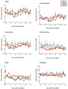
Complete blood count data for Egyptian rousette bats inoculated with Sudan virus (n = 15, green triangles), Marburg virus (n = 3, red squares) and mock-inoculated controls (n = 3, open circles/dashed line) in a serial euthanasia study. WBC = white blood cell count, RBC = red blood cell count, MARV = Marburg virus, SUDV = Sudan virus.
Figure 6
Blood chemistry measurements for Egyptian rousette bats inoculated with Sudan virus. Three Sudan virus-inoculated bats were euthanized on each of days 3, 6, 9, 12 and 15 post-inoculation. Mock-inoculated bats were euthanized on day 15. Mock = mock-inoculated controls, SUDV = Sudan virus, ALT = alanine aminotransferase, AST = aspartate aminotransferase, ALP = alkaline phosphatase, ALB = albumin, BUN = blood urea nitrogen.
Figure 7
Serology results for Egyptian rousette bats inoculated with Sudan virus in a serial euthanasia study. Results for anti-SUDV IgG measured by enzyme linked immunosorbent assay are shown as adjusted sum optical densities (OD) by day post-inoculation for 15 SUDV inoculated bats (black circles) and 3 mock-inoculated control bats (open squares). Numeric labels represent individual animal identification numbers for two bats that seroconverted.
Figure 8
Comparison of Sudan virus and Marburg virus RNA levels in Egyptian rousette tissues (skin at the inoculation site, liver, spleen, and kidney) compared at days 3, 6, 9, 12, and 15 post-infection. Viral loads are expressed as 50% tissue culture infective dose (TCID50) equivalents per gram, derived from quantitative reverse-transcriptase PCR. Data for days 3-12 for Marburg virus-inoculated bats are from Amman et al. [36].
Similar articles
- Clinical, Histopathologic, and Immunohistochemical Characterization of Experimental Marburg Virus Infection in A Natural Reservoir Host, the Egyptian Rousette Bat (Rousettus aegyptiacus).
Jones MEB, Amman BR, Sealy TK, Uebelhoer LS, Schuh AJ, Flietstra T, Bird BH, Coleman-McCray JD, Zaki SR, Nichol ST, Towner JS. Jones MEB, et al. Viruses. 2019 Mar 2;11(3):214. doi: 10.3390/v11030214. Viruses. 2019. PMID: 30832364 Free PMC article. - Broad and Temperature Independent Replication Potential of Filoviruses on Cells Derived From Old and New World Bat Species.
Miller MR, McMinn RJ, Misra V, Schountz T, Müller MA, Kurth A, Munster VJ. Miller MR, et al. J Infect Dis. 2016 Oct 15;214(suppl 3):S297-S302. doi: 10.1093/infdis/jiw199. Epub 2016 Jun 26. J Infect Dis. 2016. PMID: 27354372 Free PMC article. - Innate Immune Responses of Bat and Human Cells to Filoviruses: Commonalities and Distinctions.
Kuzmin IV, Schwarz TM, Ilinykh PA, Jordan I, Ksiazek TG, Sachidanandam R, Basler CF, Bukreyev A. Kuzmin IV, et al. J Virol. 2017 Mar 29;91(8):e02471-16. doi: 10.1128/JVI.02471-16. Print 2017 Apr 15. J Virol. 2017. PMID: 28122983 Free PMC article. - Characteristics of Filoviridae: Marburg and Ebola viruses.
Beer B, Kurth R, Bukreyev A. Beer B, et al. Naturwissenschaften. 1999 Jan;86(1):8-17. doi: 10.1007/s001140050562. Naturwissenschaften. 1999. PMID: 10024977 Review. - Recent advances in marburgvirus research.
Olejnik J, Mühlberger E, Hume AJ. Olejnik J, et al. F1000Res. 2019 May 21;8:F1000 Faculty Rev-704. doi: 10.12688/f1000research.17573.1. eCollection 2019. F1000Res. 2019. PMID: 31131088 Free PMC article. Review.
Cited by
- Ecological indicators of mammal exposure to Ebolavirus.
Schmidt JP, Maher S, Drake JM, Huang T, Farrell MJ, Han BA. Schmidt JP, et al. Philos Trans R Soc Lond B Biol Sci. 2019 Sep 30;374(1782):20180337. doi: 10.1098/rstb.2018.0337. Epub 2019 Aug 12. Philos Trans R Soc Lond B Biol Sci. 2019. PMID: 31401967 Free PMC article. - Marburg and Ebola Virus Infections Elicit a Complex, Muted Inflammatory State in Bats.
Jayaprakash AD, Ronk AJ, Prasad AN, Covington MF, Stein KR, Schwarz TM, Hekmaty S, Fenton KA, Geisbert TW, Basler CF, Bukreyev A, Sachidanandam R. Jayaprakash AD, et al. Viruses. 2023 Jan 26;15(2):350. doi: 10.3390/v15020350. Viruses. 2023. PMID: 36851566 Free PMC article. - Clinical, Histopathologic, and Immunohistochemical Characterization of Experimental Marburg Virus Infection in A Natural Reservoir Host, the Egyptian Rousette Bat (Rousettus aegyptiacus).
Jones MEB, Amman BR, Sealy TK, Uebelhoer LS, Schuh AJ, Flietstra T, Bird BH, Coleman-McCray JD, Zaki SR, Nichol ST, Towner JS. Jones MEB, et al. Viruses. 2019 Mar 2;11(3):214. doi: 10.3390/v11030214. Viruses. 2019. PMID: 30832364 Free PMC article. - De novo transcriptome reconstruction and annotation of the Egyptian rousette bat.
Lee AK, Kulcsar KA, Elliott O, Khiabanian H, Nagle ER, Jones ME, Amman BR, Sanchez-Lockhart M, Towner JS, Palacios G, Rabadan R. Lee AK, et al. BMC Genomics. 2015 Dec 7;16:1033. doi: 10.1186/s12864-015-2124-x. BMC Genomics. 2015. PMID: 26643810 Free PMC article. - Peripheral immune responses to filoviruses in a reservoir versus spillover hosts reveal transcriptional correlates of disease.
Guito JC, Arnold CE, Schuh AJ, Amman BR, Sealy TK, Spengler JR, Harmon JR, Coleman-McCray JD, Sanchez-Lockhart M, Palacios GF, Towner JS, Prescott JB. Guito JC, et al. Front Immunol. 2024 Jan 8;14:1306501. doi: 10.3389/fimmu.2023.1306501. eCollection 2023. Front Immunol. 2024. PMID: 38259437 Free PMC article.
References
- Kuhn J.H., Becker S., Ebihara H., Geisbert T.W., Johnson K.M., Kawaoka Y., Lipkin W.I., Negredo A.I., Netesov S.V., Nichol S.T., et al. Proposal for a revised taxonomy of the family Filoviridae: Classification, names of taxa and viruses, and virus abbreviations. Arch. Virol. 2010;155:2083–2103. doi: 10.1007/s00705-010-0814-x. - DOI - PMC - PubMed
- Towner J.S., Khristova M.L., Sealy T.K., Vincent M.J., Erickson B.R., Bawiec D.A., Hartman A.L., Comer J.A., Zaki S.R., Ströher U., et al. Marburgvirus genomics and association with a large hemorrhagic fever outbreak in Angola. J. Virol. 2006;80:6497–6516. doi: 10.1128/JVI.00069-06. - DOI - PMC - PubMed
MeSH terms
LinkOut - more resources
Full Text Sources
Other Literature Sources
Medical


