What to do With Wake-Up Stroke - PubMed (original) (raw)
Review
What to do With Wake-Up Stroke
Mark N Rubin et al. Neurohospitalist. 2015 Jul.
Abstract
Wake-up stroke, defined as the situation where a patient awakens with stroke symptoms that were not present prior to falling asleep, represents roughly 1 in 5 acute ischemic strokes and remains a therapeutic dilemma. Patients with wake-up stroke were excluded from most ischemic stroke treatment trials and are often not eligible for acute reperfusion therapy in clinical practice, leading to poor outcomes. Studies of neuroimaging with standard noncontrast computed tomography (CT), magnetic resonance imaging (MRI), and multimodal perfusion-based CT and MRI suggest wake-up stroke may occur shortly before awakening and may assist in selecting patients for acute reperfusion therapies. Pilot studies of wake-up stroke treatment based on these neuroimaging features are promising but have limited generalizability. Ongoing randomized treatment trials using neuroimaging-based patient selection may identify a subset of patients with wake-up stroke that can safely benefit from acute reperfusion therapies.
Keywords: acute stroke; hemorrhage; outcome; tPA; thrombolysis; wake-up stroke.
Conflict of interest statement
Declaration of Conflicting Interests: The authors declared no potential conflicts of interest with respect to the research, authorship, and/or publication of this article.
Figures
Figure 1.
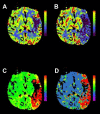
Multimodal CT mismatch (or “penumbra”). Panel (A) is a cerebral blood volume (CBV) map. The dark area noted in the left frontal operculum suggests low contrast volume in the region and is considered a surrogate for infarcted tissue, or the “infarct core.” The other maps—cerebral blood flow (CBF) in panel (B), time to peak (TTP) in panel (C), and mean transit time (MTT) in panel (D)—are different measures of contrast movement through cerebral vasculature (see Table 3) and clearly involve much more of the left hemisphere than the CBV map. This discordance is referred to as a multimodal CT mismatch or “penumbra” and may represent tissue at risk of infarction but potentially salvageable by reperfusion therapy. Siemens SOMATOM, syngo perfusion software. CT indicates computed tomography.
Figure 2.
The DWI/FLAIR mismatch. These 2 axial images of the brain at a level just above the lateral ventricles represent the so-called DWI/FLAIR mismatch that can be seen in the early hours after symptom onset when DWI (left) hyperintensity—which can arise in minutes from symptom onset—occurs in the absence of T2-based FLAIR (right) hyperintensity, which takes 3 to 6 hours to develop. DWI indicates diffusion-weighted imaging; FLAIR, fluid attenuated inversion recovery.
Similar articles
- Trial design and reporting standards for intra-arterial cerebral thrombolysis for acute ischemic stroke.
Higashida RT, Furlan AJ, Roberts H, Tomsick T, Connors B, Barr J, Dillon W, Warach S, Broderick J, Tilley B, Sacks D; Technology Assessment Committee of the American Society of Interventional and Therapeutic Neuroradiology; Technology Assessment Committee of the Society of Interventional Radiology. Higashida RT, et al. Stroke. 2003 Aug;34(8):e109-37. doi: 10.1161/01.STR.0000082721.62796.09. Epub 2003 Jul 17. Stroke. 2003. PMID: 12869717 - Wake-up stroke and CT perfusion: effectiveness and safety of reperfusion therapy.
Caruso P, Naccarato M, Furlanis G, Ajčević M, Stragapede L, Ridolfi M, Polverino P, Ukmar M, Manganotti P. Caruso P, et al. Neurol Sci. 2018 Oct;39(10):1705-1712. doi: 10.1007/s10072-018-3486-z. Epub 2018 Jul 10. Neurol Sci. 2018. PMID: 29987433 - Safety and cost-effectiveness thrombolysis by diffusion-weighted imaging and fluid attenuated inversion recovery mismatch for wake-up stroke.
Sun T, Xu Z, Diao SS, Zhang LL, Fang Q, Cai XY, Kong Y. Sun T, et al. Clin Neurol Neurosurg. 2018 Jul;170:47-52. doi: 10.1016/j.clineuro.2018.04.027. Epub 2018 Apr 23. Clin Neurol Neurosurg. 2018. PMID: 29729542 Review. - Wake-up stroke: clinical characteristics, imaging findings, and treatment option - an update.
Rimmele DL, Thomalla G. Rimmele DL, et al. Front Neurol. 2014 Mar 26;5:35. doi: 10.3389/fneur.2014.00035. eCollection 2014. Front Neurol. 2014. PMID: 24723908 Free PMC article. Review. - [Wake up stroke: Overview on diagnostic and therapeutic options for ischemic stroke on awakening].
Breuer L, Huttner HB, Dörfler A, Schellinger PD, Köhrmann M. Breuer L, et al. Fortschr Neurol Psychiatr. 2010 Feb;78(2):101-6. doi: 10.1055/s-0028-1109985. Epub 2010 Feb 9. Fortschr Neurol Psychiatr. 2010. PMID: 20146154 Review. German.
Cited by
- APIS: a paired CT-MRI dataset for ischemic stroke segmentation - methods and challenges.
Gómez S, Rangel E, Mantilla D, Ortiz A, Camacho P, de la Rosa E, Seia J, Kirschke JS, Li Y, El Habib Daho M, Martínez F. Gómez S, et al. Sci Rep. 2024 Sep 4;14(1):20543. doi: 10.1038/s41598-024-71273-x. Sci Rep. 2024. PMID: 39232010 Free PMC article. - Acute ischemic stroke care in Germany - further progress from 2016 to 2019.
Richter D, Weber R, Eyding J, Bartig D, Misselwitz B, Grau A, Hacke W, Krogias C. Richter D, et al. Neurol Res Pract. 2021 Apr 1;3(1):14. doi: 10.1186/s42466-021-00115-2. Neurol Res Pract. 2021. PMID: 33789773 Free PMC article. - Are we ready for perfusion imaging guided thrombolysis of wake-up strokes?
Drew D, Shamy M, Eagles D. Drew D, et al. CJEM. 2021 Nov;23(6):752-754. doi: 10.1007/s43678-021-00195-8. Epub 2021 Aug 21. CJEM. 2021. PMID: 34420197 No abstract available. - Ischemic stroke with unknown onset of symptoms: current scenario and perspectives for the future.
Lopes RP, Gagliardi VDB, Pacheco FT, Gagliardi RJ. Lopes RP, et al. Arq Neuropsiquiatr. 2022 Dec;80(12):1262-1273. doi: 10.1055/s-0042-1755342. Epub 2022 Dec 29. Arq Neuropsiquiatr. 2022. PMID: 36580965 Free PMC article. - Mechanical Thrombectomy- Where Do We Stand Now ?
Chakraborty D, Bhaumik S. Chakraborty D, et al. Ann Indian Acad Neurol. 2022 Mar-Apr;25(2):184. doi: 10.4103/aian.aian_263_22. Epub 2022 May 17. Ann Indian Acad Neurol. 2022. PMID: 35693687 Free PMC article. No abstract available.
References
- Saver JL. Time is brain—quantified. Stroke J Cereb Circ. 2006;37 (1):263–266. - PubMed
- Meretoja A, Keshtkaran M, Saver JL, et al. Stroke thrombolysis: save a minute, save a day. Stroke J Cereb Circ. 2014;45 (4):1053–1058. - PubMed
- Tissue plasminogen activator for acute ischemic stroke. The National Institute of Neurological Disorders and Stroke rt-PA Stroke Study Group. N Engl J Med. 1995;333 (24):1581–1588. - PubMed
- Albers GW, Clark WM, Madden KP, Hamilton SA, Davis SM, Donnan GA. ATLANTIS trial: results for patients treated within 3 hours of stroke onset * editorial comment: results for patients treated within 3 hours of stroke onset. Stroke. 2002;33 (2):493–496. - PubMed
- Hacke W, Kaste M, Bluhmki E, et al. Thrombolysis with alteplase 3 to 4.5 hours after acute ischemic stroke. N Engl J Med. 2008;359 (13):1317–1329. - PubMed
Publication types
LinkOut - more resources
Full Text Sources
Other Literature Sources

