Morphofunctional Analysis of the Quadrate of Spinosauridae (Dinosauria: Theropoda) and the Presence of Spinosaurus and a Second Spinosaurine Taxon in the Cenomanian of North Africa - PubMed (original) (raw)
Morphofunctional Analysis of the Quadrate of Spinosauridae (Dinosauria: Theropoda) and the Presence of Spinosaurus and a Second Spinosaurine Taxon in the Cenomanian of North Africa
Christophe Hendrickx et al. PLoS One. 2016.
Abstract
Six quadrate bones, of which two almost certainly come from the Kem Kem beds (Cenomanian, Upper Cretaceous) of south-eastern Morocco, are determined to be from juvenile and adult individuals of Spinosaurinae based on phylogenetic, geometric morphometric, and phylogenetic morphometric analyses. Their morphology indicates two morphotypes evidencing the presence of two spinosaurine taxa ascribed to Spinosaurus aegyptiacus and? Sigilmassasaurus brevicollis in the Cenomanian of North Africa, casting doubt on the accuracy of some recent skeletal reconstructions which may be based on elements from several distinct species. Morphofunctional analysis of the mandibular articulation of the quadrate has shown that the jaw mechanics was peculiar in Spinosauridae. In mature spinosaurids, the posterior parts of the two mandibular rami displaced laterally when the jaw was depressed due to a lateromedially oriented intercondylar sulcus of the quadrate. Such lateral movement of the mandibular ramus was possible due to a movable mandibular symphysis in spinosaurids, allowing the pharynx to be widened. Similar jaw mechanics also occur in some pterosaurs and living pelecanids which are both adapted to capture and swallow large prey items. Spinosauridae, which were engaged, at least partially, in a piscivorous lifestyle, were able to consume large fish and may have occasionally fed on other prey such as pterosaurs and juvenile dinosaurs.
Conflict of interest statement
Competing Interests: The authors have declared that no competing interests exist.
Figures
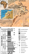
Fig 1. Geographical location and stratigraphy of the Kem Kem beds.
A, Location of Morocco (in black) in Africa (left corner), the Kem Kem and Tafilalt regions (in red) in Morocco (middle left), and the Kem Kem beds (in black) in the Kem Kem plateau (right). The dark blue star indicates the site of Jorf from which two quadrates were found; B, Stratigraphic column of the Kem Kem beds of South-Eastern Morocco. Stratigraphic position of the type remains of 1, Carcharodontosaurus saharicus (neotype; [3,23]); 2, Spinosaurus aegyptiacus (neotype; [22]); and 3, Deltadromeus agilis (holotype; [3]); and 4, probable stratigraphic position of two spinosaurid quadrates. Modified from Sereno et al. [3] and Ibrahim et al. [32].
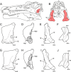
Fig 2. Quadrate position and quadrate morphotypes in Spinosaurinae from the Kem Kem beds.
A–B, Position of the quadrate bone in the Spinosaurus aegyptiacus skull in A, left lateral; and B, occipital views; C–F, Morphotype 1; and G–J, reconstructed Morphotype 2 of an idealized left quadrate in C, E, G, I, articulation with the quadratojugal (dotted line) of C–F, Spinosaurus aegyptiacus (Morphotype 1); and G–J,? Sigilmassasaurus brevicollis (Morphotype 2), in C, G, anterior; D, H, lateral; E, I, posterior; and F, J, ventral views. Abbreviations: an, angular; bo, basioccipital; bs, basisphenoid; d, dentary; dqjc, dorsal quadratojugal contact; ecc, ectocondyle; ecd, ectocondyle depression; enc, entocondyle; ics, intercondylar sulcus; j, jugal; l, lacrimal; m, maxilla; n, nasal; oc, occipital condyle; p, parietal; pm, premaxilla; pop, paroccipital process; pt, pterygoid; q, quadrate; qf, quadrate foramen; qj, quadratojugal; qr, quadrate ridge; sa, surangular; so, supraoccipital; sq, squamosal; vqjc, ventral quadratojugal contact.
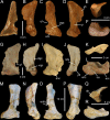
Fig 3. Quadrates of Morphotype 1 referred to Spinosaurus aegyptiacus.
A–N, Left quadrates of specimens A–F, MHNM.KK374; G–L, MHNM.KK375; and M–N, MSNM V6896, in A, G, M, anterior; B, H, N, lateral; C, O, posterior; I, posteromedial; D, posterolateral; J, P, lateral; E, K, P, dorsal; and F, L, R, ventral views. Abbreviations: dqjc, dorsal quadratojugal contact; ecc, ectocondyle; enc, entocondyle, ics, intercondylar sulcus; mfq, medial fossa; pfl, pterygoid flange; pgq, posterior groove; qf, quadrate foramen; qh, quadrate head; qr, quadrate ridge; vpdq, ventral projection of the dorsal quadratojugal contact; vqjc, ventral quadratojugal contact.
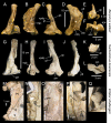
Fig 4. Quadrate of Morphotype 2 referred to Sigilmassasaurus brevicollis.
A–F, Right quadrate MHNM.KK376 in A, anterior; B, lateral; C, posterior; D, medial; E, ventral; F, ventromedial; and G, dorsal views. Abbreviations: dqjc, dorsal quadratojugal contact; ecc, ectocondyle; ecd, depression of the ectocondyle; enc, entocondyle, ics, intercondylar sulcus; mfq, medial fossa; pfl, pterygoid flange; qf, quadrate foramen; qr, quadrate ridge; vpdq, ventral projection of the dorsal quadratojugal contact; vqjc, ventral quadratojugal contact.
Fig 5. Measurements.
A–D, Measurements taken on the six spinosaurine quadrates from the Kem Kem beds of Morocco in A, lateral; B, posterior, C, medial; and D, ventral views; E, location of the ten landmarks used in the morphometric analyses in an idealized mandibular articulation of a non-avian theropod in ventral view. Abbreviations: ecc, ectocondyle; enc, entocondyle, ics, intercondylar sulcus.
Fig 6. Quadrate-based phylogeny of non-avian theropods.
Strict consensus cladogram from most parsimonious trees after the a posteriori deletion of Monolophosaurus. Initial analysis was a New Technology Search using TNT v.1.1 of a supermatrix comprising 98 quadrate-based characters combined with seven recent datasets (i.e., [,–71,73,132]) based on the whole skeleton, for one outgroup (Eoraptor lunensis) and 18 non-avian theropod taxa. Tree length = 4994; CI = 0.522, RI = 0.611. Dinosaur silhouettes by Scott Hartman (all but Coelophysoidea, Shuvuuia and Therizinosauria) and Funkmonk (Coelophysoidea, Shuvuuia, Therizinosauria) from Phylopic, used with permission.
Fig 7. Results of the geometric morphometric analysis performed on the mandibular articulation of non-avian theropods.
PCA plot of the principal component analysis performed on 37 theropod taxa and 10 landmarks along the first two principal axes explaining 35.8% and 20.04% of the variation in the sample. Colors refer to theropod clades and correspond to those in Fig 6. Major groupings at family level are delimited and outline images are associated with taxa of hypothetical extremes.
Fig 8. Results of the phylogenetic morphometric analysis.
A–B, Phylogenetic morphometric analysis of the mandibular articulation of 36 non-avian taxa performed with a degree of thoroughness of one, and using A, 10 landmarks on the quadrate in ventral view (Tree score = 5.18); and B, combination of the phylogenetic morphometric character based on 10 landmarks of the mandibular articulation in ventral view and 2377 discrete characters from the supermatrix (Tree score = 6.61).
Fig 9. Quadrate morphology in Baryonychinae and Irritator.
A–L, Left quadrates of A–F, Baryonyx walkeri (NHM R9951); and G–L, Suchomimus tenerensis (MNN GAD 502) in A, G, anterior; B, H, lateral; C, I, posterior; D, J, medial; E, K, dorsal; and F, L, ventral views. M–N, Right and O–Q, left quadrates of Irritator challengeri (SMNS 58022) with M, close up on the lateral portion of the quadrate body; N, quadrate foramen; O, anteromedial surface of the pterygoid flange; and P–Q, quadrate head in M–N, P, posterolateral, O, anterior; Q, and dorsal views. Abbreviations: ecc, ectocondyle; enc, entocondyle; lpq, lateral process; mfq, medial fossa; pfl, pterygoid flange; pin, posterior intercondylar notch; qf, quadrate foramen; qh, quadrate head; qjp, quadratojugal process; qr, quadrate ridge; qs, quadrate shaft; vpdq, ventral projection of the dorsal quadratojugal contact; vsh, ventral shelf of the pterygoid flange.
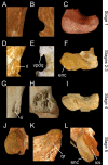
Fig 10. Ontogenetic changes in the quadrates of Spinosaurus aegyptiacus (Morphotype 1).
A–C, Left quadrate MHNM.KK374 representing ontogenetic stage 1 (juvenile) with A, close up on the smooth lateral surface of the dorsal quadratojugal contact in lateral view; B, quadrate foramen and absence of a ventral projection of the dorsal quadratojugal contact in posterior view; C, and non-delimited mandibular condyles in ventral view. D–E, Left quadrates of specimens D, F, MHNM.KK377; and E, MSNM V6896 representing ontogenetic stage 2 and 3 (non-juvenile immature individuals) with D, close up on the ridged dorsal quadratojugal contact in lateral view; E, quadrate foramen and ventral projection of the dorsal quadratojugal contact in posterior view; and F, poorly delimited mandibular condyles in ventral view. G–I, Left quadrate MHNM.KK375 representing ontogenetic stage 4 (subadult) with G, close up on the irregular and ridged lateral surface of the dorsal quadratojugal contact in lateral view; H, deeply excavated ventral quadratojugal contact in lateral view; and I, well-delimited mandibular condyles in ventral view. J–L, Left quadrate MHNM.KK378 representing ontogenetic stage 4 (fully grown individual) with J, close up on the protuberant squamosal capitulum in ventral view; K, second dorsal quadrate ridge extending to the quadrate head in posterior view; and L, well-delimited entocondyle with deep intercondylar sulcus in ventral view. Abbreviations: enc, entocondyle; ics, intercondylar sulcus; qr, quadrate ridge; ri, ridge of the dorsal quadratojugal contact; vpdq, ventral projection of the dorsal quadratojugal contact. Quadrates not to scale.
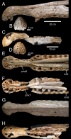
Fig 11. Comparison of the snout of two specimens of Spinosaurus from the ‘Continental intercalaire’ of Northwestern Africa.
A–H, Fused maxillae and premaxillae of A–B, E, G, MSNM V4047 referred to Spinosaurus aegyptiacus by Dal Sasso et al. [21] (courtesy of Simone Maganuco); and C–D, F, H, MNHM SAM 124 referred to Spinosaurus maroccanus (nomen dubium) by Taquet and Russell [93] in A, C, lateral; B, D, anterior; E, F, ventral; and G, H, dorsal views. Abbreviation: mx9, ninth maxillary alveolus; pmx6, sixth premaxillary alveolus, pmx7, seventh premaxillary alveolus. Scale = 20 cm (A, E, G), 10 cm (C, F, H), 5 cm (B), 2 cm (D).
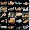
Fig 12. Morphological diversity of the mandibular articulation in non-avetheropod theropods.
A–P, R–T, right quadrate (unless indicated) in ventral view in; A, Herrerasaurus ischigualastensis (formerly ‘_Frenguellisaurus_’ ischigualastensis; PVSJ 053; left reversed); B, Eodromaeus murphi (PVSJ 562); C, Tawa hallae (GR 241; courtesy of Sterling Nesbitt); D, Dilophosaurus wetherilli (UCMP 37302; left reversed; courtesy of Juan Canale); E, Ceratosaurus nasicornis (MWC 1; left reversed); F, Masiakasaurus knopfleri (FMNH PR 2496); G, Noasaurus leali (PVL 4061); H, Ilokelesia aguadagrandensis (MCF-PVPH 35); I, Abelisaurus comahuensis (MPCA 11098; left reversed; in posteroventral view); J, Aucasaurus garridoi (MCF-PVPH 236); K, Majungasaurus crenatissimus (FMNH PR 2100; left reversed); L, Carnotaurus sastrei (MACN-CH 894; left reversed; courtesy of Pablo Asaroff); M, Baryonyx walkeri (NHM R.9951; left reversed); N, Suchomimus tenerensis (MNN GAD502; left reversed); O, Spinosaurus aegyptiacus (MHNM.KK375; left reversed); P,? Sigilmassasaurus brevicollis (Morphotype 2; MHNM.KK376; left reversed); Q, anatomy and orientation of an idealized right quadrate in ventral view; R, Eustreptospondylus oxoniensis (OUMNH J.13558; courtesy of Paul Barrett); S, Afrovenator abakensis (MNN UBA1; left reversed; courtesy of Roger Benson); T, Torvosaurus tanneri (BYU-VP 5110). Abbreviations: ecc, ectocondyle; enc, entocondyle; ics, intercondylar sulcus. Taxa framed in blue are those belonging to morphoclade II retrieved in the phylogenetic morphometric analysis (Fig 8A). Quadrates not to scale.
Fig 13. Morphological diversity of the mandibular articulation in non-avian Avetheropoda.
A–T, Right quadrate (unless indicated) in ventral view in; A, Allosaurus ‘_jimmadseni_’ (SMA 05/002); B, Aerosteon riocoloradensis (MCNA-PV-3137; left reversed; courtesy of Martin Ezcurra); C, Acrocanthosaurus atokensis (NCSM 14345); D, Giganotosaurus carolinii (MUCPv-CH-1; left reversed); E, Guanlong wucaii (IVPP V14531; left reversed; courtesy of Oliver Rauhut); F, Eotyrannus lengi (MIWG 1997.550); G, Qianzhousaurus sinensis (GM F10004-1; left reversed; courtesy of Stephen Brusatte); H, Tyrannosaurus rex (BHI l013; modified from Larson [133]); I, Bicentenaria argentina (MPCA 865; left reversed); J, Ornitholestes hermanni (AMNH FARB 619); K, Shuvuuia deserti (IGM 100–1001; left reversed); L, Gallimimus bullatus (IGM 100–1133; left reversed); M, Falcarius utahensis (UMNH VP 14559; left reversed; courtesy of Lindsay Zanno); N, Avimimus portentosus (cast of PIN 3907–3; left reversed; courtesy of Lawrence Witmer); O, Indeterminate Oviraptoridae (?Ajancingenia yanshini or? Conchoraptor gracilis; IGM A; left reversed; [85]); P, Citipati osmolskae (IGM 100–978); Q, Indeterminate Oviraptoridae (?Saurornithoides mongoliensis; IGM 100–1083; [134], modified); R, Dromaeosaurus albertensis (AMNH FARB 5356); S, Bambiraptor feinbergi (AMNH FARB 30556; left reversed); T, Tsaagan mangas (IGM 100–1015; [135]; courtesy of Mick Ellison). Taxa framed in blue are those belonging to morphoclade II retrieved in the phylogenetic morphometric analysis (Fig 8A). Quadrates not to scale.
Fig 14. Morphology of the left articular in Spinosauridae.
A–B, Baryonyx walkeri (NHM R.9951); and C, Irritator challengeri (SMNS 58022) in A, C, medial; and B, dorsal views. Abbreviations: igr, interglenoid ridge; gfo, glenoid fossa; lgd, lateral glenoid depression; mgd, medial glenoid depression; retp, retroarticular process.
Fig 15. Jaw mechanics in the spinosaurid Spinosaurus.
A–D, Mandibular articulation; and F, G, skull in A, C, F–G, lateral; and B, D, anterior views; when A–B, F, the mouth is closed; and C–D, G, fully open, illustrating the lateral movement (in red) of the mandibular ramus for a 45° rotation of the lower jaw (courtesy of Jaime A. Headden); E, skeletal reconstruction of Spinosaurus aegyptiacus by Ibrahim et al. [22]) in swimming position in lateral view with a human (1.8 m) as a scale (modified from Ibrahim et al. [22]). This model is based on spinosaurid cranial and postcranial remains (colored in red) from the Albian-Cenomanian of Northern Africa which possibly belong to two spinosaurine taxa (see also Evers et al. [27]); H, reconstruction of a semi-aquatic Spinosaurus in fishing position (i.e., jaws wide open) in anterolateral view (courtesy of Jason Poole). Abbreviations: an, angular; ar, articular; d, dentary; ecc, ectocondyle; enc, entocondyle; j, jugal; m, maxilla; n, nasal; p, parietal; pm, premaxilla; po, postorbital; pt, pterygoid; ptf, pterygoid flange; q, quadrate; qf, quadrate foramen; qj, quadratojugal; retp, retroarticular process of the articular; sa, surangular; sq, squamosal.
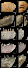
Fig 16. Morphological diversity of the mandibular symphysis in non-avian theropods in medial view.
A–B, left dentary of Spinosaurus cf. aegyptiacus (NHM R.16421); A, Anterior portion; and B, close up on the well-developed anterior ridges of the mandibular symphysis. C–E, left dentary of Baryonyx walkeri (NHM R.9951 and ML 1190); C, anterior portion; and D–E, close up on the weakly developed anterior ridges of the mandibular symphysis in D, NHM R.9951; and E, ML 1190. F–G, right dentary of Majungasaurus crenatissimus (FMNH PR 2100; reversed); F, anterior portion; and G, close up on the irregular surface of the mandibular symphysis. H–I, right dentary of Megalosaurus bucklandii (OUMNH J13505; reversed); H, anterior portion; and I, close up on the smooth surface of the mandibular symphysis. J–K, right dentary of Tyrannosaurus rex (NHM R.7994); J, anterior portion; and K, close up on the poorly developed anterodorsal ridges of the mandibular symphysis. The symphyseal surface is colored in light grey. Abbreviation: ri, anteroposterior ridges of the mandibular symphysis.
Similar articles
- A reappraisal of the morphology and systematic position of the theropod dinosaur Sigilmassasaurus from the "middle" Cretaceous of Morocco.
Evers SW, Rauhut OW, Milner AC, McFeeters B, Allain R. Evers SW, et al. PeerJ. 2015 Oct 20;3:e1323. doi: 10.7717/peerj.1323. eCollection 2015. PeerJ. 2015. PMID: 26500829 Free PMC article. - A new dinosaur (Theropoda, Spinosauridae) from the Cretaceous (Cenomanian) Alcântara Formation, Cajual Island, Brazil.
Kellner AW, Azevedo SA, Machado EB, Carvalho LB, Henriques DD. Kellner AW, et al. An Acad Bras Cienc. 2011 Mar;83(1):99-108. doi: 10.1590/s0001-37652011000100006. An Acad Bras Cienc. 2011. PMID: 21437377 - An abelisaurid (Dinosauria: Theropoda) ilium from the Upper Cretaceous (Cenomanian) of the Kem Kem beds, Morocco.
Zitouni S, Laurent C, Dyke G, Jalil NE. Zitouni S, et al. PLoS One. 2019 Apr 2;14(4):e0214055. doi: 10.1371/journal.pone.0214055. eCollection 2019. PLoS One. 2019. PMID: 30939139 Free PMC article. - The first definitive Asian spinosaurid (Dinosauria: Theropoda) from the Early Cretaceous of Laos.
Allain R, Xaisanavong T, Richir P, Khentavong B. Allain R, et al. Naturwissenschaften. 2012 May;99(5):369-77. doi: 10.1007/s00114-012-0911-7. Epub 2012 Apr 18. Naturwissenschaften. 2012. PMID: 22528021 - A large abelisaurid (Dinosauria, Theropoda) from Morocco and comments on the Cenomanian theropods from North Africa.
Chiarenza AA, Cau A. Chiarenza AA, et al. PeerJ. 2016 Feb 29;4:e1754. doi: 10.7717/peerj.1754. eCollection 2016. PeerJ. 2016. PMID: 26966675 Free PMC article.
Cited by
- Macroevolutionary patterns in the pelvis, stylopodium and zeugopodium of megalosauroid theropod dinosaurs and their importance for locomotor function.
Lacerda MBS, Bittencourt JS, Hutchinson JR. Lacerda MBS, et al. R Soc Open Sci. 2023 Aug 16;10(8):230481. doi: 10.1098/rsos.230481. eCollection 2023 Aug. R Soc Open Sci. 2023. PMID: 37593714 Free PMC article. - Isolated tooth reveals hidden spinosaurid dinosaur diversity in the British Wealden Supergroup (Lower Cretaceous).
Barker CT, Naish D, Gostling NJ. Barker CT, et al. PeerJ. 2023 May 31;11:e15453. doi: 10.7717/peerj.15453. eCollection 2023. PeerJ. 2023. PMID: 37273543 Free PMC article. - A new spinosaurid dinosaur species from the Early Cretaceous of Cinctorres (Spain).
Santos-Cubedo A, de Santisteban C, Poza B, Meseguer S. Santos-Cubedo A, et al. Sci Rep. 2023 May 18;13(1):6471. doi: 10.1038/s41598-023-33418-2. Sci Rep. 2023. PMID: 37202441 Free PMC article. - A new theropod dinosaur from the early cretaceous (Barremian) of Cabo Espichel, Portugal: Implications for spinosaurid evolution.
Mateus O, Estraviz-López D. Mateus O, et al. PLoS One. 2022 Feb 16;17(2):e0262614. doi: 10.1371/journal.pone.0262614. eCollection 2022. PLoS One. 2022. PMID: 35171930 Free PMC article. - New spinosaurids from the Wessex Formation (Early Cretaceous, UK) and the European origins of Spinosauridae.
Barker CT, Hone DWE, Naish D, Cau A, Lockwood JAF, Foster B, Clarkin CE, Schneider P, Gostling NJ. Barker CT, et al. Sci Rep. 2021 Sep 29;11(1):19340. doi: 10.1038/s41598-021-97870-8. Sci Rep. 2021. PMID: 34588472 Free PMC article.
References
- Evans DC, Barrett PM, Brink KS, Carrano MT. Osteology and bone microstructure of new, small theropod dinosaur material from the early Late Cretaceous of Morocco. Gondwana Research. 2014; 10.1016/j.gr.2014.03.016 - DOI
- Russell DA. Isolated dinosaur bones from the Middle Cretaceous of the Tafilalt, Morocco. Bulletin du Muséum National d’Histoire Naturelle. 1996;4: 349–402.
- Ibrahim N. Too many theropods? The diversity of predatory dinosaurs in the mid-Cretaceous of Morocco. 56th Annual Symposium of Vertebrate Palaeontology and Comparative Anatomy Dublin (September 2nd-6th 2008). 2008; 29.
- Cavin L, Tong H, Boudad L, Meister C, Piuz A, Tabouelle J, et al. Vertebrate assemblages from the early Late Cretaceous of southeastern Morocco: an overview. Journal of African Earth Sciences. 2010;57: 391–412.
Publication types
MeSH terms
Grants and funding
The Association des Géologues Amateurs de Belgique (AGAB), granted to C .H. for this research project at the Ulg. The funders had no role in study design, data collection and analysis, decision to publish, or preparation of the manuscript.
LinkOut - more resources
Full Text Sources
Other Literature Sources
