The CASTOR Proteins Are Arginine Sensors for the mTORC1 Pathway - PubMed (original) (raw)
The CASTOR Proteins Are Arginine Sensors for the mTORC1 Pathway
Lynne Chantranupong et al. Cell. 2016.
Abstract
Amino acids signal to the mTOR complex I (mTORC1) growth pathway through the Rag GTPases. Multiple distinct complexes regulate the Rags, including GATOR1, a GTPase activating protein (GAP), and GATOR2, a positive regulator of unknown molecular function. Arginine stimulation of cells activates mTORC1, but how it is sensed is not well understood. Recently, SLC38A9 was identified as a putative lysosomal arginine sensor required for arginine to activate mTORC1 but how arginine deprivation represses mTORC1 is unknown. Here, we show that CASTOR1, a previously uncharacterized protein, interacts with GATOR2 and is required for arginine deprivation to inhibit mTORC1. CASTOR1 homodimerizes and can also heterodimerize with the related protein, CASTOR2. Arginine disrupts the CASTOR1-GATOR2 complex by binding to CASTOR1 with a dissociation constant of ~30 μM, and its arginine-binding capacity is required for arginine to activate mTORC1 in cells. Collectively, these results establish CASTOR1 as an arginine sensor for the mTORC1 pathway.
Copyright © 2016 Elsevier Inc. All rights reserved.
Figures
Figure 1. CASTOR1 and CASTOR2 are ACT domain-containing proteins that interact with GATOR2
(A) Endogenous GATOR2, FAM164A, and CASTOR2 co-immunoprecipitate with stably expressed CASTOR1. The schematic is adapted from the BioPlex database (Huttlin et al., 2015). Solid blue lines denote proteins that were detected by mass spectrometric analysis of CASTOR1 immunoprecipitates, and dashed purple lines indicate interactions between GATOR2 subunits that were present in Bioplex. (B) Schematic alignment of human CASTOR1 and CASTOR2 proteins with annotated ACT domains. (C) The ACT domains of CASTOR1 and CASTOR2 display sequence similarity with the ACT domains of fungal aspartate kinases and putative amino acid binding proteins in bacteria. Amino acid positions are colored from white to blue in order of increasing sequence identity. The red star denotes the position of the I280 residue in CASTOR1. (D) Recombinant CASTOR1 and CASTOR2 co-immunoprecipitate endogenous GATOR2, as detected by the presence of mios. Anti-HA immunoprecipitates and lysates were prepared from HEK-293T cells cotransfected with the indicated cDNAs in expression vectors. Cell lysates and immunoprecipitates were analyzed by immunoblotting for levels of indicated proteins. HA-metap2 served as a negative control.
Figure 2. CASTOR1 and CASTOR2 form homo- and heterodimeric complexes
(A) Recombinant CASTOR1 and CASTOR2 coimmunoprecipitate both themselves and each other. HEK-293T cells were cotransfected with the indicated cDNAs in expression vectors and cell lysates and anti-HA immunoprecipitates were analyzed by immunoblotting for the indicated proteins as in Figure 1D. (B) Recombinant CASTOR2 coimmunoprecipitates endogenous CASTOR1. HEK-293T cells were cotransfected with the indicated cDNAs in expression vectors and anti-HA immunoprecipitates were collected and analyzed as in Figure 1D. The arrow denotes the band corresponding to CASTOR1. (C) Recombinant CASTOR1 coimmunoprecipitates endogenous CASTOR2. HEK-293T cells were cotransfected with the indicated cDNAs in expression vectors and anti-HA immunoprecipitates were collected and analyzed as in (A). (D) Recombinant CASTOR1 and CASTOR2 are present in approximately equal ratios within the heterodimeric complex. SDS-polyacrylamide gel electrophoresis (PAGE), followed by Coomassie blue staining, was used to analyze the indicated protein preparations from HEK-293T cells. The asterisk denotes a common protein contaminant present in these purifications.
Figure 3. Arginine regulates the interaction of GATOR2 with CASTOR1-homodimers and CASTOR1-CASTOR2 heterodimers in cells and in vitro
(A) Amino acids differentially regulate the interaction of GATOR2 with the three CASTOR complexes. HEK-293T cells cotransfected with the indicated cDNAs were deprived of amino acids for 50 min or starved and restimulated with amino acids for 10 min. Anti-HA immunoprecipitates and cell lysates were analyzed by immunoblotting for levels of the indicated proteins. (B) Endogenous CASTOR1, but not CASTOR2, associates with GATOR2 in an amino acid-sensitive manner. A HEK-293T cell line expressing endogenously FLAG-tagged WDR59 was treated as in (A) and anti-FLAG immunoprecipitates were analyzed by immunoblotting for the indicated proteins. (C) Deprivation of arginine, but not leucine, promotes the interaction between the CASTOR1 homodimer and endogenous GATOR2. Cells were deprived of leucine, arginine, or all amino acids for 50 min, and restimulated for 10 min with the respective amino acids where indicated. Anti-HA immunoprecipitates were prepared and analyzed as in (A). (D) Arginine disrupts the interaction between GATOR2 and CASTOR1-containing dimers in vitro. Anti-HA immunoprecipitates were prepared from HEK-293T cells expressing the indicated cDNAs and deprived of amino acids for 50 min. Indicated amino acids were added directly to the immunoprecipitates, which after re-washing, were analyzed as in (A). (E) Arginine dose-dependently disrupts the interaction between GATOR2 and CASTOR1-containing dimers in vitro. The experiment was performed and analyzed as in (D), except using the indicated concentrations of arginine. (F) Arginine regulates the interaction between the ACT domains of CASTOR1 but not CASTOR2 in cells. HEK-293T cells cotransfected with the indicated cDNAs in expression vectors were either deprived of arginine in the cell media for 50 min or starved and restimulated with arginine for 10 min. Anti-HA immunoprecipitates were prepared and analyzed as in (A).
Figure 4. The CASTOR1 homodimer and CASTOR1-CASTOR2 heterodimer bind arginine with a Kd of around 30 μM
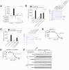
(A) Radiolabelled arginine, but not radiolabelled leucine or lysine, binds to CASTOR1 homodimers. FLAG-immunoprecipitates were prepared from HEK-293T cells cotransfected with the indicated cDNAs, and binding assays were performed with these immunoprecipitates as described in the methods. Unlabelled amino acids were added where indicated. Values are mean ± SD of three technical replicates from one representative experiment (n.s., not significant). (B) Arginine binds to CASTOR1-containing homo- and heterodimers, but not the CASTOR2 homodimer. FLAG immunoprecipitates of the indicated complexes were prepared from HEK-293T cells and analyzed as in (A). Equal volumes of eluants from immunoprecipitates of the denoted complexes were loaded and analyzed in SDS-PAGE, followed by Coomassie blue staining. (C) Arginine binds to bacterially produced CASTOR1-containing complexes, but not the CASTOR2 homodimer or the control protein Sestrin2. Proteins purified from bacteria were analyzed as in (A) and (B). (D) Arginine binds to the CASTOR1 homodimer with a dissociation constant of 34.8 μM. Binding assays were performed as in (A) with the indicated concentrations of unlabelled arginine. A representative experiment is shown, and each point represents the mean ± SD for three experiments. The Kd was calculated from four experiments. (E) Arginine binds to the CASTOR1-CASTOR2 heterodimer with a dissociation constant of 24.2 μM. FLAG-immunoprecipitates were prepared from HEK-293T cells and analyzed as in (D). (F) The concentration of arginine that half-maximally activates the mTORC1 pathway correlates with the concentration of arginine that disrupts half of the complexes of GATOR2 and CASTOR1 homodimers. HEK-293T cells were transfected with the indicated cDNAs and immunoprecipitates and lysates analyzed as in Figure 3C.
Figure 5. CASTOR1 functions in parallel with SLC38A9 to regulate arginine signaling to mTORC1
(A) Transient overexpression of recombinant CASTOR2 and CASTOR1 inhibits mTORC1 activation in response to amino acids. HEK-293T cells were cotransfected with the indicated cDNAs. Cells were treated as in Figure 3A and anti-FLAG immunoprecipitates analyzed by immunoblotting for the indicated proteins. (B) RNAi-mediated depletion of CASTOR1 in HEK-293T cells renders the mTORC1 pathway partially insensitive to arginine deprivation. HEK-293T cells stably expressing the indicated shRNAs were starved of arginine in the cell media for 50 min or starved and restimulated with arginine for 10 min. Lysates were analyzed via immunoblotting for the indicated proteins and phosphorylation states. (C) CRISPR/Cas9 mediated depletion of CASTOR1 in HEK-293T cells confers resistance of the mTORC1 pathway to arginine deprivation. HEK-293T cells stably coexpressing Cas9 with the indicated guide RNAs were treated as in (B) and lysates were analyzed by immunoblotting for indicated proteins. (D) Loss of CASTOR2 slightly increases mTORC1 activity in response to arginine. HEK-293T cells stably expressing the indicated shRNAs were treated as in (B) and lysates were analyzed by immunoblotting for indicated proteins. The normalized phosphorylated S6K1 signal under arginine stimulation for shCASTOR2_1 and shCASTOR2_2 expressing cells is 1.4 fold and 1.1 fold of shGFP expressing cells, respectively, as quantified with ImageJ. (E) CASTOR1 and SLC38A9 likely function in parallel to signal arginine availability to the mTORC1 pathway. Wild type or SLC38A9 knockout HEK-293T cells expressing the indicated shRNAs were treated as in (B) and lysates were analyzed by immunoblotting for indicated proteins.
Figure 6. Arginine must be able to bind to CASTOR1 for it to activate mTORC1
(A) The CASTOR1 I280A mutant does not bind arginine. Binding assays were performed with FLAG immunoprecipitates of the indicated complexes as in Figure 4A. (B) Arginine does not regulate the interaction of CASTOR1 I280A with GATOR2. HEK-293T cells cotransfected with the indicated cDNAs in expression vectors were treated as in Figure 5B and anti-HA immunoprecipitates were analyzed by immunoblotting for levels of the indicated proteins. (C) Reintroduction of the CASTOR1 I280A mutant into CASTOR1 knockdown cells renders the mTORC1 pathway unable to sense the presence of arginine. HEK-293T cells stably expressing the indicated shRNAs and cDNA constructs were treated as in Figure 5B and lysates analyzed by immunoblotting for indicated proteins. (D) A model depicting how the cytosolic and lysosomal amino acid inputs impinge on CASTORs, Sestrins, and SLC38A9 to regulate mTORC1 activity.
Comment in
- CASTORing New Light on Amino Acid Sensing.
Hallett JEH, Manning BD. Hallett JEH, et al. Cell. 2016 Mar 24;165(1):15-17. doi: 10.1016/j.cell.2016.03.002. Cell. 2016. PMID: 27015302
Similar articles
- Mechanism of arginine sensing by CASTOR1 upstream of mTORC1.
Saxton RA, Chantranupong L, Knockenhauer KE, Schwartz TU, Sabatini DM. Saxton RA, et al. Nature. 2016 Aug 11;536(7615):229-33. doi: 10.1038/nature19079. Epub 2016 Aug 3. Nature. 2016. PMID: 27487210 Free PMC article. - Sestrin2 is a leucine sensor for the mTORC1 pathway.
Wolfson RL, Chantranupong L, Saxton RA, Shen K, Scaria SM, Cantor JR, Sabatini DM. Wolfson RL, et al. Science. 2016 Jan 1;351(6268):43-8. doi: 10.1126/science.aab2674. Epub 2015 Oct 8. Science. 2016. PMID: 26449471 Free PMC article. - Metabolism. Lysosomal amino acid transporter SLC38A9 signals arginine sufficiency to mTORC1.
Wang S, Tsun ZY, Wolfson RL, Shen K, Wyant GA, Plovanich ME, Yuan ED, Jones TD, Chantranupong L, Comb W, Wang T, Bar-Peled L, Zoncu R, Straub C, Kim C, Park J, Sabatini BL, Sabatini DM. Wang S, et al. Science. 2015 Jan 9;347(6218):188-94. doi: 10.1126/science.1257132. Epub 2015 Jan 7. Science. 2015. PMID: 25567906 Free PMC article. - Recent Advances in Understanding Amino Acid Sensing Mechanisms that Regulate mTORC1.
Zheng L, Zhang W, Zhou Y, Li F, Wei H, Peng J. Zheng L, et al. Int J Mol Sci. 2016 Sep 29;17(10):1636. doi: 10.3390/ijms17101636. Int J Mol Sci. 2016. PMID: 27690010 Free PMC article. Review. - Rheb and Rags come together at the lysosome to activate mTORC1.
Groenewoud MJ, Zwartkruis FJ. Groenewoud MJ, et al. Biochem Soc Trans. 2013 Aug;41(4):951-5. doi: 10.1042/BST20130037. Biochem Soc Trans. 2013. PMID: 23863162 Review.
Cited by
- Principles and functions of metabolic compartmentalization.
Bar-Peled L, Kory N. Bar-Peled L, et al. Nat Metab. 2022 Oct;4(10):1232-1244. doi: 10.1038/s42255-022-00645-2. Epub 2022 Oct 20. Nat Metab. 2022. PMID: 36266543 Free PMC article. Review. - Metabolic signaling in T cells.
Shyer JA, Flavell RA, Bailis W. Shyer JA, et al. Cell Res. 2020 Aug;30(8):649-659. doi: 10.1038/s41422-020-0379-5. Epub 2020 Jul 24. Cell Res. 2020. PMID: 32709897 Free PMC article. Review. - Amino acid-dependent control of mTORC1 signaling: a variety of regulatory modes.
Takahara T, Amemiya Y, Sugiyama R, Maki M, Shibata H. Takahara T, et al. J Biomed Sci. 2020 Aug 17;27(1):87. doi: 10.1186/s12929-020-00679-2. J Biomed Sci. 2020. PMID: 32799865 Free PMC article. Review. - Coordinated Regulation of Myelination by Growth Factor and Amino-acid Signaling Pathways.
Yang Z, Yu Z, Xiao B. Yang Z, et al. Neurosci Bull. 2023 Mar;39(3):453-465. doi: 10.1007/s12264-022-00967-x. Epub 2022 Nov 9. Neurosci Bull. 2023. PMID: 36352321 Free PMC article. Review. - Lysosomes as coordinators of cellular catabolism, metabolic signalling and organ physiology.
Settembre C, Perera RM. Settembre C, et al. Nat Rev Mol Cell Biol. 2024 Mar;25(3):223-245. doi: 10.1038/s41580-023-00676-x. Epub 2023 Nov 24. Nat Rev Mol Cell Biol. 2024. PMID: 38001393 Review.
References
- Aravind L, Koonin EV. Gleaning non-trivial structural, functional and evolutionary information about proteins by iterative database searches. Journal of Molecular Biology. 1999;287:1023–1040. - PubMed
- Ban H, Shigemitsu K, Yamatsuji T, Haisa M, Nakajo T, Takaoka M, Nobuhisa T, Gunduz M, Tanaka N, Naomoto Y. Arginine and Leucine regulate p70 S6 kinase and 4E-BP1 in intestinal epithelial cells. International journal of molecular medicine. 2004;13:537–543. - PubMed
- Blommaart EFC, Luiken JJFP, Blommaart PJE, van Woerkom GM, Meijer AJ. Phosphorylation of Ribosomal Protein S6 Is Inhibitory for Autophagy in Isolated Rat Hepatocytes. Journal of Biological Chemistry. 1995;270:2320–2326. - PubMed
Publication types
MeSH terms
Substances
Grants and funding
- R01 AI047389/AI/NIAID NIH HHS/United States
- R01 CA103866/CA/NCI NIH HHS/United States
- T32 GM007287/GM/NIGMS NIH HHS/United States
- P30 CA014051/CA/NCI NIH HHS/United States
- F31 CA180271/CA/NCI NIH HHS/United States
- R01 GM095567/GM/NIGMS NIH HHS/United States
- R37 AI047389/AI/NIAID NIH HHS/United States
- U41 HG006673/HG/NHGRI NIH HHS/United States
- R01CA103866/CA/NCI NIH HHS/United States
- AI47389)/AI/NIAID NIH HHS/United States
- F31 CA189437/CA/NCI NIH HHS/United States
LinkOut - more resources
Full Text Sources
Other Literature Sources
Molecular Biology Databases
Research Materials
Miscellaneous


