Melatonin: A Mitochondrial Targeting Molecule Involving Mitochondrial Protection and Dynamics - PubMed (original) (raw)
Review
Melatonin: A Mitochondrial Targeting Molecule Involving Mitochondrial Protection and Dynamics
Dun-Xian Tan et al. Int J Mol Sci. 2016.
Abstract
Melatonin has been speculated to be mainly synthesized by mitochondria. This speculation is supported by the recent discovery that aralkylamine _N_-acetyltransferase/serotonin _N_-acetyltransferase (AANAT/SNAT) is localized in mitochondria of oocytes and the isolated mitochondria generate melatonin. We have also speculated that melatonin is a mitochondria-targeted antioxidant. It accumulates in mitochondria with high concentration against a concentration gradient. This is probably achieved by an active transportation via mitochondrial melatonin transporter(s). Melatonin protects mitochondria by scavenging reactive oxygen species (ROS), inhibiting the mitochondrial permeability transition pore (MPTP), and activating uncoupling proteins (UCPs). Thus, melatonin maintains the optimal mitochondrial membrane potential and preserves mitochondrial functions. In addition, mitochondrial biogenesis and dynamics is also regulated by melatonin. In most cases, melatonin reduces mitochondrial fission and elevates their fusion. Mitochondrial dynamics exhibit an oscillatory pattern which matches the melatonin circadian secretory rhythm in pinealeocytes and probably in other cells. Recently, melatonin has been found to promote mitophagy and improve homeostasis of mitochondria.
Keywords: antioxidant; melatonin; mitochondria; mitochondrial dynamics; mitophagy.
Conflict of interest statement
The authors declare no conflict of interest.
Figures
Figure 1
Large amounts of mitochondria are present in pinealocytes of the Syrian hamster (34,000×). Inset shows a longitudinal section of mitochondrion with cristae arranged like a string of beads (44,500×). Modified from Bucana et al. [89].
Figure 2
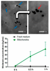
Upper panel: The localization of the SNAT. Blue arrows: The mitochondria isolated from the oocytes of mice. White arrow: enlarged image from left side of the arrow tail. Red arrow: SNAT staining (black dot). SNAT: serotonin _N_-acetyltransferase; Low panel: Melatonin concentrations in mitochondrial culture media with 10−4 M serotonin (mean ± SEM) modified from He et al. [112].
Figure 3
The similarities of mitochondrial dynamics in pinealocytes, brain neurons and cultured SH-SY5Y cells. Upper panel: Mitochondrial dynamics in pinealocytes (27,000×). (A) Condensed state; (B) Second intermediate state; (C) Third intermediate state; Middle panel: Mitochondrial dynamics in brain neurons of mice. (E) Mitochondrial fission induced by cadmium treatment; (F) The transition of mitochondrial fission and fusion in the animal treated with cadmium plus melatonin; (G) Mitochondrial fusion in control healthy animal; Lower panel: Mitochondrial dynamics in cultured SH-SY5Y cells (60,000×). (H) Mitochondrial fission induced by methamphetamine; (I) The transition of mitochondrial fission and fusion in cells treated with methamphetamine plus melatonin; (J) Mitochondrial fusion in control cells. The similarities of A, E and H; B, F and I; C, G and J are obvious. Mordified from [90,184,185].
Figure 4
A summary of the potential effects of melatonin on a mitochondrion. MPTP: mitochondrial permeability transition pore; UCP: uncoupling protein; ROS: reactive oxygen species; ETC: electron transport chain; Cyto C: cytochrome C; AFMK: a melatonin metabolite, _N_1-acetyl-_N_2-fomyl-5-methoxykynuramine, which is also a potent antioxidant. Melatonin is metabolized to AFMK by cytochrome C via pseudo-enzymatic process [77]. Upper panel: The targeting sites of melatonin on mitochondrion; green lines: inhibition; red arrows: activation; red dash arrows: the directions of the multiple steps of reactions; black arrows: directions; Lower panel: Summary of the outcomes induced by melatonin’s action on mitochondrion; upward arrowhead: activation; downward arrowhead: inhibition; horizontal arrowhead: preservation; connecting lines indicate hierarches of the events.
Similar articles
- Mitophagy in yeast: Molecular mechanisms and physiological role.
Kanki T, Furukawa K, Yamashita S. Kanki T, et al. Biochim Biophys Acta. 2015 Oct;1853(10 Pt B):2756-65. doi: 10.1016/j.bbamcr.2015.01.005. Epub 2015 Jan 17. Biochim Biophys Acta. 2015. PMID: 25603537 Review. - Melatonin: an ancient molecule that makes oxygen metabolically tolerable.
Manchester LC, Coto-Montes A, Boga JA, Andersen LP, Zhou Z, Galano A, Vriend J, Tan DX, Reiter RJ. Manchester LC, et al. J Pineal Res. 2015 Nov;59(4):403-19. doi: 10.1111/jpi.12267. Epub 2015 Sep 11. J Pineal Res. 2015. PMID: 26272235 Review. - Mitochondria Synthesize Melatonin to Ameliorate Its Function and Improve Mice Oocyte's Quality under in Vitro Conditions.
He C, Wang J, Zhang Z, Yang M, Li Y, Tian X, Ma T, Tao J, Zhu K, Song Y, Ji P, Liu G. He C, et al. Int J Mol Sci. 2016 Jun 14;17(6):939. doi: 10.3390/ijms17060939. Int J Mol Sci. 2016. PMID: 27314334 Free PMC article. - Mitophagy and mitochondrial dynamics in Saccharomyces cerevisiae.
Müller M, Lu K, Reichert AS. Müller M, et al. Biochim Biophys Acta. 2015 Oct;1853(10 Pt B):2766-74. doi: 10.1016/j.bbamcr.2015.02.024. Epub 2015 Mar 6. Biochim Biophys Acta. 2015. PMID: 25753536 Review. - PM2.5 induces liver fibrosis via triggering ROS-mediated mitophagy.
Qiu YN, Wang GH, Zhou F, Hao JJ, Tian L, Guan LF, Geng XK, Ding YC, Wu HW, Zhang KZ. Qiu YN, et al. Ecotoxicol Environ Saf. 2019 Jan 15;167:178-187. doi: 10.1016/j.ecoenv.2018.08.050. Epub 2018 Oct 15. Ecotoxicol Environ Saf. 2019. PMID: 30336408
Cited by
- Clinical Trials for Use of Melatonin to Fight against COVID-19 Are Urgently Needed.
Kleszczyński K, Slominski AT, Steinbrink K, Reiter RJ. Kleszczyński K, et al. Nutrients. 2020 Aug 24;12(9):2561. doi: 10.3390/nu12092561. Nutrients. 2020. PMID: 32847033 Free PMC article. Review. - Unveiling the Protective Role of Melatonin in Osteosarcoma: Current Knowledge and Limitations.
Al-Ansari N, Samuel SM, Büsselberg D. Al-Ansari N, et al. Biomolecules. 2024 Jan 24;14(2):145. doi: 10.3390/biom14020145. Biomolecules. 2024. PMID: 38397382 Free PMC article. Review. - Antioxidants in Down Syndrome: From Preclinical Studies to Clinical Trials.
Rueda Revilla N, Martínez-Cué C. Rueda Revilla N, et al. Antioxidants (Basel). 2020 Aug 3;9(8):692. doi: 10.3390/antiox9080692. Antioxidants (Basel). 2020. PMID: 32756318 Free PMC article. Review. - Effects of inflammation and oxidative stress on postoperative delirium in cardiac surgery.
Pang Y, Li Y, Zhang Y, Wang H, Lang J, Han L, Liu H, Xiong X, Gu L, Wu X. Pang Y, et al. Front Cardiovasc Med. 2022 Nov 22;9:1049600. doi: 10.3389/fcvm.2022.1049600. eCollection 2022. Front Cardiovasc Med. 2022. PMID: 36505383 Free PMC article. Review. - Neuroprotective Effects of Melatonin during Demyelination and Remyelination Stages in a Mouse Model of Multiple Sclerosis.
Abo Taleb HA, Alghamdi BS. Abo Taleb HA, et al. J Mol Neurosci. 2020 Mar;70(3):386-402. doi: 10.1007/s12031-019-01425-6. Epub 2019 Nov 11. J Mol Neurosci. 2020. PMID: 31713152
References
Publication types
MeSH terms
Substances
LinkOut - more resources
Full Text Sources
Other Literature Sources


