Follow-up chest radiographic findings in patients with MERS-CoV after recovery - PubMed (original) (raw)
Follow-up chest radiographic findings in patients with MERS-CoV after recovery
Karuna M Das et al. Indian J Radiol Imaging. 2017 Jul-Sep.
Abstract
Purpose: To evaluate the follow-up chest radiographic findings in patients with Middle East respiratory syndrome coronavirus (MERS-CoV) who were discharged from the hospital following improved clinical symptoms.
Materials and methods: Thirty-six consecutive patients (9 men, 27 women; age range 21-73 years, mean ± SD 42.5 ± 14.5 years) with confirmed MERS-CoV underwent follow-up chest radiographs after recovery from MERS-CoV. The 36 chest radiographs were obtained at 32 to 230 days with a median follow-up of 43 days. The reviewers systemically evaluated the follow-up chest radiographs from 36 patients for lung parenchymal, airway, pleural, hilar and mediastinal abnormalities. Lung parenchyma and airways were assessed for consolidation, ground-glass opacity (GGO), nodular opacity and reticular opacity (i.e., fibrosis). Follow-up chest radiographs were also evaluated for pleural thickening, pleural effusion, pneumothorax and lymphadenopathy. Patients were categorized into two groups: group 1 (no evidence of lung fibrosis) and group 2 (chest radiographic evidence of lung fibrosis) for comparative analysis. Patient demographics, length of ventilations days, number of intensive care unit (ICU) admission days, chest radiographic score, chest radiographic deterioration pattern (Types 1-4) and peak lactate dehydrogenase level were compared between the two groups using the student _t_-test, Mann-Whitney U test and Fisher's exact test.
Results: Follow-up chest radiographs were normal in 23 out of 36 (64%) patients. Among the patients with abnormal chest radiographs (13/36, 36%), the following were found: lung fibrosis in 12 (33%) patients GGO in 2 (5.5%) patients, and pleural thickening in 2 (5.5%) patients. Patients with lung fibrosis had significantly greater number of ICU admission days (19 ± 8.7 days; P value = 0.001), older age (50.6 ± 12.6 years; P value = 0.02), higher chest radiographic scores [10 (0-15.3); P value = 0.04] and higher peak lactate dehydrogenase levels (315-370 U/L; P value = 0.001) when compared to patients without lung fibrosis.
Conclusion: Lung fibrosis may develop in a substantial number of patients who have recovered from Middle East respiratory syndrome coronavirus (MERS-CoV). Significantly greater number of ICU admission days, older age, higher chest radiographic scores, chest radiographic deterioration patterns and peak lactate dehydrogenase levels were noted in the patients with lung fibrosis on follow-up chest radiographs after recovery from MERS-CoV.
Keywords: Chest radiograph; Lung fibrosis; MERS-CoV.
Conflict of interest statement
There are no conflicts of interest.
Figures
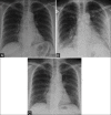
Figure 1 (A-C)
A 58-year-old male with Middle East respiratory syndrome coronavirus (MERS-CoV), serial radiographs showing irregular reticular lines of fibrosis (A) Frontal chest radiograph obtained on two days before illness shows a normal chest radiograph. (B) A follow-up frontal chest radiograph obtained at day 5, shows ground-glass opacities in the right lower zone and left mid and lower zones. (C) A follow-up frontal chest radiograph obtained at day 33 shows unilateral multiple irregular reticular lines of fibrosis in the right lower and left mid zones
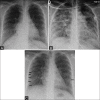
Figure 2 (A-C)
A 33-year-old female with Middle East respiratory syndrome coronavirus (MERS-CoV), serial radiographs showing multiple irregular reticular lines of fibrosis on follow-up chest radiographs. The rest of the lung is completely free of any irregular reticular lines of fibrosis (A) Frontal chest radiograph obtained on the initial presentation shows the area of ground-glass opacity at the right cardio-phrenic angle. (B) A follow-up frontal chest radiograph obtained at day 20, shows bilateral diffuse ground-glass opacities with occasional airspace consolidations. (C) A follow-up frontal chest radiograph obtained at day 230, shows bilateral multiple irregular reticular lines of fibrosis (arrows)
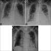
Figure 3 (A-C)
A 73-year-old female with Middle East respiratory syndrome coronavirus (MERS-CoV), serial radiographs showing multiple thick reticular lines of fibrosis and sub-pleural reticular opacities on follow-up chest radiographs. (A) A frontal chest radiograph obtained on day 3, of the initial presentation, shows bilateral ill-defined ground-glass opacities with air space disease at both lung bases. (B) A follow-up frontal chest radiograph obtained at day 19, shows bilateral sub-pleural ground-glass opacities with occasional air space consolidations. (C) A follow-up frontal chest radiograph obtained at day 130, shows unilateral thick multiple linear fibrotic parenchymal bands (arrows) in the right side along with sub-pleural reticulations, pleural thickening, and bilateral ground-glass opacities
Figure 4 (A and B)
A 24-year-old female with Middle East respiratory syndrome coronavirus (MERS-CoV), serial radiographs showing ground-glass opacities on follow-up chest radiographs. (A) A frontal chest radiograph obtained on the second day of the presentation shows the area of air space consolidation involving the lingual and left lower lobe. (B) Follow-up frontal chest radiograph obtained at 210 days shows only a small area of ground-glass opacity obscuring the left cardiac border
Figure 5
A 52-year-old female with Middle East respiratory syndrome coronavirus (MERS-CoV). A frontal chest radiograph obtained at day 168, shows bilateral multiple irregular reticular lines of fibrosis along with obscured lateral aspect of the right hemidiaphragm and costo-phrenic angle in the same side due to pleural thickening
Similar articles
- Acute Middle East Respiratory Syndrome Coronavirus: Temporal Lung Changes Observed on the Chest Radiographs of 55 Patients.
Das KM, Lee EY, Al Jawder SE, Enani MA, Singh R, Skakni L, Al-Nakshabandi N, AlDossari K, Larsson SG. Das KM, et al. AJR Am J Roentgenol. 2015 Sep;205(3):W267-74. doi: 10.2214/AJR.15.14445. Epub 2015 Jun 23. AJR Am J Roentgenol. 2015. PMID: 26102309 - Swine-origin influenza a (H1N1) viral infection in children: initial chest radiographic findings.
Lee EY, McAdam AJ, Chaudry G, Fishman MP, Zurakowski D, Boiselle PM. Lee EY, et al. Radiology. 2010 Mar;254(3):934-41. doi: 10.1148/radiol.09092083. Epub 2009 Dec 23. Radiology. 2010. PMID: 20032128 - CT correlation with outcomes in 15 patients with acute Middle East respiratory syndrome coronavirus.
Das KM, Lee EY, Enani MA, AlJawder SE, Singh R, Bashir S, Al-Nakshbandi N, AlDossari K, Larsson SG. Das KM, et al. AJR Am J Roentgenol. 2015 Apr;204(4):736-42. doi: 10.2214/AJR.14.13671. Epub 2015 Jan 23. AJR Am J Roentgenol. 2015. PMID: 25615627 - Middle East Respiratory Syndrome Coronavirus: What Does a Radiologist Need to Know?
Das KM, Lee EY, Langer RD, Larsson SG. Das KM, et al. AJR Am J Roentgenol. 2016 Jun;206(6):1193-201. doi: 10.2214/AJR.15.15363. Epub 2016 Mar 21. AJR Am J Roentgenol. 2016. PMID: 26998804 Review. - COVID-19 and MERS: Are their Chest X-ray and Computed Tomography Scanning Signs Related?
Ghaderian M, Kiani M, Shahbazi-Gahrouei S, Shahbazi-Gahrouei D, Ghadimi Moghadam A, Haghani M. Ghaderian M, et al. J Med Signals Sens. 2021 Dec 28;12(1):1-7. doi: 10.4103/jmss.JMSS_84_20. eCollection 2022 Jan-Mar. J Med Signals Sens. 2021. PMID: 35265460 Free PMC article. Review.
Cited by
- Post-discharge chest CT findings and pulmonary function tests in severe COVID-19 patients.
Balbi M, Conti C, Imeri G, Caroli A, Surace A, Corsi A, Mercanzin E, Arrigoni A, Villa G, Di Marco F, Bonaffini PA, Sironi S. Balbi M, et al. Eur J Radiol. 2021 May;138:109676. doi: 10.1016/j.ejrad.2021.109676. Epub 2021 Mar 20. Eur J Radiol. 2021. PMID: 33798931 Free PMC article. - Histopathological features in fatal COVID-19 acute respiratory distress syndrome.
Merdji H, Mayeur S, Schenck M, Oulehri W, Clere-Jehl R, Cunat S, Herbrecht JE, Janssen-Langenstein R, Nicolae A, Helms J, Meziani F, Chenard MP; CRICS TRIGGERSEP Group (Clinical Research in Intensive Care, Sepsis Trial Group for Global Evaluation, Research in Sepsis). Merdji H, et al. Med Intensiva (Engl Ed). 2021 Jun-Jul;45(5):261-270. doi: 10.1016/j.medin.2021.02.007. Epub 2021 Feb 27. Med Intensiva (Engl Ed). 2021. PMID: 34054173 Free PMC article. - Histopathological features in fatal COVID-19 acute respiratory distress syndrome.
Merdji H, Mayeur S, Schenck M, Oulehri W, Clere-Jehl R, Cunat S, Herbrecht JE, Janssen-Langenstein R, Nicolae A, Helms J, Meziani F, Chenard MP; CRICS TRIGGERSEP Group (Clinical Research in Intensive Care, Sepsis Trial Group for Global Evaluation, Research in Sepsis). Merdji H, et al. Med Intensiva (Engl Ed). 2021 Jun-Jul;45(5):261-270. doi: 10.1016/j.medine.2021.02.005. Med Intensiva (Engl Ed). 2021. PMID: 34059216 Free PMC article. - Fibrotic-like abnormalities notably prevalent one year after hospitalization with COVID-19.
van Raaij BFM, Stöger JL, Hinnen C, Penfornis KM, de Jong CMM, Klok FA, Roukens AHE, Veldhuijzen DS, Arbous MS, Noordam R, Marges ER, Geelhoed JJM. van Raaij BFM, et al. Respir Med Res. 2022 Nov;82:100973. doi: 10.1016/j.resmer.2022.100973. Epub 2022 Nov 17. Respir Med Res. 2022. PMID: 36403358 Free PMC article. - Unveiling the Clinical Spectrum of Post-COVID-19 Conditions: Assessment and Recommended Strategies.
Assiri AM, Alamaa T, Elenezi F, Alsagheir A, Alzubaidi L, TIeyjeh I, Alhomod AS, Gaffas EM, Amer SA. Assiri AM, et al. Cureus. 2024 Jan 23;16(1):e52827. doi: 10.7759/cureus.52827. eCollection 2024 Jan. Cureus. 2024. PMID: 38406111 Free PMC article. Review.
References
- Zaki AM, Van Boheemen S, Bestebroer TM, Osterhaus AD, Fouchier RA. Isolation of a novel coronavirus from a man with pneumonia in Saudi Arabia. N Engl J Med. 2012;367:1814–20. - PubMed
- World Health Organization. Global Alert and Response (GAR). Corona virus infections. http://www.who.int/csr/disease/coronavirus_infections/en/
- World Health Organization. Preliminary clinical description of severe acute respiratory syndrome. [Accessed 2003 March 21]. www.who.int/csr/sars/clinical/en/
- Das KM, Lee EY, Jawder SE, Enani MA, Singh R, Skakni L, et al. Acute Middle East Respiratory Syndrome Coronavirus: Temporal Lung Changes Observed on the Chest Radiographs of 55 Patients. AJR Am J Roentgenol. 2015;205:W267–74. - PubMed
LinkOut - more resources
Full Text Sources
Other Literature Sources


