Host gene expression profiles in ferrets infected with genetically distinct henipavirus strains - PubMed (original) (raw)
Host gene expression profiles in ferrets infected with genetically distinct henipavirus strains
Alberto J Leon et al. PLoS Negl Trop Dis. 2018.
Abstract
Henipavirus infection causes severe respiratory and neurological disease in humans that can be fatal. To characterize the pathogenic mechanisms of henipavirus infection in vivo, we performed experimental infections in ferrets followed by genome-wide gene expression analysis of lung and brain tissues. The Hendra, Nipah-Bangladesh, and Nipah-Malaysia strains caused severe respiratory and neurological disease with animals succumbing around 7 days post infection. Despite the presence of abundant viral shedding, animal-to-animal transmission did not occur. The host gene expression profiles of the lung tissue showed early activation of interferon responses and subsequent expression of inflammation-related genes that coincided with the clinical deterioration. Additionally, the lung tissue showed unchanged levels of lymphocyte markers and progressive downregulation of cell cycle genes and extracellular matrix components. Infection in the brain resulted in a limited breadth of the host responses, which is in accordance with the immunoprivileged status of this organ. Finally, we propose a model of the pathogenic mechanisms of henipavirus infection that integrates multiple components of the host responses.
Conflict of interest statement
The authors have declared that no competing interests exist.
Figures
Fig 1. Differences in minimal infectious dose of HeV, NiV-M and NiV-B in ferrets.
A first study using different infective doses of the three viruses was performed to establish the LD50 of each virus in ferrets: A-C) Survival rates are shown for 6-week old ferrets (n = 5 per group) that were infected with different doses (101, 102, 103 and 105 TCID50) of NiV-M, NiV-B and HeV, respectively. D-F) A comparative study using a dose of 5,000 TCID50 for each of the three viruses was performed in ferrets: D) cumulative clinical scores, calculated as described in the methods section; E) % lung weight per body weight; F) average number of histological lesions in the lung tissue per field of view. Tissues were harvested from 6-week old ferrets infected with NiV-B (grey), NiV-M (black solid) and HeV (black dashed) on various days post infection as described in Materials and Methods. Samples from 5 animals per group were analyzed at each time point. * p<0.01, two-way ANOVA, Bonferroni’s multiple comparison test. Error bars represent standard deviations.
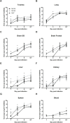
Fig 2. Viral spread across body compartments in henipavirus infected ferrets.
A comparative study using a dose of 5,000 TCID50 for each of the three viruses was performed in ferrets: viral growth in the ferret tissue, as measured by quantitative RT-PCR in the trachea (A), lung (B), olfactory bulb (C), frontal brain (D), liver (E), kidney (F), spleen (G), and blood(H); tissues were harvested from 6-week old ferrets infected with NiV-B (grey), NiV-M (black solid) and HeV (black dashed) on various days post infection as described in Materials and Methods. Samples from 5 animals per group were analyzed at each time point. * p<0.01, two-way ANOVA, Bonferroni’s multiple comparison test. Error bars represent standard deviations.
Fig 3. Gene expression profiles in the lung tissue of ferrets infected with HeV and NiV-B.
Ferrets were infected with 5,000 TCID50 of HeV and NiV-B and euthanized at 3 and 5 d.p.i. and together with an uninfected control group (n = 3 per group). Transcriptomic analysis of the lung tissue was performed by RNA-seq. A) Number of significantly up- and down-regulated genes with respect to the control group (fold-variation>±1.5 and FDR<0.05) at 3 and 5 dpi. B) Functional gene classification of differentially expressed genes. For each time-point, up- and down-regulated genes were subjected to functional classification and a selected group of gene categories from 1Gene Ontology and 2KEGG Pathways that are related with the immune responses are shown. For each functional category, the number of up- and down-regulated genes and the level of significance of the enrichment (Fisher’s exact test p-value) are indicated. C) Heatmap showing the level of activation of genes belonging to the Response to Type I Interferon category, which encompasses antiviral response genes, during the course of HeV and NiV-B infection. D) Functional enrichment of up-regulated genes that belong to protein-protein interaction (PPI) hubs of transcription factors. E-F) Evolution in the mRNA expression levels of selected macrophage markers and lymphocyte markers, respectively, during HeV and NiV-B infection. These sets of markers were selected from the literature according to their association with these two cell populations, and their coordinated upregulation reflects an increase of the associated cell population in the tissue. Dark grey shaded areas indicate no variation with respect to the control group (<±1.5 fold-change).
Fig 4. Decreased expression of genes related with cell cycle and growth signaling in the lung tissue during infection with HeV and NiV-B.
A) Evolution of gene enrichment score (Fisher’s exact test p-value) of the Cell Cycle KEGG Pathway throughout the infection. B) Functional gene classification of differentially expressed genes in the lung tissue. Regulated genes were subjected to functional classification and selected 1Gene Ontology categories and 2KEGG Pathways that are related to the immune responses and cell growth signaling are shown. For each functional category, the number of up- and down-regulated genes and the level of significance of the enrichment (Fisher’s exact test p-value) are indicated. C) Functional enrichment of down-regulated genes that belong to protein-protein interaction (PPI) hubs of transcription factors.
Fig 5. Gene expression profiles in the brain of ferrets infected with HeV and NiV-B.
Ferrets were infected with 5,000 TCID50 of HeV and NiV-B and euthanized at 3 and 5 d.p.i. (n = 3 per group, except for HeV_D5 with n = 2) and together with an uninfected control group (n = 2). Transcriptomic analysis of frontal lobe tissue was performed by RNA-seq. A) Number of significantly up- and down-regulated genes with respect to the control group (fold-variation>±1.5 and FDR<0.05) at 3 and 5 d.p.i. B) Evolution of gene enrichment score (Fisher’s exact test p-value) of the Innate Immune Response category (Gene Ontology) throughout the infection. C) Functional gene classification of differentially expressed genes in the brain tissue. Regulated genes were subjected to functional classification and a selected 1Gene Ontology categories and 2KEGG Pathways that are related to the immune responses and cell growth signaling are shown. For each functional category, the number of up- and down-regulated genes and the level of significance of the enrichment (Fisher’s exact test p-value) are indicated. D-E) Evolution in the mRNA expression levels of selected macrophage markers and lymphocyte markers, respectively, during HeV and NiV-B infection in the brain tissue. Dark grey shaded areas indicate no variation with respect to the control group (<±1.5 fold-change).
Fig 6. Sequential activation of host responses during Henipavirus infection.
A) Upon infection of the upper and lower respiratory tract, the virus quickly spreads to a myriad of organs, including the central nervous system, liver, spleen, heart, kidney, bladder and blood (schematic representation of the data from HeV infection). B) In the lung tissue, the virus presents high growth rates and only after the inflammatory responses become fully active, at around day 4, the levels of virus stabilize but without significantly decreasing. Signaling and effector molecules of the innate immunity become expressed, innate immune cells migrate to the lung tissue, but, interestingly, gene expression data strongly suggest that lymphocytes are not migrating and expanding in the affected tissues. Additionally, the expression of genes related to the cell cycle, growth factors and growth factor signaling decline through the infectious process, possibly related to the degradation of the physiological functions of the lung tissue. C) In the brain tissue, the virus grows at a slower rate as compared to the lung tissue. The brain mounts a restricted immune response in accordance with the immunoprivileged status of this tissue that includes infiltration and/or local expansion of macrophages, and possibly of lymphocytes, but without leading to a noticeable alteration of the viral growth kinetics.
Similar articles
- Transcriptome Profiling of the Virus-Induced Innate Immune Response in Pteropus vampyrus and Its Attenuation by Nipah Virus Interferon Antagonist Functions.
Glennon NB, Jabado O, Lo MK, Shaw ML. Glennon NB, et al. J Virol. 2015 Aug;89(15):7550-66. doi: 10.1128/JVI.00302-15. Epub 2015 May 13. J Virol. 2015. PMID: 25972557 Free PMC article. - A Functional Genomics Approach to Henipavirus Research: The Role of Nuclear Proteins, MicroRNAs and Immune Regulators in Infection and Disease.
Stewart CR, Deffrasnes C, Foo CH, Bean AGD, Wang LF. Stewart CR, et al. Curr Top Microbiol Immunol. 2018;419:191-213. doi: 10.1007/82_2017_28. Curr Top Microbiol Immunol. 2018. PMID: 28674944 Free PMC article. Review. - Pathogenesis of Hendra and Nipah virus infection in humans.
Escaffre O, Borisevich V, Rockx B. Escaffre O, et al. J Infect Dev Ctries. 2013 Apr 17;7(4):308-11. doi: 10.3855/jidc.3648. J Infect Dev Ctries. 2013. PMID: 23592639 Review. - Type I interferon signaling protects mice from lethal henipavirus infection.
Dhondt KP, Mathieu C, Chalons M, Reynaud JM, Vallve A, Raoul H, Horvat B. Dhondt KP, et al. J Infect Dis. 2013 Jan 1;207(1):142-51. doi: 10.1093/infdis/jis653. Epub 2012 Oct 22. J Infect Dis. 2013. PMID: 23089589 Free PMC article. - Acute experimental infection of bats and ferrets with Hendra virus: Insights into the early host response of the reservoir host and susceptible model species.
Woon AP, Boyd V, Todd S, Smith I, Klein R, Woodhouse IB, Riddell S, Crameri G, Bingham J, Wang LF, Purcell AW, Middleton D, Baker ML. Woon AP, et al. PLoS Pathog. 2020 Mar 30;16(3):e1008412. doi: 10.1371/journal.ppat.1008412. eCollection 2020 Mar. PLoS Pathog. 2020. PMID: 32226041 Free PMC article.
Cited by
- Interferons-Implications in the Immune Response to Respiratory Viruses.
Bergeron HC, Hansen MR, Tripp RA. Bergeron HC, et al. Microorganisms. 2023 Aug 29;11(9):2179. doi: 10.3390/microorganisms11092179. Microorganisms. 2023. PMID: 37764023 Free PMC article. Review. - Improving immunological insights into the ferret model of human viral infectious disease.
Wong J, Layton D, Wheatley AK, Kent SJ. Wong J, et al. Influenza Other Respir Viruses. 2019 Nov;13(6):535-546. doi: 10.1111/irv.12687. Epub 2019 Oct 3. Influenza Other Respir Viruses. 2019. PMID: 31583825 Free PMC article. Review. - Recent advances in the understanding of Nipah virus immunopathogenesis and anti-viral approaches.
Pelissier R, Iampietro M, Horvat B. Pelissier R, et al. F1000Res. 2019 Oct 16;8:F1000 Faculty Rev-1763. doi: 10.12688/f1000research.19975.1. eCollection 2019. F1000Res. 2019. PMID: 31656582 Free PMC article. Review. - Henipavirus Immune Evasion and Pathogenesis Mechanisms: Lessons Learnt from Natural Infection and Animal Models.
Lawrence P, Escudero-Pérez B. Lawrence P, et al. Viruses. 2022 Apr 29;14(5):936. doi: 10.3390/v14050936. Viruses. 2022. PMID: 35632678 Free PMC article. Review. - Ferret Models for Henipavirus Infection.
Rockx B, Mire CE. Rockx B, et al. Methods Mol Biol. 2023;2682:205-217. doi: 10.1007/978-1-0716-3283-3_15. Methods Mol Biol. 2023. PMID: 37610584
References
- Ksiazek TG, Rota PA, Rollin PE. A review of Nipah and Hendra viruses with an historical aside. Virus Res. 2011;162(1–2):173–83. Epub 2011/10/04. doi: S0168-1702(11)00379-0 [pii] doi: 10.1016/j.virusres.2011.09.026 . - DOI - PubMed
- Luby SP, Gurley ES. Epidemiology of henipavirus disease in humans. Curr Top Microbiol Immunol. 2012;359:25–40. Epub 2012/07/04. doi: 10.1007/82_2012_207 . - DOI - PubMed
- Luby SP. The pandemic potential of Nipah virus. Antiviral Res. 2013;100(1):38–43. Epub 2013/08/06. doi: S0166-3542(13)00198-8 [pii] doi: 10.1016/j.antiviral.2013.07.011 . - DOI - PubMed
- Kulkarni DD, Tosh C, Venkatesh G, Senthil Kumar D. Nipah virus infection: current scenario. Indian J Virol. 2013;24(3):398–408. Epub 2014/01/16. doi: 10.1007/s13337-013-0171-y ; PubMed Central PMCID: PMC3832692. - DOI - PMC - PubMed
- Harcourt BH, Tamin A, Ksiazek TG, Rollin PE, Anderson LJ, Bellini WJ, et al. Molecular characterization of Nipah virus, a newly emergent paramyxovirus. Virology. 2000;271(2):334–49. Epub 2000/06/22. doi: 10.1006/viro.2000.0340 S0042-6822(00)90340-4 [pii]. . - DOI - PubMed
Publication types
MeSH terms
Substances
LinkOut - more resources
Full Text Sources
Other Literature Sources


