Imaging Patterns Are Associated with Interstitial Lung Abnormality Progression and Mortality - PubMed (original) (raw)
. 2019 Jul 15;200(2):175-183.
doi: 10.1164/rccm.201809-1652OC.
Gunnar Gudmundsson 2 3, Gisli Thor Axelsson 3 4, Tomoyuki Hida 5 6, Osamu Honda 7, Tetsuro Araki 5 6, Masahiro Yanagawa 7, Mizuki Nishino 5 6, Ezra R Miller 1, Gudny Eiriksdottir 4, Elías F Gudmundsson 4, Noriyuki Tomiyama 7, Hiroshi Honda 8, Ivan O Rosas 1, George R Washko 1 6, Michael H Cho 1 9, David A Schwartz 10, Vilmundur Gudnason 4, Hiroto Hatabu 5 6, Gary M Hunninghake 1 6
Affiliations
- PMID: 30673508
- PMCID: PMC6635786
- DOI: 10.1164/rccm.201809-1652OC
Imaging Patterns Are Associated with Interstitial Lung Abnormality Progression and Mortality
Rachel K Putman et al. Am J Respir Crit Care Med. 2019.
Abstract
Rationale: Interstitial lung abnormalities (ILA) are radiologic abnormalities on chest computed tomography scans that have been associated with an early or mild form of pulmonary fibrosis. Although ILA have been associated with radiologic progression, it is not known if specific imaging patterns are associated with progression or risk of mortality. Objectives: To determine the role of imaging patterns on the risk of death and ILA progression. Methods: ILA (and imaging pattern) were assessed in 5,320 participants from the AGES-Reykjavik Study, and ILA progression was assessed in 3,167 participants. Multivariable logistic regression was used to assess factors associated with ILA progression, and Cox proportional hazards models were used to assess time to mortality. Measurements and Main Results: Over 5 years, 327 (10%) had ILA on at least one computed tomography, and 1,435 (45%) did not have ILA on either computed tomography. Of those with ILA, 238 (73%) had imaging progression, whereas 89 (27%) had stable to improved imaging; increasing age and copies of MUC5B genotype were associated with imaging progression. The definite fibrosis pattern was associated with the highest risk of progression (odds ratio, 8.4; 95% confidence interval, 2.7-25; P = 0.0003). Specific imaging patterns were also associated with an increased risk of death. After adjustment, both a probable usual interstitial pneumonia and usual interstitial pneumonia pattern were associated with an increased risk of death when compared with those indeterminate for usual interstitial pneumonia (hazard ratio, 1.7; 95% confidence interval, 1.2-2.4; P = 0.001; hazard ratio, 3.9; 95% confidence interval, 2.3-6.8;P < 0.0001), respectively. Conclusions: In those with ILA, imaging patterns can be used to help predict who is at the greatest risk of progression and early death.
Keywords: idiopathic pulmonary fibrosis; imaging pattern; interstitial lung abnormalities; mortality; progression.
Figures
Figure 1.
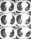
Serial chest computed tomography scans from three participants with interstitial lung abnormalities. (A1_–_C1) Representative axial images from computed tomography scan 1. (A2_–_C2) Representative axial images from computed tomography scan 2. Participant A is indeterminate for usual interstitial pneumonia (UIP) on scan 1 (A1) and progressed to a probable UIP pattern (A2). Participant B has a probable UIP pattern on scan 1 (B1) and progressed to a UIP pattern (B2). Participant C had a UIP pattern on scan 1 (C1) and had imaging progression that remained consistent with a UIP pattern (C2).
Figure 2.
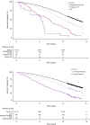
Kaplan-Meier survival curves showing percent survival, comparing participants without interstitial lung abnormalities (ILA) with those with ILA, first with ILA subset based on consistency with an idiopathic pulmonary fibrosis diagnosis, and then by the presence of definite fibrosis (pulmonary parenchymal architectural distortion). UIP = usual interstitial pneumonia.
Comment in
- Subclinical Interstitial Lung Abnormalities: Lumping and Splitting Revisited.
Walsh SLF, Richeldi L. Walsh SLF, et al. Am J Respir Crit Care Med. 2019 Jul 15;200(2):121-123. doi: 10.1164/rccm.201901-0180ED. Am J Respir Crit Care Med. 2019. PMID: 30699307 Free PMC article. No abstract available.
Similar articles
- The MUC5B promoter polymorphism is associated with specific interstitial lung abnormality subtypes.
Putman RK, Gudmundsson G, Araki T, Nishino M, Sigurdsson S, Gudmundsson EF, Eiríksdottír G, Aspelund T, Ross JC, San José Estépar R, Miller ER, Yamada Y, Yanagawa M, Tomiyama N, Launer LJ, Harris TB, El-Chemaly S, Raby BA, Cho MH, Rosas IO, Washko GR, Schwartz DA, Silverman EK, Gudnason V, Hatabu H, Hunninghake GM. Putman RK, et al. Eur Respir J. 2017 Sep 11;50(3):1700537. doi: 10.1183/13993003.00537-2017. Print 2017 Sep. Eur Respir J. 2017. PMID: 28893869 Free PMC article. - Development and Progression of Radiologic Abnormalities in Individuals at Risk for Familial Interstitial Lung Disease.
Salisbury ML, Hewlett JC, Ding G, Markin CR, Douglas K, Mason W, Guttentag A, Phillips JA 3rd, Cogan JD, Reiss S, Mitchell DB, Wu P, Young LR, Lancaster LH, Loyd JE, Humphries SM, Lynch DA, Kropski JA, Blackwell TS. Salisbury ML, et al. Am J Respir Crit Care Med. 2020 May 15;201(10):1230-1239. doi: 10.1164/rccm.201909-1834OC. Am J Respir Crit Care Med. 2020. PMID: 32011901 Free PMC article. - Radiologic-pathologic correlation of interstitial lung abnormalities and predictors for progression and survival.
Chae KJ, Chung MJ, Jin GY, Song YJ, An AR, Choi H, Goo JM. Chae KJ, et al. Eur Radiol. 2022 Apr;32(4):2713-2723. doi: 10.1007/s00330-021-08378-8. Epub 2022 Jan 5. Eur Radiol. 2022. PMID: 34984519 - Spectrum of Pulmonary Fibrosis from Interstitial Lung Abnormality to Usual Interstitial Pneumonia: Importance of Identification and Quantification of Traction Bronchiectasis in Patient Management.
Hino T, Lee KS, Han J, Hata A, Ishigami K, Hatabu H. Hino T, et al. Korean J Radiol. 2021 May;22(5):811-828. doi: 10.3348/kjr.2020.1132. Epub 2020 Dec 21. Korean J Radiol. 2021. PMID: 33543848 Free PMC article. Review. - Interstitial Lung Abnormalities: State of the Art.
Hata A, Schiebler ML, Lynch DA, Hatabu H. Hata A, et al. Radiology. 2021 Oct;301(1):19-34. doi: 10.1148/radiol.2021204367. Epub 2021 Aug 10. Radiology. 2021. PMID: 34374589 Free PMC article. Review.
Cited by
- Interstitial lung abnormalities detected incidentally on CT: a Position Paper from the Fleischner Society.
Hatabu H, Hunninghake GM, Richeldi L, Brown KK, Wells AU, Remy-Jardin M, Verschakelen J, Nicholson AG, Beasley MB, Christiani DC, San José Estépar R, Seo JB, Johkoh T, Sverzellati N, Ryerson CJ, Graham Barr R, Goo JM, Austin JHM, Powell CA, Lee KS, Inoue Y, Lynch DA. Hatabu H, et al. Lancet Respir Med. 2020 Jul;8(7):726-737. doi: 10.1016/S2213-2600(20)30168-5. Lancet Respir Med. 2020. PMID: 32649920 Free PMC article. - Risk factors for pneumonitis in advanced extrapulmonary cancer patients treated with immune checkpoint inhibitors.
Uchida Y, Kinose D, Nagatani Y, Tanaka-Mizuno S, Nakagawa H, Fukunaga K, Yamaguchi M, Nakano Y. Uchida Y, et al. BMC Cancer. 2022 May 16;22(1):551. doi: 10.1186/s12885-022-09642-w. BMC Cancer. 2022. PMID: 35578210 Free PMC article. - Diagnosis and treatment of pneumonia, a common cause of respiratory failure in patients with neuromuscular disorders.
Carannante N, Annunziata A, Coppola A, Simioli F, Marotta A, Bernardo M, Piscitelli E, Imitazione P, Fiorentino G. Carannante N, et al. Acta Myol. 2021 Sep 30;40(3):124-131. doi: 10.36185/2532-1900-053. eCollection 2021 Sep. Acta Myol. 2021. PMID: 34632294 Free PMC article. - Traction Bronchiectasis/Bronchiolectasis on CT Scans in Relationship to Clinical Outcomes and Mortality: The COPDGene Study.
Hata A, Hino T, Putman RK, Yanagawa M, Hida T, Menon AA, Honda O, Yamada Y, Nishino M, Araki T, Valtchinov VI, Jinzaki M, Honda H, Ishigami K, Johkoh T, Tomiyama N, Christiani DC, Lynch DA, San José Estépar R, Washko GR, Cho MH, Silverman EK, Hunninghake GM, Hatabu H; COPDGene Investigators. Hata A, et al. Radiology. 2022 Sep;304(3):694-701. doi: 10.1148/radiol.212584. Epub 2022 May 31. Radiology. 2022. PMID: 35638925 Free PMC article. - Interstitial Lung Disease in Connective Tissue Disease: A Common Lesion With Heterogeneous Mechanisms and Treatment Considerations.
Shao T, Shi X, Yang S, Zhang W, Li X, Shu J, Alqalyoobi S, Zeki AA, Leung PS, Shuai Z. Shao T, et al. Front Immunol. 2021 Jun 7;12:684699. doi: 10.3389/fimmu.2021.684699. eCollection 2021. Front Immunol. 2021. PMID: 34163483 Free PMC article. Review.
References
- Putman RK, Hatabu H, Araki T, Gudmundsson G, Gao W, Nishino M, et al. Evaluation of COPD Longitudinally to Identify Predictive Surrogate Endpoints (ECLIPSE) Investigators; COPDGene Investigators. Association between interstitial lung abnormalities and all-cause mortality. JAMA. 2016;315:672–681. - PMC - PubMed
- Miller ER, Putman RK, Vivero M, Hung Y, Araki T, Nishino M, et al. Interstitial lung abnormalities and histopathologic correlates in patients undergoing lung nodule resection [abstract] Am J Respir Crit Care Med. 2017;195:A1120.
Publication types
MeSH terms
Substances
Grants and funding
- R01 HL113264/HL/NHLBI NIH HHS/United States
- R01 HL130974/HL/NHLBI NIH HHS/United States
- U01 HL133232/HL/NHLBI NIH HHS/United States
- R01 HL116473/HL/NHLBI NIH HHS/United States
- R01 HL135142/HL/NHLBI NIH HHS/United States
- R01 HL137927/HL/NHLBI NIH HHS/United States
- T32 HL007085/HL/NHLBI NIH HHS/United States
- R01 HL097163/HL/NHLBI NIH HHS/United States
- R33 HL120770/HL/NHLBI NIH HHS/United States
- R01 CA203636/CA/NCI NIH HHS/United States
- P01 HL114501/HL/NHLBI NIH HHS/United States
- R01 HL122464/HL/NHLBI NIH HHS/United States
- P01 HL092870/HL/NHLBI NIH HHS/United States
- K08 HL140087/HL/NHLBI NIH HHS/United States
- R01 HL111024/HL/NHLBI NIH HHS/United States
LinkOut - more resources
Full Text Sources
Other Literature Sources
Medical
Research Materials

