The Use of Large-Particle Aerosol Exposure to Nipah Virus to Mimic Human Neurological Disease Manifestations in the African Green Monkey - PubMed (original) (raw)
. 2020 May 11;221(Suppl 4):S419-S430.
doi: 10.1093/infdis/jiz502.
Dima A Hammoud 2, Yu Cong 1, Louis M Huzella 1, Marcelo A Castro 1, Jeffrey Solomon 3, Joseph Laux 1, Matthew Lackemeyer 1, J Kyle Bohannon 1, Oscar Rojas 1, Russ Byrum 1, Ricky Adams 1, Danny Ragland 1, Marisa St Claire 1, Vincent Munster 4, Michael R Holbrook 1
Affiliations
- PMID: 31687756
- PMCID: PMC7368178
- DOI: 10.1093/infdis/jiz502
The Use of Large-Particle Aerosol Exposure to Nipah Virus to Mimic Human Neurological Disease Manifestations in the African Green Monkey
Ji Hyun Lee et al. J Infect Dis. 2020.
Abstract
Nipah virus (NiV) is an emerging virus associated with outbreaks of acute respiratory disease and encephalitis. To develop a neurological model for NiV infection, we exposed 6 adult African green monkeys to a large-particle (approximately 12 μm) aerosol containing NiV (Malaysian isolate). Brain magnetic resonance images were obtained at baseline, every 3 days after exposure for 2 weeks, and then weekly until week 8 after exposure. Four of six animals showed abnormalities reminiscent of human disease in brain magnetic resonance images. Abnormalities ranged from cytotoxic edema to vasogenic edema. The majority of lesions were small infarcts, and a few showed inflammatory or encephalitic changes. Resolution or decreased size in some lesions resembled findings reported in patients with NiV infection. Histological lesions in the brain included multifocal areas of encephalomalacia, corresponding to known ischemic foci. In other regions of the brain there was evidence of vasculitis, with perivascular infiltrates of inflammatory cells and rare intravascular fibrin thrombi. This animal model will help us better understand the acute neurological features of NiV infection and develop therapeutic approaches for managing disease caused by NiV infection.
Keywords: Computed tomography; Magnetic resonance imaging; Nipah virus; Paramyxovirus; pathology.
Published by Oxford University Press for the Infectious Diseases Society of America 2019.
Figures
Figure 1.
Incidence of survival in African green monkeys after Nipah virus (NiV) challenge and resulting viremia and cerebrospinal fluid (CSF) concentrations. (A), Kaplan-Meier survival curve. B–F, Viral RNA copy concentrations in whole blood (B), serum (C), CSF (D), oral swab (E), and nasal swab (F) samples. Viral RNA was detected in 4 of 6 animals, with no detectable viral RNA in oral (E) and nasal (F) mucosa in animal 8168, which succumbed on day 5 after exposure, or in 1 survivor (8212). The other survivor (8671) had peak viral replication in mucosa on day 9 after exposure, which then dropped to normal on day 12 after exposure.
Figure 2.
(A), A small focal lesion (arrows) appeared on day 15 after exposure in the left frontal subcortical white matter in 1 animal (8078). (B), Progression of disease is noted on day 17 (terminal day), with a total of 7 abnormal signal intensities now seen bilaterally. Incidental note is made of a preexisting congenital abnormality in this animal (colpocephaly, dilatation of the left posterior occipital horn). Abbreviations: DW, diffusion weighted; FLAIR, fluid-attenuated inversion-recovery; trace (DT), trace of diffusion tensor.
Figure 3.
Magnetic resonance imaging of animal 8233 shows a focal lesion with increased T2-weighted and fluid-attenuated inversion-recovery (FLAIR) signal intensity on the right side of the midbrain on day 20 after exposure. This lesion demonstrated facilitated (non-restricted) diffusion (white arrows). Four other smaller lesions with similar characteristics were seen in this animal (not shown). Abbreviations: DW, diffusion weighted; trace (DT), trace of diffusion tensor.
Figure 4.
Longitudinal changes on fluid-attenuated inversion-recovery images in a survivor (8671). Focal area of increased signal in the left cerebellar hemisphere (arrows) appeared on day 15 after exposure and decreased in size over time. The lesion was still noted, but to a lesser extent, on day 56 after exposure, the study end point.
Figure 5.
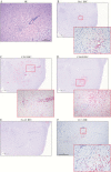
Histopathological findings showing encephalitis (8233). (A), Hematoxylin-eosin (HE) staining of brain with perivascular infiltrates of inflammatory cells extending into surrounding parenchyma. (B–F), Immunohistochemistry (IHC) signal changes_, (B),_ Focal ionized calcium-binding adaptor molecule 1 (Iba1) IHC signal indicating microglial response (inset; original magnification ×200). (C), Perivascular CD3-positive T-cell infiltrates, extending into surrounding parenchyma (inset; original magnification ×200). (D), CD68 positive monocyte/macrophage infiltrates penetrating the surrounding parenchyma in conjunction with T-cell infiltrates (inset; original magnification ×200). (E), Locally absent neuronal nuclei (NeuN) IHC signal. (F), Focal extensive areas of positivity for cleaved caspase 3 (CC3) (inset; original magnification ×200) indicate areas with cellular apoptosis.
Figure 6.
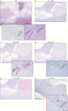
Ischemia-related encephalomalacia within the frontal lobe of animal 8078. (A), Large area of encephalomalacia with parenchymal loss with adjacent, nonsuppurative encephalitis (inset; original magnification ×200). Left inset shows specific NiV antigen staining of neurons; right inset, perivascular inflammatory infiltrates and vasculitis. (B–F), Immunohistochemistry (IHC) signal changes. (B), No significant ionized calcium-binding adaptor molecule 1 (Iba1) IHC signal. (C), Perivascular CD3 positive T-cell infiltrates, extending into surrounding parenchyma (inset; original magnification ×200). (D), Minimal CD68 positive monocyte/macrophage infiltrates (inset; original magnification ×200). (E), locally absent neuronal nuclei (NeuN) IHC signal in the area of the lesion, strongly present in areas not affected. (F), focal extensive areas of positivity for cleaved caspase (CC3) (inset; original magnification ×200) indicate areas with cellular apoptosis. Abbreviation: HE, hematoxylin-eosin.
Similar articles
- Aerosol exposure to intermediate size Nipah virus particles induces neurological disease in African green monkeys.
Hammoud DA, Lentz MR, Lara A, Bohannon JK, Feuerstein I, Huzella L, Jahrling PB, Lackemeyer M, Laux J, Rojas O, Sayre P, Solomon J, Cong Y, Munster V, Holbrook MR. Hammoud DA, et al. PLoS Negl Trop Dis. 2018 Nov 21;12(11):e0006978. doi: 10.1371/journal.pntd.0006978. eCollection 2018 Nov. PLoS Negl Trop Dis. 2018. PMID: 30462637 Free PMC article. - A Lethal Aerosol Exposure Model of Nipah Virus Strain Bangladesh in African Green Monkeys.
Prasad AN, Agans KN, Sivasubramani SK, Geisbert JB, Borisevich V, Mire CE, Lawrence WS, Fenton KA, Geisbert TW. Prasad AN, et al. J Infect Dis. 2020 May 11;221(Suppl 4):S431-S435. doi: 10.1093/infdis/jiz469. J Infect Dis. 2020. PMID: 31665351 - An Intranasal Exposure Model of Lethal Nipah Virus Infection in African Green Monkeys.
Geisbert JB, Borisevich V, Prasad AN, Agans KN, Foster SL, Deer DJ, Cross RW, Mire CE, Geisbert TW, Fenton KA. Geisbert JB, et al. J Infect Dis. 2020 May 11;221(Suppl 4):S414-S418. doi: 10.1093/infdis/jiz391. J Infect Dis. 2020. PMID: 31665362 Free PMC article. - Animal challenge models of henipavirus infection and pathogenesis.
Geisbert TW, Feldmann H, Broder CC. Geisbert TW, et al. Curr Top Microbiol Immunol. 2012;359:153-77. doi: 10.1007/82_2012_208. Curr Top Microbiol Immunol. 2012. PMID: 22476556 Free PMC article. Review. - Emerging trends of Nipah virus: A review.
Sharma V, Kaushik S, Kumar R, Yadav JP, Kaushik S. Sharma V, et al. Rev Med Virol. 2019 Jan;29(1):e2010. doi: 10.1002/rmv.2010. Epub 2018 Sep 24. Rev Med Virol. 2019. PMID: 30251294 Free PMC article. Review.
Cited by
- Henipavirus Immune Evasion and Pathogenesis Mechanisms: Lessons Learnt from Natural Infection and Animal Models.
Lawrence P, Escudero-Pérez B. Lawrence P, et al. Viruses. 2022 Apr 29;14(5):936. doi: 10.3390/v14050936. Viruses. 2022. PMID: 35632678 Free PMC article. Review. - Nonhuman Primate Models for Nipah and Hendra Virus Countermeasure Evaluation.
Mire CE, Satterfield BA, Geisbert TW. Mire CE, et al. Methods Mol Biol. 2023;2682:159-173. doi: 10.1007/978-1-0716-3283-3_12. Methods Mol Biol. 2023. PMID: 37610581 - Nipah Virus Bangladesh Infection Elicits Organ-Specific Innate and Inflammatory Responses in the Marmoset Model.
Stevens CS, Lowry J, Juelich T, Atkins C, Johnson K, Smith JK, Panis M, Ikegami T, tenOever B, Freiberg AN, Lee B. Stevens CS, et al. J Infect Dis. 2023 Aug 31;228(5):604-614. doi: 10.1093/infdis/jiad053. J Infect Dis. 2023. PMID: 36869692 Free PMC article. - Detailed analysis of the pathologic hallmarks of Nipah virus (Malaysia) disease in the African green monkey infected by the intratracheal route.
Cline C, Bell TM, Facemire P, Zeng X, Briese T, Lipkin WI, Shamblin JD, Esham HL, Donnelly GC, Johnson JC, Hensley LE, Honko AN, Johnston SC. Cline C, et al. PLoS One. 2022 Feb 10;17(2):e0263834. doi: 10.1371/journal.pone.0263834. eCollection 2022. PLoS One. 2022. PMID: 35143571 Free PMC article. - Toward the determination of sensitive and reliable whole-lung computed tomography features for robust standard radiomics and delta-radiomics analysis in a nonhuman primate model of coronavirus disease 2019.
Castro MA, Reza S, Chu WT, Bradley D, Lee JH, Crozier I, Sayre PJ, Lee BY, Mani V, Friedrich TC, O'Connor DH, Finch CL, Worwa G, Feuerstein IM, Kuhn JH, Solomon J. Castro MA, et al. J Med Imaging (Bellingham). 2022 Nov;9(6):066003. doi: 10.1117/1.JMI.9.6.066003. Epub 2022 Dec 8. J Med Imaging (Bellingham). 2022. PMID: 36506838 Free PMC article.
References
- Chua KB, Bellini WJ, Rota PA, et al. . Nipah virus: a recently emergent deadly paramyxovirus. Science 2000; 288:1432–5. - PubMed
- Centers for Disease Control and Prevention. Outbreak of Hendra-like virus—Malaysia and Singapore, 1998–1999. MMWR Morb Mortal Wkly Rep 1999; 48:265–9. - PubMed
- Paton NI, Leo YS, Zaki SR, et al. . Outbreak of Nipah virus infection among abattoir workers in Singapore. Lancet 1999; 354:1253–6. - PubMed
- Sahani M, Parashar UD, Ali R, et al. . Nipah virus infection among abattoir workers in Malaysia, 1998–1999. Int J Epidemiol 2001; 30:1017–20. - PubMed
- World Health Organization Regional Office for South-East Asia. Nipah virus outbreaks in the WHO South-East Asia region http://www.searo.who.int/entity/emerging_diseases/links/nipah_virus_outb.... Accessed 14 June 2019.


