Role of donor genotype in RT-QuIC seeding activity of chronic wasting disease prions using human and bank vole substrates - PubMed (original) (raw)
Role of donor genotype in RT-QuIC seeding activity of chronic wasting disease prions using human and bank vole substrates
Soyoun Hwang et al. PLoS One. 2020.
Abstract
Chronic wasting disease is a transmissible spongiform encephalopathy of cervids. This fatal neurodegenerative disease is caused by misfolding of the cellular prion protein (PrPC) to pathogenic conformers (PrPSc), and the pathogenic forms accumulate in the brain and other tissues. Real-time Quaking Induced Conversion (RT-QuIC) can be used for the detection of prions and for prion strain discrimination in a variety of biological tissues from humans and animals. In this study, we evaluated how either PrPSc from cervids of different genotypes or PrPSc from different sources of CWD influence the fibril formation of recombinant bank vole (BV) or human prion proteins using RT-QuIC. We found that reaction mixtures seeded with PrPSc from different genotypes of white-tailed deer or reindeer brains have similar conversion efficiency with both substrates. Also, we observed similar results when assays were seeded with different sources of CWD. Thus, we conclude that the genotypes of all sources of CWD used in this study do not influence the level of conversion of PrPC to PrPSc.
Conflict of interest statement
The authors have declared that no competing interests exist.
Figures
Fig 1. RT-QuIC reactions seeded with CWD infected or not infected white tailed deer brains using BV rPrP as a substrate.
RT-QuIC reactions were seeded with EIA normalized stock (a), 10−1 (b), 10−2 (c), and 10−3 (d) dilutions of three CWD infected WTD brains and one negative animal. All reactions were seeded with brain homogenates of WTD with the addition of 0.001% of SDS. Shown are the average ThT fluorescence readings (thick lines) with standard deviations (thin lines) determined from all replicates (four replicate reactions per each animal).
Fig 2
Comparison of seeding activity of RT-QuIC reactions with BV rPrP which contain seeds from WTD inoculated with brains of infected WTD (a and b), elk (c and d) or mule deer (e and f). Left panel shows RT-QuIC reactions seeded with EIA normalized brains and right panel shows RT-QuIC reactions seeded with 10−2 dilutions of brains. Data are presented as mean ThT fluorescence of 4 repeated reactions.
Fig 3. Comparison of averaged seeding activity of RT-QuIC reactions with BV rPrP seeded with brains from WTD inoculated with brains from different cervid species.
(WTD: blue line, elk: red line, MD: green line). All assays seeded with (a) EIA normalized stock and (b) 10−2 dilution of WTD brains inoculated with each CWD source was averaged and compared to assays seeded with two other CWD inocula. Data are presented as mean ThT fluorescence of 4 repeated reactions.
Fig 4
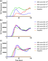
Comparison of seeding activity of RT-QuIC reactions with human rPrP which contain brain seeds from WTD inoculated with infected brains from WTD (a), elk (b) or mule deer (c). This shows RT-QuIC reactions seeded with 10−2 dilution of EIA normalized brains in the presence of 0.002% SDS. Data are presented as mean ThT fluorescence of 4 repeated reactions. Data are presented as mean ThT fluorescence of 4 repeated reactions.
Fig 5
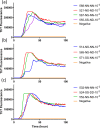
Comparison of seeding activity of RT-QuIC reactions with human rPrP which contain brain seeds from WTD inoculated with brains of infected white-tailed deer (a), elk (b) or mule deer (c) in the presence of 0.001% SDS. This shows RT-QuIC reactions seeded with 10−2 dilution of EIA normalized brains. Data are presented as mean ThT fluorescence of 4 repeated reactions. Data are presented as mean ThT fluorescence of 4 repeated reactions.
Fig 6. Comparison of averaged seeding activity of RT-QuIC reactions with human rPrP seeded with brains from WTD inoculated with brains of infected white-tailed deer, elk, or mule deer.
(WTD: blue line, elk: red line, MD: green line). All assays from each CWD source was averaged and compared to assays seeded with different CWD inocula. Data are presented as mean ThT fluorescence of 4 repeated reactions.
Fig 7
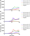
Comparison of seeding activity of RT-QuIC reactions with BV rPrP which contain seeds from reindeer inoculated with brains of infected WTD (a and b), elk (c and d) or mule deer (e and f). Left panel shows RT-QuIC reactions seeded with 10−2 brain dilution of EIA normalized and right panel shows RT-QuIC reactions seeded with 10−3 dilutions of brains. Data are presented as mean ThT fluorescence of 4 repeated reactions.
Fig 8. Comparison of averaged seeding activity of RT-QuIC reactions with BV rPrP seeded with brains from reindeer inoculated with brains of infectious cervid species.
(WTD: blue line, elk: red line, MD: green line). All assays seeded with (a) 10−2 or (b) 10−3 dilution of EIA normalized stock dilutions of reindeer brains inoculated with each CWD source was averaged and compared to assays seeded with two other CWD inocula. Data are presented as mean ThT fluorescence of 4 repeated reactions.
Fig 9
Comparison of seeding activity of RT-QuIC reactions with human rPrP which contain brain seeds from reindeer inoculated with brains of infectious WTD (a), elk (b) or mule deer (c). This shows RT-QuIC reactions seeded with 10−2 dilution of EIA normalized brains in the presence of 0.002% SDS. Data are presented as mean ThT fluorescence of 4 repeated reactions. Data are presented as mean ThT fluorescence of 4 repeated reactions.
Fig 10. Comparison of averaged seeding activity of RT-QuIC reactions with human rPrP seeded with 10−2 dilution of EIA normalized brains from reindeer inoculated with brains of infectious cervid species.
(WTD: blue line, elk: red line, MD: green line). All assays from each CWD source was averaged and compared to assays seeded with different CWD inocula. Data are presented as mean ThT fluorescence of 4 repeated reactions.
References
Publication types
MeSH terms
Substances
LinkOut - more resources
Full Text Sources


