CT features of SARS-CoV-2 pneumonia according to clinical presentation: a retrospective analysis of 120 consecutive patients from Wuhan city - PubMed (original) (raw)
CT features of SARS-CoV-2 pneumonia according to clinical presentation: a retrospective analysis of 120 consecutive patients from Wuhan city
Rui Zhang et al. Eur Radiol. 2020 Aug.
Abstract
Objectives: To characterize the chest computed tomography (CT) findings of severe acute respiratory syndrome coronavirus 2 (SARS-CoV-2) according to clinical severity. We compared the CT features of common cases and severe cases, symptomatic patients and asymptomatic patients, and febrile and afebrile patients.
Methods: This was a retrospective analysis of the clinical and thoracic CT features of 120 consecutive patients with confirmed SARS-CoV-2 pneumonia admitted to a tertiary university hospital between January 10 and February 10, 2020, in Wuhan city, China.
Results: On admission, the patients generally complained of fever, cough, shortness of breath, and myalgia or fatigue, with diarrhea often present in severe cases. Severe patients were 20 years older on average and had comorbidities and an elevated lactate dehydrogenase (LDH) level. There were no differences in the CT findings between asymptomatic and symptomatic common type patients or between afebrile and febrile patients, defined according to Chinese National Health Commission guidelines.
Conclusions: The clinical and CT features at admission may enable clinicians to promptly evaluate the prognosis of patients with SARS-CoV-2 pneumonia. Clinicians should be aware that clinically silent cases may present with CT features similar to those of symptomatic common patients.
Key points: • The clinical features and predominant patterns of abnormalities on CT for asymptomatic, typic common, and severe cases were summarized. These findings may help clinicians to identify severe patients quickly at admission. • Clinicians should be cautious that CT findings of afebrile/asymptomatic patients are not better than the findings of other types of patients. These patients should also be quarantined. • The use of chest CT as the main screening method in epidemic areas is recommended.
Keywords: Chest; Fever; SARS-CoV-2; Tomography.
Conflict of interest statement
The authors of this manuscript declare no relationships with any companies whose products or services may be related to the subject matter of the article.
Figures
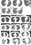
Fig. 1
(a) Unenhanced axial CT images of an afebrile 37-year-old male doctor with a history of exposure to confirmed SARS-CoV-2 patients. (a1) Patchy ground-glass opacities (GGOs) in the left upper lobe. (a2) Small GGO nodule in the contralateral lower lobe. (A3) Enlarged image of the right lower lobe. (b) Unenhanced axial CT images of an afebrile 28-year-old female with a history of exposure to confirmed SARS-CoV-2 patients presenting with a mild sore throat. (b1) A rounded, ground-glass nodular opacity (GGO) is seen in a subpleural location in the right lower lobe. (b2) Another focal GGO is seen in a subpleural location, in the posterobasal segment of the left lower lobe. (c) Unenhanced axial CT images of a 27-year-old male doctor with a history of exposure to confirmed SARS-CoV-2 patients, initially presenting with fever (39 °C), non-productive cough, dyspnea, and myalgia (c1) who progressed to a severe case requiring oxygen supplementation (c2). (c1) Multifocal, limited GGO is seen in the peripheral zone of both lungs. (c2) Six days later, while oxygen supplementation has been instore, diffuse, bilateral, and ill-defined GGO has developed. Superimposed linear consolidations can be observed, consistent with areas of organizing pneumonia. (d) Unenhanced axial CT images of a 52-year-old male doctor with asthma and exposure to confirmed SARS-CoV-2 patients, initially presenting with fever (39 °C), non-productive cough, dyspnea, and myalgia who rapidly progressed to a severe form requiring mechanical ventilation. (d1) Multifocal, limited GGO in the periphery of both lungs, predominantly affecting left lung. (d2) Two days later, focal GGO has increased in size and density, and new diffuse ill-defined GGO has developed. (d3) After 4 days of mask oxygen supplementation, disease progressed further, with more patchy consolidations and linear densities observed in nearly all lung zones except the anterior part of both lungs. (e) Unenhanced axial CT images of a 57-year-old male with an exposure history initially presenting with fever (38 °C), non-productive cough, dyspnea, myalgia, and headache, being treated for hypertension for 12 years. Diffuse consolidation with air bronchograms is seen in both lungs, with relative sparing of peri-hilar and anterior lung areas, extending from the lung apices to the lung bases. These findings are consistent with a “white lung” appearance
References
- World Health Organization (2020) Statement on the second meeting of the International Health Regulations (2005) Emergency Committee regarding the outbreak of novel coronavirus (2019-nCoV). WHO, Geneva, Switzerland
- World Health Organization (2020) WHO Director-General’s opening remarks at the media briefing on COVID-19 - 11 March 2020. https://www.who.int/dg/speeches/detail/whodirector-general-s-opening-rem...
MeSH terms
LinkOut - more resources
Full Text Sources
Miscellaneous
