Relationships among lymphocyte subsets, cytokines, and the pulmonary inflammation index in coronavirus (COVID-19) infected patients - PubMed (original) (raw)
. 2020 May;189(3):428-437.
doi: 10.1111/bjh.16659. Epub 2020 Apr 20.
Qingjie Yi 1 3, Shibing Fan 1 4, Jinglong Lv 1 5, Xianxiang Zhang 1 6, Lian Guo 1 6, Chunhui Lang 1 7, Qing Xiao 8, Kaihu Xiao 1 9, Zhengjun Yi 10, Mao Qiang 11 12, Jianglin Xiang 1 13, Bangshuo Zhang 1 5, Yongping Chen 1 5, Cailiang Gao 1 14
Affiliations
- PMID: 32297671
- PMCID: PMC7262036
- DOI: 10.1111/bjh.16659
Relationships among lymphocyte subsets, cytokines, and the pulmonary inflammation index in coronavirus (COVID-19) infected patients
Suxin Wan et al. Br J Haematol. 2020 May.
Abstract
We explored the relationships between lymphocyte subsets, cytokines, pulmonary inflammation index (PII) and disease evolution in patients with (corona virus disease 2019) COVID-19. A total of 123 patients with COVID-19 were divided into mild and severe groups. Lymphocyte subsets and cytokines were detected on the first day of hospital admission and lung computed tomography results were quantified by PII. Difference analysis and correlation analysis were performed on the two groups. A total of 102 mild and 21 severe patients were included in the analysis. There were significant differences in cluster of differentiation 4 (CD4+ T), cluster of differentiation 8 (CD8+ T), interleukin 6 (IL-6), interleukin 10 (IL-10) and PII between the two groups. There were significant positive correlations between CD4+ T and CD8+ T, IL-6 and IL-10 in the mild group (r2 = 0·694, r 2 = 0·633, respectively; P < 0·01). After 'five-in-one' treatment, all patients were discharged with the exception of the four who died. Higher survival rates occurred in the mild group and in those with IL-6 within normal values. CD4+ T, CD8+ T, IL-6, IL-10 and PII can be used as indicators of disease evolution, and the PII can be used as an independent indicator for disease progression of COVID-19.
Keywords: coronavirus infected disease; cytokines; disease classification; lymphocyte subsets; pulmonary inflammatory index.
© 2020 British Society for Haematology and John Wiley & Sons Ltd.
Conflict of interest statement
All authors declare no conflicts of interest.
Figures
Fig 1
Typical CT images of patients with mild 2019 novel coronavirus pneumonia (COVID‐19). Patchy ground glass opacities can be seen in the apical and posterior segments of the right upper lobe, the posterior basal segment of the right lower lobe, the medial segment of the right middle lobe, the lingual segment of the left upper lobe, the anterior inner basal segment of the left lower lobe, the posterior basal segment, and the subpleural of outer basal segment of the left lower lobe. [Colour figure can be viewed at
http://www.wileyonlinelibrary.com/
]
Fig 2
Typical CT images of patients with severe 2019 novel coronavirus pneumonia (COVID‐19). Strip‐like consolidation shadows and patchy ground glass‐like density shadows can be seen in the apical and posterior segment of the upper lobe of both lungs, tongue segment of the left upper lobe, and the lower lobe of both lungs with a fuzzy boundary, mainly distributed under the pleura of both lungs when the patient was in severe condition. [Colour figure can be viewed at
http://www.wileyonlinelibrary.com/
]
Fig 3
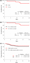
Kaplan–Meier curves of mild and severe patients, within normal and above normal values IL‐6 patients. (A) Kaplan–Meier curve of mild and severe group; (B) Kaplan–Meier curve of within normal and above normal IL‐6 group; (C) Progression‐free curve of the mild group and within normal IL‐6 group. [Colour figure can be viewed at
http://www.wileyonlinelibrary.com/
]
Similar articles
- Prediction Model Based on the Combination of Cytokines and Lymphocyte Subsets for Prognosis of SARS-CoV-2 Infection.
Luo Y, Mao L, Yuan X, Xue Y, Lin Q, Tang G, Song H, Wang F, Sun Z. Luo Y, et al. J Clin Immunol. 2020 Oct;40(7):960-969. doi: 10.1007/s10875-020-00821-7. Epub 2020 Jul 13. J Clin Immunol. 2020. PMID: 32661797 Free PMC article. - The changes of the peripheral CD4+ lymphocytes and inflammatory cytokines in Patients with COVID-19.
Sun HB, Zhang YM, Huang LG, Lai QN, Mo Q, Ye XZ, Wang T, Zhu ZZ, Lv XL, Luo YJ, Gao SD, Xu JS, Zhu HH, Li T, Wang ZK. Sun HB, et al. PLoS One. 2020 Sep 25;15(9):e0239532. doi: 10.1371/journal.pone.0239532. eCollection 2020. PLoS One. 2020. PMID: 32976531 Free PMC article. - The role of cytokine profile and lymphocyte subsets in the severity of coronavirus disease 2019 (COVID-19): A systematic review and meta-analysis.
Akbari H, Tabrizi R, Lankarani KB, Aria H, Vakili S, Asadian F, Noroozi S, Keshavarz P, Faramarz S. Akbari H, et al. Life Sci. 2020 Oct 1;258:118167. doi: 10.1016/j.lfs.2020.118167. Epub 2020 Jul 29. Life Sci. 2020. PMID: 32735885 Free PMC article. - Biochemical indicators of coronavirus disease 2019 exacerbation and the clinical implications.
An PJ, Zhu YZ, Yang LP. An PJ, et al. Pharmacol Res. 2020 Sep;159:104946. doi: 10.1016/j.phrs.2020.104946. Epub 2020 May 23. Pharmacol Res. 2020. PMID: 32450346 Free PMC article. Review. - Hematological findings in coronavirus disease 2019: indications of progression of disease.
Liu X, Zhang R, He G. Liu X, et al. Ann Hematol. 2020 Jul;99(7):1421-1428. doi: 10.1007/s00277-020-04103-5. Epub 2020 Jun 3. Ann Hematol. 2020. PMID: 32495027 Free PMC article. Review.
Cited by
- Targeting the Complement-Sphingolipid System in COVID-19 and Gaucher Diseases: Evidence for a New Treatment Strategy.
Trivedi VS, Magnusen AF, Rani R, Marsili L, Slavotinek AM, Prows DR, Hopkin RJ, McKay MA, Pandey MK. Trivedi VS, et al. Int J Mol Sci. 2022 Nov 18;23(22):14340. doi: 10.3390/ijms232214340. Int J Mol Sci. 2022. PMID: 36430817 Free PMC article. Review. - COVID-19: age, Interleukin-6, C-reactive protein, and lymphocytes as key clues from a multicentre retrospective study.
Jurado A, Martín MC, Abad-Molina C, Orduña A, Martínez A, Ocaña E, Yarce O, Navas AM, Trujillo A, Fernández L, Vergara E, Rodríguez B, Quirant B, Martínez-Cáceres E, Hernández M, Perurena-Prieto J, Gil J, Cantenys S, González-Martínez G, Martínez-Saavedra MT, Rojo R, Marco FM, Mora S, Ontañón J, López-Hoyos M, Ocejo-Vinyals G, Melero J, Aguilar M, Almeida D, Medina S, Vegas MC, Jiménez Y, Prada Á, Monzón D, Boix F, Cunill V, Molina J. Jurado A, et al. Immun Ageing. 2020 Aug 14;17:22. doi: 10.1186/s12979-020-00194-w. eCollection 2020. Immun Ageing. 2020. PMID: 32802142 Free PMC article. - Can COVID-19 Vaccines Induce Premature Non-Communicable Diseases: Where Are We Heading to?
Hromić-Jahjefendić A, Barh D, Uversky V, Aljabali AA, Tambuwala MM, Alzahrani KJ, Alzahrani FM, Alshammeri S, Lundstrom K. Hromić-Jahjefendić A, et al. Vaccines (Basel). 2023 Jan 17;11(2):208. doi: 10.3390/vaccines11020208. Vaccines (Basel). 2023. PMID: 36851087 Free PMC article. Review. - The Robustness of Cellular Immunity Determines the Fate of SARS-CoV-2 Infection.
Moga E, Lynton-Pons E, Domingo P. Moga E, et al. Front Immunol. 2022 Jun 27;13:904686. doi: 10.3389/fimmu.2022.904686. eCollection 2022. Front Immunol. 2022. PMID: 35833134 Free PMC article. Review. - Rapid and Effective Vitamin D Supplementation May Present Better Clinical Outcomes in COVID-19 (SARS-CoV-2) Patients by Altering Serum INOS1, IL1B, IFNg, Cathelicidin-LL37, and ICAM1.
Gönen MS, Alaylıoğlu M, Durcan E, Özdemir Y, Şahin S, Konukoğlu D, Nohut OK, Ürkmez S, Küçükece B, Balkan İİ, Kara HV, Börekçi Ş, Özkaya H, Kutlubay Z, Dikmen Y, Keskindemirci Y, Karras SN, Annweiler C, Gezen-Ak D, Dursun E. Gönen MS, et al. Nutrients. 2021 Nov 12;13(11):4047. doi: 10.3390/nu13114047. Nutrients. 2021. PMID: 34836309 Free PMC article.
References
- General Office of the National Health and Family Planning Commission . Diagnosis and treatment of pneumonia with a new coronavirus infection (trial version 5). Chin J Integr Tradit West Med. 2020:1–3.
Publication types
MeSH terms
Substances
LinkOut - more resources
Full Text Sources
Research Materials


