Dendrites | Structure, History, Types & Development (original) (raw)
Introduction
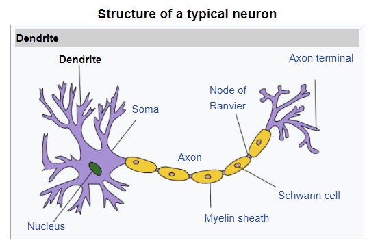
Coordination among the different organs and organ systems in the body of animals is brought about by the nervous system. It is comprised of millions of neurons that are responsible for carrying messages in the form of nerve impulses from one part of the body to the other. These neurons have a cell body with radiating cellular processes. Nerve impulses travel along with these cellular processes of the neurons.
The cellular processes of neurons are divided into two categories based on the direction of nerve impulses. These are dendrites and axons. Dendrites are the cellular processes that carry nerve impulses towards the cell bodies of neurons.
In this article, we will talk about the structure of dendrites, their embryologic development, their metabolism, and different scheme of dendrites arborization. We will also study different functions performed by dendrites as well as their clinical importance.
Dendrites are the cytoplasmic projections of neurons. They have varying shapes and structures among different neurons found in the central as well as the peripheral nervous system.
The cytoplasm of dendrites has the same composition as found in the cell body of neurons. It is rich in cytoskeletal components that provide structural support to the dendrites. The membrane of dendrites has abundant receptors where the neurotransmitters released at the synapse attach and initiate an action potential in the neuron.
The size and thickness of dendrites vary among different types of neurons. They show multiple branching or arborization. The branching pattern of dendrites is also different among different neurons. As the dendrites branch, their thickness goes on decreasing. Unlike axons, they have a variable diameter or thickness that decreases as the branching proceeds.
Dendritic Spines
These are the short blunt structures associated with dendrites. They occur at regular intervals along the length of dendrites. Most of the synapses impinge on these dendritic spines.
These structures are visible on dendrites upon silver staining. They are composed of actin filaments and are highly plastic.
Dendritic spines are abundantly present in the neurons of the cerebral cortex. These serve as the initial sites for the processing of information received by the synapse. These structures are considered to play an important role in neural plasticity for adaptation, learning, and memory.
Historical Facts
Dendrites were first studied by Golgi in 1873 who proposed them to be the projections of protoplasm found in the neurons. They were named protoplasmic projections by Golgi. The function of these projections was not known at that time. It was thought that the protoplasmic projections played a nutritive role in the functioning of neurons. The term dendrites were first introduced and used in 1889.
Later, they were discovered to be a part of synapses by which nerve impulse travels from one neuron to the other. The electrical activity in the dendrites was recorded in 1930.
Soon, they were recognized to be an input component of the neurons. Initially, it was thought that the action potential generated simultaneously at the dendrites did not add or cancel each other. However, now it is a known fact that neural summation of action potentials can take place in the dendrites.
Types of Neurons
Based on the number of cellular processes, neurons are divided into the following types. The shape and orientation of dendrites in these neurons are discussed below.
- Multipolar Neurons: These neurons have multiple dendrites and a single axon. The dendrites show different varieties of branching or arborization based on the location of neurons. Most of the neurons in the human body are multiple neurons. Any neuron having two or more dendrites with one axon is included in this category.
- Bipolar Neurons: These neurons have a single axon and a single dendrite. The dendrite in these neurons also shows extensive branching soon after leaving the cell body. Like multipolar neurons, dendrites in bipolar neurons are devoid of the myelin sheath or any other insulation. They are found in the retina, olfactory epithelium, and organs of the inner ear; cochlea and vestibule.
- Unipolar Neuron: These neurons lack original dendrites. They have a single axon that leaves the cell body and divides into two processes. One process that carries the impulses towards the cell body is called the peripheral process. The other process carrying nerve impulses away from the cell body is called the central process. The peripheral process shows some branching like dendrites, so it is called the dendritic process. However, it is also insulated by myelin sheath as in axons. These neurons are found in the dorsal root ganglion of the spinal nerves.
- Anaxonic Neurons: These neurons lack a true axon. They have multiple dendrites originating from the cell body. These neurons are unable to generate any action potential. They are not involved in conducting the nerve impulses from one cell to the other. Their only function is to regulate the electrical charges of the nearby neurons.
Development
Dendrites are the sites of synapses. They are necessary to take information to the cell bodies of neurons. The development of dendrites determines the number and pattern of synapses that will be impinging on neurons.
Not only this, but the arborization pattern of dendrites also determines the synaptic range or field through which the neuron will receive input information.
Dendritic arborization patterns are formed during the migration of neurons. During the development of the brain, once the neurons become specified, they migrate to their specific location in the brain. These neurons undergo polarization that determines the dendritic and axonal ends of the neurons. As a result, highly polarized neurons are formed having a characteristic dendritic tree and a specific axonal fibre.
The distinctions of axons and dendrites that are formed during the polarization process are permanent. The axons and dendrites have distinctive features that remain throughout life.
In the early time, it was a great challenge in neurobiology to determine how this polarization occurs. Now, it is evident that certain intrinsic and extrinsic factors play a role in the polarization of dendrites and axons. Certain molecular signalling pathways have been recognized that are responsible for neuronal polarity.
Factors Controlling Dendritic Development
The process of dendritic development has been studied in detail in the last two decades. It has been found that dendritic development is controlled by several environmental and body factors. The following are the important factors that can influence the development of dendrites.
- Sensory input modulation
- Neuronal activity
- Body temperature
- Presence of environmental pollutants
- Drugs and toxins
During certain experiments, it was found that the rats developed in the dark environments had fewer dendrites as compared to the control group. These rats had less number of the dendritic spine in the neurons of pyramids in the cerebral cortex.
Dendritic Arborization
It is a step-wise process by which dendrites undergo branching to form a dendritic tree. This process is also called dendritic branching.
The branching pattern is distinct for each type of neuron. The dendritic arborization process plays an important role in determining the function of neurons. Different types of arborization found in different neurons are mentioned below;
- Pyramidal cells:These are the pyramid-shaped cells present in the cerebral cortex. They are in the form of a triangle having three corners. Dendrites arise from all the three corners of these cells. The dendrites synapse in all three directions and receive information for processing from the surrounding neurons.
- Basket cells: These are the basket-shaped cells present in the cortex of the cerebellum. These cells have tree-like dendritic branching that arises from the top0 surface of the basket. The dendrites synapse with the nerve fibers present in the upper layer of the cerebellar cortex.
- Satellite cells: These are the star-shaped cells present in the cerebral cortex. They have star-shaped arborization of the dendritic tree. As a result, they form synapses in all the directions and thus, are capable of receiving information from all the surrounding neurons.
Physiologic Properties
In this section, we will discuss the properties of dendrites that help them in performing their function effectively.
Generation of Action Potential
These properties enable the dendrites to conduct nerve impulses to the cell body of the neurons. These cellular processes can conduct action nerve impulses because of the availability of voltage-gated ion channels.
Just like the axons, the plasma membrane of dendrites has abundant voltage-gated sodium and potassium channels. Most of the dendrites become activated at a synapse. It occurs when a neurotransmitter released by a presynaptic neuron diffuses through the synapse and binds to the receptors present on the dendrites. This activates the receptors.
The receptors on dendrites are coupled with ion channels. The activation of the receptor causes the opening of sodium channels with sodium ions entering the dendrites down the concentration gradient.
In this way, dendrites become depolarized and an action potential is generated that travels towards the cell body of the neuron.
Summation
It is an important property of dendrites to generate a cumulative effect. In most cases, one dendrite synapses with multiple neurons. What happens when all the synapses activate the dendrite at the same time?
The answer to this question lies in the property of summation. Dendrites have the property to generate one collective response when activated by multiple synapses. The action potential thus generated is the algebraic sum of all the excitatory and inhibitory signals arriving at the dendrite simultaneously. This property is called spatial summation.
Besides, dendrites can integrate the excitatory signals arriving at a single synapse in quick succession. This property of dendrites is called temporal summation.
Back Propagation
Dendrites have the ability to conduct action potential in a backward direction i.e. from the cell body to the dendritic arborization. This property is called dendritic arborization.
It can occur when a dendrite is stimulated near the soma or a synaptic stimulation at the cell body of the neuron. This property plays an important role in processes like synapse modulation and potentiation.
Plasticity
Synaptic plasticity or simply plasticity is a phenomenon by which synapses can strengthen or weaken over time. Dendrites can undergo plastic changes even during the adult phase of life. This property can affect the arborization pattern of dendrites.
Dendrites of all the cells have a specific arborization pattern that is determined during embryologic development. The plastic changes during adult life can disturb this natural arborization pattern and affect the normal functioning of the brain.
Read more about Nerve Impulses
Summary
- Dendrites are the afferent cellular processes of neurons that carry the nerve impulses towards the cell body.
- They have the same composition of cytoplasm as present in the cell body of neurons. They undergo branching with a decreasing diameter. Unlike axons, they are not insulated by the myelin sheath.
- Certain spike-like processes are present along the length of dendrites called dendritic spines. These spines are the sites for synaptic communication.
- They were first described as protoplasmic projections by Golgi. The term dendrites was coined in 1889. The electrical activity of dendrites was measured in the 1930s.
- The number of dendritic processes in different types of neurons is as follows.
- Multipolar neurons have multiple dendrites
- Bipolar neurons have one dendritic process that undergoes branches
- Unipolar neurons lack true dendrites and have an axon-like false dendritic process
- Anaxonic neurons have multiple dendrites
- The development of dendrites occurs as a result of neuronal polarization, a process by which axonal and dendritic poles are established. The polarization process occurs during the migration of neurons to their location in the brain or spinal cord. The arborization of the dendritic tree also occurs during the same time.
- The dendritic arborization depends on several internal and external factors like synaptic modulation, temperature, neuronal activity, etc.
- The dendritic arborization pattern determines the major functional properties of neurons.
- Dendrites can conduct action potentials due to the presence of voltage-gated ion channels. They undergo depolarization upon stimulation by a synapse.
- They exhibit the properties of temporal and spatial summation.
- Dendrites can also backpropagate the action potential when stimulated near the cell body.
- Dendrites have high plastic properties that can alter arborization patterns in the adult age.
Frequently Asked Questions
What are dendrites?
Dendrites are the appendages of neurons that carry nerve impulses towards the cell body of the neuron.
What is the function of dendrites?
Dendrites carry signals towards the cell body of neurons. Dendrites receive signals from other neurons or receptors and carry them in the form of nerve impulses towards the cell body of neurons.
What is the difference between axons and dendrites?
Axons are the projections of neurons that carry nerve impulses away from the cell body. On the other hand, dendrites are cytoplasmic projections of a neuron that transmit nerve impulses toward the cell body. In simple words, dendrites are afferent nerve fibres while axons are efferent nerve fibres.
What happens when dendrites are damaged?
If dendrites are damaged, no mechanism will be present to transmit nerve impulses toward the cell body of a neuron. Thus, the cell will not be able to receive any information from other cells.
References
- Urbanska, M.; Blazejczyk, M.; Jaworski, J. (2008). “Molecular basis of dendritic arborization”. Acta Neurobiologiae Experimentalis. 68 (2): 264–288. PMID18511961.
- Tavosanis, G. (2012). “Dendritic structural plasticity”. Developmental Neurobiology. 72 (1): 73–86. doi:10.1002/dneu.20951. PMID21761575.
- Koch, C.; Zador, A. (February 1993). “The Function of Dendritic Spines: Devices Subserving Biochemical Rather Than Electrical Compartmentalization”. The Journal of Neuroscience. 13 (2): 413–422. doi:10.1523/JNEUROSCI.13-02-00413.1993. PMC6576662. PMID8426220.
- Alberts, Bruce (2009). Essential Cell Biology (3rd ed.). New York: Garland Science. ISBN978-0-8153-4129-1.
- Yau, K. W. (1976). “Receptive fields, geometry and conduction block of sensory neurones in the central nervous system of the leech”. The Journal of Physiology. 263 (3): 513–38. doi:10.1113/jphysiol.1976.sp011643. PMC1307715. PMID1018277.
- Carlson, Neil R. (2013). Physiology of Behavior (11th ed.). Boston: Pearson. ISBN978-0-205-23939-9.