AI – NIH Director's Blog (original) (raw)
AI Model Takes New Approach to Performing Diagnostic Tasks in Multiple Cancer Types
Posted on October 3rd, 2024 by Dr. Monica M. Bertagnolli

Credit: Donny Bliss/NIH, Adobe Stock
In recent years, medical researchers have been looking for ways to use artificial intelligence (AI) technology for diagnosing cancer. So far, most AI models have been developed to perform specific tasks in cancer diagnosis, such as detecting cancer presence or predicting a tumor’s genetic profile in certain cancer types. But what if an AI system could be more flexible, like a large language model such as ChatGPT, performing a variety of diagnostic tasks across multiple cancer types?
As reported in the journal Nature, researchers have developed an AI system that can perform a wide range of cancer evaluation tasks and outperforms current AI methods in tasks like cancer cell detection and tumor origin identification. It was tested on 19 cancer types, leading the researchers to refer to it as “ChatGPT-like” in its flexibility. According to the research team, whose work is supported in part by NIH, this is also the first AI model based on analyzing slide images to not only accurately predict if a cancer is likely to respond to treatment, but also to validate these predictions across multiple patient groups around the world.
Today, when doctors order a biopsy to find out if cancer is present, those samples are sent to a pathologist, who examines the tissues or cells under a microscope to determine if they are cancerous. The team behind this AI model, led by Kun-Hsing Yu, Harvard Medical School, Boston, recognized that pathologists must analyze a wide variety of disease samples. To make accurate diagnoses in different cancer types, they must take many subtle factors into account.
Most earlier attempts to devise an AI model to analyze tissue samples have depended on training computers to recognize one cancer type at a time. In the new work, the researchers developed a more general-purpose pathology AI system that could analyze a broader range of tissues and sample types. To develop their Clinical Histopathology Imaging Evaluation Foundation (CHIEF) model, the researchers used an AI approach known as self-supervised learning. In this method, a computer is given large volumes of data, in this case 15 million pathology images, to allow it to identify intrinsic patterns and structures. This process allows a computer to “learn” from experience to identify informative features in a vast data set.
The tool was then trained further on more than 60,500 whole-slide images in tissues collected from 19 different parts of the body—such as the lungs, breast, prostate, kidney, brain, and bladder—to bolster the model’s ability to capture similarities and differences among cancer types. This training data was in part comprised of data from The Cancer Genome Atlas (TCGA) program and the Genotype-Tissue Expression (GTEx) Project, both NIH-supported resources. The researchers directed the model to consider both the image as a whole and its finer details, enabling it to interpret the image in a broader context than one region. They then put CHIEF to the test, using another 19,491 whole-slide images from 32 independent slide sets collected from 24 hospitals around the world.
They found that CHIEF worked equally well no matter how the samples were collected (biopsy or surgical excision) and in different clinical settings. In addition to detecting cancers and predicting a cancer’s tissue of origin, CHIEF also predicted with 70% accuracy whether a tissue carried one among dozens of genetic mutations that are commonly seen in cancers. CHIEF showed an ability to predict whether a sample contained mutated copies of 18 genes that oncologists use to make treatment decisions. CHIEF could predict better than earlier models how long a patient was likely to survive following a cancer diagnosis and how aggressively a particular cancer would grow.
This is all good news, but there’s much more work ahead before an AI model like this could be used in the clinic. Next steps for the researchers include training the model on images of tissues from rare cancers, as well as from pre-cancerous and non-cancerous conditions. With continued development and validation, the researchers aim to enable the system to identify cancers most likely to benefit from targeted or experimental therapies in hopes of improving outcomes for more people with cancer in diverse clinical settings around the world.
Reference:
Wang X, et al. A pathology foundation model for cancer diagnosis and prognosis prediction. Nature. DOI: 10.1038/s41586-024-07894-z (2024).
NIH Support: National Institute of General Medical Sciences, National Cancer Institute
Posted In: Health, Science, technology
Tags: AI, artificial intelligence, cancer, cancer diagnosis, computer learning, genetic mutations, genetics, imaging, pathology, technology
Speeding the Diagnosis of Rare Genetic Disorders with the Help of Artificial Intelligence
Posted on May 16th, 2024 by Dr. Monica M. Bertagnolli

Credit: Donny Bliss/NIH, Qpt/Shutterstock, taka/Adobe Stock
Millions of children around the world are born each year with severe genetic disorders. Many of these are Mendelian disorders, which are rare genetic conditions caused by mutations in a single gene. But pinpointing the specific gene responsible for a disorder to get a clear diagnosis for an individual can be labor-intensive, and reanalysis of undiagnosed cases is also difficult. As a result, only about 30% of people with a rare genetic disorder get a definitive diagnosis, and on average, it takes 6 years from symptom onset to diagnosis.
Progress is needed to get accurate diagnoses to individuals and families more often and faster, and to create more efficient ways to update genetic diagnoses as new discoveries are made. As an important step in this direction, a team funded in part by NIH has developed a new artificial intelligence (AI) system called AI-MARRVEL (AI-Model organism Aggregated Resources for Rare Variant ExpLoration).1
As reported in NEJM AI by a research team led by Pengfei Liu, Hugo Bellen, and Zhandong Liu at the Baylor College of Medicine and the Jan and Dan Duncan Neurological Research Institute at Texas Children’s Hospital in Houston, AI-MARRVEL relies on a machine learning approach. Machine learning involves using vast quantities of data to train computer systems to become increasingly better at recognizing patterns.
The AI-MARRVEL system was trained using a compendium of data called MARRVEL, previously developed by the research team. MARRVEL integrates genetic information from six human databases and seven model organism databases into one site and includes more than 3.5 million known genetic variants from thousands of healthy individuals as well as those with diagnosed cases of genetic disorders. Using what it has learned from that compendium of data, AI-MARRVEL uses a person’s symptoms and protein-coding genome sequences to narrow down the most likely variants responsible for that person’s genetic condition.
To find out how well it works, the researchers compared the results from AI-MARRVEL to other previously published tools for genetic diagnosis based on three different databases containing established molecular diagnoses from a clinical diagnostic laboratory: Baylor Genetics, the NIH-funded Undiagnosed Diseases Network (UDN), and the Deciphering Developmental Disorders (DDD) project. Overall, the researchers found that AI-MARRVEL consistently made accurate diagnoses in twice as many cases as these other tools.
While hundreds of new disease-causing variants are discovered each year, there’s currently no streamlined way to determine which cases should be reanalyzed when previous sequencing and interpretation failed to identify the cause.2 To see how well AI-MARRVEL does at identifying diagnosable cases from pools of unsolved cases, the researchers designed a confidence metric and found the tool achieved a precision rate of 98% and correctly identified 57% of diagnosable cases out of a collection of 871 cases. The researchers also suggest that AI-MARRVEL could help identify short lists of possible gene candidates in even more potentially solvable cases and then send them on to a panel of experts for follow-up review.
There is some early evidence that AI-MARRVEL could also be put to work in making new discoveries that link novel gene variants to diseases for the first time. In fact, the model already correctly identified two recently reported disease genes in a list of top candidates.
These findings suggest a promising path forward where machine learning could one day make diagnostic decisions in a way that’s comparable to experts, only more efficiently. What’s especially exciting is AI-MARRVEL could have the potential for solving rare disease cases, including those that have remained a mystery for years. The hope is that, by combining the power of AI tools together with the latest sequencing data in the years to come, doctors will be able to get faster diagnoses to many more people with rare genetic disorders.
References:
[1] Mao, D, et al. AI-MARRVEL: A Knowledge-Driven Artificial Intelligence for Molecular Diagnostics of Mendelian Disorders. NEJM AI. DOI: 10.1056/AIoa2300009 (2024).
[2] Liu, P, et al. Reanalysis of Clinical Exome Sequencing Data. N Engl J Med. DOI: 10.1056/NEJMc1812033 (2019).
NIH Support: NIH Common Fund, National Human Genome Research Institute, National Institute of Neurological Disorders and Stroke, Eunice Kennedy Shriver National Institute of Child Health and Human Development
AI Tool Using Single-Cell Data Has Promise for Optimally Matching Cancer Drugs to Patients
Posted on May 9th, 2024 by Dr. Monica M. Bertagnolli

Credit: Donny Bliss/NIH, NicoElNino/Adobe Stock
Precision oncology, in which doctors choose cancer treatment options based on the underlying molecular or genetic signature of individual tumors, has come a long way. The Food and Drug Administration has approved a growing number of tests that look for specific genetic changes that drive cancer growth to match patients to targeted treatments. The NCI-MATCH trial, supported by the National Cancer Institute, in which participants with advanced or rare cancer had their tumors sequenced in search of genetic changes that matched them to a treatment, has also suggested benefits for guiding treatment through genetic sequencing. But there remains a need to better predict treatment responses for people with cancer.
A promising approach is to analyze a tumor’s RNA in addition to its DNA. The idea is to not only better understand underlying genetic changes, but also learn how those changes impact gene activity as measured by RNA sequencing data. A recent study introduces an artificial intelligence (AI)-driven tool, dubbed PERCEPTION (PERsonalized single-Cell Expression-based Planning for Treatments In ONcology), developed by an NIH-led team to do just this.1 This proof-of-concept study, published in Nature Cancer, shows that it’s possible to fine-tune predictions of a patient’s treatment responses from bulk RNA data by zeroing in on what’s happening inside single cells.
One of the challenges in relying on bulk data from tumor samples is they typically include mixtures of like cells known as clones. Because different clones may respond differently to specific drugs, averaging what’s happening in cells across a particular patient’s tumor may not provide a clear picture of how that cancer will respond to a drug. Being able to capture gene activity patterns all the way down to the single-cell level might be a better way to target clones with specific alterations and therefore see better drug responses, but so far, single-cell gene expression data haven’t been widely available.
To explore the potential of single-cell RNA data, a team led by Eytan Ruppin and Alejandro Schäffer at NCI’s Cancer Data Science Laboratory at the NIH Clinical Center in Bethesda, MD, and Sanju Sinha, now at Sanford Burnham Prebys in San Diego, used a technique called transfer learning to train an AI model to predict drug responses. They first used existing bulk RNA sequencing data and then fine-tuned those models using single-cell RNA sequencing data from cell lines and large-scale drug screens. All told, they built AI models for 44 drugs approved by the FDA.
They found that PERCEPTION predicts the success of targeted treatments against cell lines with an accuracy reflected by an AUC score of about 0.8. AUC measures how well a model can distinguish between drug-sensitive and drug-resistant cell lines, with 0.5 being no better than a random guess and 1.0 being perfect accuracy. While there’s room for improvement, the findings show that PERCEPTION works better than earlier methods. The results also extended to single drugs and combination treatments in cultured cells and in cells isolated from patient tumors.
But would the tool accurately predict responses to treatments for patients? To find out, the researchers used their models to predict treatment responses based on clinical trial data for 41 patients with multiple myeloma treated with a combination of four drugs and 33 patients with breast cancer treated with a combination of two drugs. Their findings showed that the model could successfully predict treatment responses in patients, again with an AUC score of about 0.8.
Interestingly, their research shows that having just one clone in a tumor that is resistant to a particular drug is enough to thwart a response to that drug. As a result, the clone with the worst response in a tumor will best explain a person’s overall treatment response. Further study revealed that the model could also predict the development of resistance to treatment in published data from 24 people treated with targeted therapies for non-small cell lung cancer.
The researchers note that the accuracy of their technique will only improve as single-cell RNA sequencing data becomes more widely available for more patients with additional cancer types. To aid in this endeavor, they’ve developed a research website and guide to enable other researchers to use PERCEPTION to build AI models that predict treatment responses. Their hope is, as these findings suggest, that single-cell RNA sequencing data could one day help doctors more precisely match patients to their optimal cancer treatments.
Reference:
[1] Sinha S, et al. PERCEPTION predicts patient response and resistance to treatment using single-cell transcriptomics of their tumors. Nature Cancer. DOI 10.1038/s43018-024-00756-7 (2024).
NIH Support: National Cancer Institute
Welcoming Internet Pioneer Vint Cerf for Rall Cultural Lecture on AI in Biomedical Research
Posted on March 26th, 2024 by Dr. Monica M. Bertagnolli
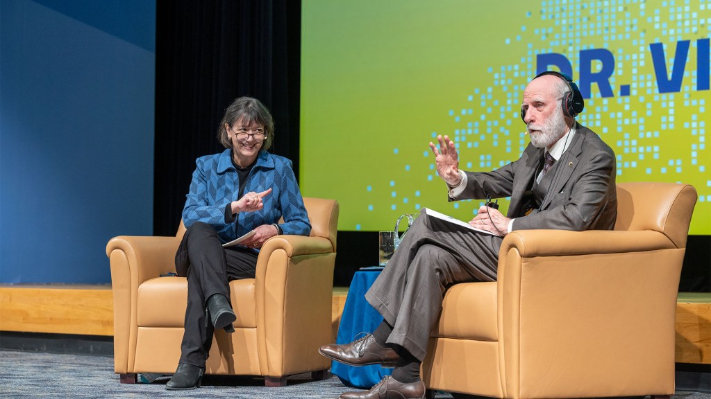
NIH Director Monica Bertagnolli in a fireside chat with Google Chief Internet Evangelist Vint Cerf at the Rall Cultural Lecture on March 19, 2024, at Masur Auditorium. Credit (all photos): Leslie Kossoff
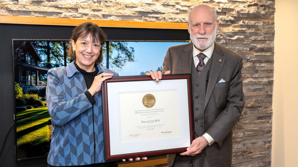
Last week it was my pleasure to welcome to NIH Vinton “Vint” Cerf, a pioneer of the digital world widely known as one of the “fathers of the internet,” to speak at our annual J. Edward Rall Cultural Lecture. We had a lively fireside chat focusing on “The Promises and Perils of AI in Biomedical Research and Health Care Delivery” that included discussions on topics like improving data collection, ensuring broad representativeness of the data being used to train AI and machine learning, expanding clinical research, and offering advice for scientists just starting out. It was an insightful, eye-opening conversation on a topic close to my heart. As NIH Director, I very much hope to deliver evidence-based health care to all people, and I’m excited about how AI and machine learning can help us advance toward that goal.
Dr. Cerf, who is vice president and Chief Internet Evangelist for Google, has too many awards to list, but just a few include the U.S. National Medal of Technology, the Turing Award, and the Presidential Medal of Freedom. He serves as an advisor to several government agencies, including the National Science Foundation, NASA, and the Departments of Defense, Energy, and Commerce.
The NIH Rall Cultural Lecture is held in honor of Dr. J. Edward Rall, who helped define the modern intramural research program at NIH. In the 1950s, he helped establish a stable, academic-minded, and culturally rich community within our rapidly expanding government agency. In 1984, he recommended that NIH add a cultural lecture to its Director’s Lecture series to enrich our scientific community.
As I said at the event, I’m grateful to Dr. Cerf for his visit and this conversation, and I can’t think of a more spectacular Rall Cultural Lecture than what he provided for all of us. You can watch the full talk here.
Aided by AI, Study Uncovers Hidden Sex Differences in Dynamic Brain Function
Posted on February 29th, 2024 by Dr. Monica M. Bertagnolli

Credit: Adobe/Feodora
We’re living in an especially promising time for biomedical discovery and advances in the delivery of data-driven health care for everyone. A key part of this is the tremendous progress made in applying artificial intelligence to study human health and ultimately improve clinical care in many important and sometimes surprising ways.1 One new example of this comes from a fascinating study, supported in part by NIH, that uses AI approaches to reveal meaningful sex differences in the way the brain works.
As reported in the Proceedings of the National Academy of Sciences, researchers led by Vinod Menon at Stanford Medicine, Stanford, CA, have built an AI model that can—nine times out of ten—tell whether the brain in question belongs to a female or male based on scans of brain activity alone.2 These findings not only help resolve long-term debates about whether reliable differences between sexes exist in the human brain, but they’re also a step toward improving our understanding of why some psychiatric and neurological disorders affect women and men differently.
The prevalence of certain psychiatric and neurological disorders in men and women can vary significantly, leading researchers to suspect that sex differences in brain function likely exist. For example, studies have found that females are more likely to experience depression, anxiety, and eating disorders, while autism, Attention-Deficit/Hyperactivity Disorder, and schizophrenia are seen more often in males. But earlier research to understand sex differences in the brain have focused mainly on anatomical and structural studies of brain regions and their connections. Much less is known about how those structural differences translate into differences in brain activity and function.
To help fill those gaps in the new study, Menon’s team took advantage of vast quantities of brain activity data from MRI scans from the NIH-supported Human Connectome Project. The data was captured from hundreds of healthy young adults with the goal of studying the brain and how it changes with growth, aging, and disease. To use this data to explore sex differences in brain function, the researchers developed what’s known as a deep neural network model in which a computer “learned” how to recognize patterns in brain activity data that could distinguish a male from a female brain.
This approach doesn’t rely on any preconceived notions about what features might be important. A computer is simply shown many examples of brain activity belonging to males and females and, over time, can begin to pick up on otherwise hidden differences that are useful for making such classifications accurately. One of the things that made this work different from earlier attempts was it relied on dynamic scans of brain activity, which capture the interplay among brain regions.
After analyzing about 1,500 brain scans, a computer could usually (although not always) tell whether a scan came from a male or female brain. The findings also showed the model worked reliably well in different datasets and in brain scans for people in different places in the U.S. and Europe. Overall, the findings confirm that reliable sex differences in brain activity do exist.
Where did the model find those differences? To get an idea, the researchers turned to an approach called explainable AI, which allowed them to dig deeper into the specific features and brain areas their model was using to pick up on sex differences. It turned out that one set of areas the model was relying on to distinguish between male and female brains is what’s known as the default mode network. This area is responsible for processing self-referential information and constructing a coherent sense of the self and activates especially when people let their minds wander.3 Other important areas included the striatum and limbic network, which are involved in learning and how we respond to rewards, respectively.
Many questions remain, including whether such differences arise primarily due to inherent biological differences between the sexes or what role societal circumstances play. But the researchers say that the discovery already shows that sex differences in brain organization and function may play important and overlooked roles in mental health and neuropsychiatric disorders. Their AI model can now also be applied to begin to explain other kinds of brain differences, including those that may affect learning or social behavior. It’s an exciting example of AI-driven progress and good news for understanding variations in human brain functions and their implications for our health.
References:
[1] Bertagnolli, MM. Advancing health through artificial intelligence/machine learning: The critical importance of multidisciplinary collaboration. PNAS Nexus. DOI: 10.1093/pnasnexus/pgad356 (2023).
[2] Ryali S, et al. Deep learning models reveal replicable, generalizable, and behaviorally relevant sex differences in human functional brain organization. Proc Natl Acad Sci. DOI: 10.1073/pnas.2310012121 (2024).
[3] Menon, V. 20 years of the default mode network: A review and synthesis. Neuron.DOI: 10.1016/j.neuron.2023.04.023 (2023).
NIH Support: National Institute of Mental Health, National Institute of Biomedical Imaging and Bioengineering, Eunice Kennedy Shriver National Institute of Child Health and Human Development, National Institute on Aging
NIH HEAL Initiative Meets People Where They Are
Posted on April 18th, 2023 by Rebecca Baker, Ph.D., NIH Helping to End Addiction Long-term® (HEAL) Initiative

Credit: NIH HEAL Initiative
The opioid crisis continues to devastate communities across America. Dangerous synthetic opioids, like fentanyl, have flooded the illicit drug supply with terrible consequences. Tragically, based on our most-recent data, about 108,000 people in the U.S. die per year from overdoses of opioids or stimulants [1]. Although this complex public health challenge started from our inability to treat pain effectively, chronic pain remains a life-altering problem for 50 million Americans.
To match the size and complexity of the crisis, in 2018 NIH developed the NIH Helping to End Addiction Long-term® (HEAL) Initiative, an aggressive effort involving nearly all of its 27 institutes and centers. Through more than 1,000 research projects, including basic science, clinical testing of new and repurposed drugs, research with communities, and health equity research, HEAL is dedicated to building a new future built on hope.
In this future:
- A predictive tool used during a health visit personalizes treatment for back pain. The tool estimates the probability that a person will benefit from physical therapy, psychotherapy, or surgery.
- Visits to community health clinics and emergency departments serve as routine opportunities to prevent and treat opioid addiction.
- Qualified school staff and pediatricians screen all children for behavioral and other mental health conditions that increase risk for harmful developmental outcomes, including opioid misuse.
- Infants born exposed to opioids during a mother’s pregnancy receive high-quality care—setting them up for a healthy future.
Five years after getting started (and interrupted by a global pandemic), HEAL research is making progress toward achieving this vision. I’ll highlight three ways in which scientific solutions are meeting people where they are today.
A Window of Opportunity for Treatment in the Justice System
Sadly, jails and prisons are “ground zero” for the nation’s opioid crisis. Eighty-five percent of people who are incarcerated have a substance use disorder or a history of substance use. Our vision at HEAL is that every person in jail, prison, or a court-supervised program receives medical care, which includes effective opioid use disorder treatment.
Some research results already are in supporting this approach: A recent HEAL study learned that individuals who had received addiction treatment while in one Massachusetts jail were about 30 percent less likely to be arrested, arraigned, or incarcerated again compared with those incarcerated during the same time period in a neighboring jail that did not offer treatment [2]. Research from the HEAL-supported Justice Community Opioid Innovation Network also is exploring public perceptions about opioid addiction. One such survey showed that most U.S. adults see opioid use disorder as a treatable medical condition rather than as a criminal matter [3]. That’s hopeful news for the future.
A Personalized Treatment Plan for Chronic Back Pain
Half of American adults live with chronic back pain, a major contributor to opioid use. The HEAL-supported Back Pain Consortium (BACPAC) is creating a whole-system model for comprehensive testing of everything that contributes to chronic low back pain, from anxiety to tissue damage. It also includes comprehensive testing of promising pain-management approaches, including psychotherapy, antidepressants, or surgery.
Refining this whole-system model, which is nearing completion, includes finding computer-friendly ways to describe the relationship between the different elements of pain and treatment. That might include developing mathematical equations that describe the physical movements and connections of the vertebrae, discs, and tendons.
Or it might include an artificial intelligence technique called machine learning, in which a computer looks for patterns in existing data, such as electronic health records or medical images. In keeping with HEAL’s all-hands-on-deck approach, BACPAC also conducts clinical trials to test new (or repurposed) treatments and develop new technologies focused on back pain, like a “wearable muscle” to help support the back.
Harnessing Innovation from the Private Sector
The HEAL research portfolio spans basic science to health services research. That allows us to put many shots on goal that will need to be commercialized to help people. Through its research support of small businesses, HEAL funding offers a make-or-break opportunity to advance a great idea to the marketplace, providing a bridge to venture capital or other larger funding sources needed for commercialization.
This bridge also allows HEAL to invest directly in the heart of innovation. Currently, HEAL funds nearly 100 such companies across 20 states. While this is a relatively small portion of all HEAL research, it is science that will make a difference in our communities, and these researchers are passionate about what they do to build a better future.
A couple of current examples of this research passion include: delivery of controlled amounts of non-opioid pain medications after surgery using a naturally absorbable film or a bone glue; immersive virtual reality to help people with opioid use disorder visualize the consequences of certain personal choices; and mobile apps that support recovery, taking medications, or sensing an overdose.
In 2023, HEAL is making headway toward its mission to accelerate development of safe, non-addictive, and effective strategies to prevent and treat pain, opioid misuse, and overdose. We have 314 clinical trials underway and 41 submissions to the Food and Drug Administration to begin clinical testing of investigational new drugs or devices: That number has doubled in the last year. More than 100 projects alone are addressing back pain, and more than 200 projects are studying medications for opioid use disorder.
The nation’s opioid crisis is profoundly difficult and multifaceted—and it won’t be solved with any single approach. Our research is laser-focused on its vision of ending addiction long-term, including improving pain management and expanding access to underused, but highly effective, addiction medications. Every day, we imagine a better future for people with physical and emotional pain and communities that are hurting. Hundreds of researchers and community members across the country are working to achieve a future where people and communities have the tools they need to thrive.
References:
[1] Provisional drug overdose death counts. Ahmad FB, Cisewski JA, Rossen LM, Sutton P. National Center for Health Statistics. 2023.
[2] Recidivism and mortality after in-jail buprenorphine treatment for opioid use disorder. Evans EA, Wilson D, Friedmann PD. Drug Alcohol Depend. 2022 Feb 1;231:109254.
[3] Social stigma toward persons with opioid use disorder: Results from a nationally representative survey of U.S. adults. Taylor BG, Lamuda PA, Flanagan E, Watts E, Pollack H, Schneider J. Subst Use Misuse. 2021;56(12):1752-1764.
Links:
SAMHSA’s National Helpline (Substance Abuse and Mental Health Services Administration, Rockville, MD)
NIH Helping to End Addiction Long-term® (HEAL) Initiative
Video: The NIH HEAL Initiative–HEAL Is Hope
Justice Community Opioid Innovation Network (HEAL)
Back Pain Consortium Research Program (HEAL)
NIH HEAL Initiative 2023 Annual Report (HEAL)
Small Business Programs (HEAL)
Rebecca Baker (HEAL)
Note: Dr. Lawrence Tabak, who performs the duties of the NIH Director, has asked the heads of NIH’s Institutes, Centers, and Offices to contribute occasional guest posts to the blog to highlight some of the interesting science that they support and conduct. This is the 28th in the series of NIH guest posts that will run until a new permanent NIH director is in place.
Posted In: Generic
Tags: AI, artificial intelligence, Back Pain, Back Pain Consortium, BACPAC, child health, Chronic Pain, clinical trial, drug repurposing, drugs, FDA, HEAL, health equity, Helping to End Addiction Long-term, jails, Justice Community Opioid Innovation Network, machine learning, opioid addiction, opioid crisis, pain, prisons, psychotherapy, small business, substance use disorders, wearable muscle
A Look Back at Science’s 2022 Breakthroughs
Posted on January 3rd, 2023 by Lawrence Tabak, D.D.S., Ph.D.
Credit: National Institute of Allergy and Infectious Diseases, NIH; Centers for Disease Control and Prevention; Shutterstock/tobe24, Midjourney Inc.
Happy New Year! I hope everyone finished 2022 with plenty to celebrate, whether it was completing a degree or certification, earning a promotion, attaining a physical fitness goal, or publishing a hard-fought scientific discovery.
If the latter, you are in good company. Last year produced some dazzling discoveries, and the news and editorial staff at the journal Science kept a watchful eye on the most high-impact advances of 2022. In December, the journal released its list of the top 10 advances across the sciences, from astronomy to zoology. In case you missed it, Science selected NASA’s James Webb Space Telescope (JWST) as the 2022 Breakthrough of the Year [1].
This unique space telescope took 20 years to complete, but it has turned out to be time well spent. Positioned 1.5-million-kilometers from Earth, the JWST and its unprecedented high-resolution images of space have unveiled the universe anew for astronomers and wowed millions across the globe checking in online. The telescope’s image stream, beyond its sheer beauty, will advance study of the early Universe, allowing astronomers to discover distant galaxies, explore the early formation of stars, and investigate the possibility of life on other planets.
While the biomedical sciences didn’t take home the top prize, they were well represented among _Science_’s runner-up breakthroughs. Some of these biomedical top contenders also have benefited, directly or indirectly, from NIH efforts and support. Let’s take a look:
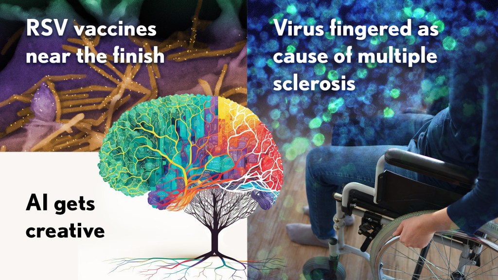
RSV vaccines nearing the finish line: It’s been one of those challenging research marathons. But scientists last year started down the homestretch with the first safe-and-effective vaccine for respiratory syncytial virus (RSV), a leading cause of severe respiratory illness in the very young and the old.
In August, the company Pfizer presented evidence that its experimental RSV vaccine candidate offered protection for those age 60 and up. Later, they showed that the same vaccine, when administered to pregnant women, helped to protect their infants against RSV for six months after birth. Meanwhile, in October, the company GSK announced encouraging results from its late-stage phase III trial of an RSV vaccine in older adults.
As Science noted, the latest clinical progress also shows the power of basic science. For example, researchers have been working with chemically inactivated versions of the virus to develop the vaccine. But these versions have a key viral surface protein that changes its shape after fusing with a cell to start an infection. In this configuration, the protein elicits only weak levels of needed protective antibodies.
Back in 2013, Barney Graham, then with NIH’s National Institute of Allergy and Infectious Diseases (NIAID), and colleagues, solved the problem [2]. Graham’s NIH team discovered a way to lock the protein into its original prefusion state, which the immune system can better detect. This triggers higher levels of potent antibodies, and the discovery kept the science—and the marathon—moving forward.
These latest clinical advances come as RSV and other respiratory viruses, including SARS-CoV-2, the cause of COVID-19, are sending an alarming number of young children to the hospital. The hope is that researchers will cross the finish line this year or next, and we’ll have the first approved RSV vaccine.
Virus fingered as cause of multiple sclerosis: Researchers have long thought that multiple sclerosis, or MS, has a viral cause. Pointing to the right virus with the required high degree of certainty has been the challenge, slowing progress on the treatment front for those in need. As published in Science last January, Alberto Ascherio, Harvard T.H. Chan School of Public Health, Boston, and colleagues produced the strongest evidence yet that MS is caused by the Epstein-Barr virus (EBV), a herpesvirus also known for causing infectious mononucleosis [3].
The link between EBV and MS had long been suspected. But it was difficult to confirm because EBV infections are so widespread, and MS is so disproportionately rare. In the recent study, the NIH-supported researchers collected blood samples every other year from more than 10 million young adults in the U.S. military, including nearly 1,000 who were diagnosed with MS during their service. The evidence showed that the risk of an MS diagnosis increased 32-fold after EBV infection, but it held steady following infection with any other virus. Levels in blood serum of a biomarker for MS neurodegeneration also went up only after an EBV infection, suggesting that the viral illness is a leading cause for MS.
Further evidence came last year from a discovery published in the journal Nature by William Robinson, Stanford University School of Medicine, Stanford, CA, and colleagues. The NIH-supported team found a close resemblance between an EBV protein and one made in the healthy brain and spinal cord [4]. The findings suggest an EBV infection may produce antibodies that mistakenly attack the protective sheath surrounding our nerve cells. Indeed, the study showed that up to one in four people with MS had antibodies that bind both proteins.
This groundbreaking research suggests that an EBV vaccine and/or antiviral drugs that thwart this infection might ultimately prevent or perhaps even cure MS. Of note, NIAID launched last May an early-stage clinical trial for an experimental EBV vaccine at the NIH Clinical Center, Bethesda, MD.
AI Gets Creative: _Science_’s 2021 Breakthrough of the Year was AI-powered predictions of protein structure. In 2022, AI returned to take another well-deserved bow. This time, Science singled out AI’s now rapidly accelerating entry into once uniquely human attributes, such as artistic expression and scientific discovery.
On the scientific discovery side, Science singled out AI’s continued progress in getting creative with the design of novel proteins for vaccines and myriad other uses. One technique, called “hallucination,” generates new proteins from scratch. Researchers input random amino acid sequences into the computer, and it randomly and continuously mutates them into sequences that other AI tools are confident will fold into stable proteins. This greatly simplifies the process of protein design and frees researchers to focus their efforts on creating a protein with a desired function.
AI research now engages scientists around world, including hundreds of NIH grantees. Taking a broader view of AI, NIH recently launched the Artificial Intelligence/Machine Learning Consortium to Advance Health Equity and Researcher Diversity (AIM-AHEAD) Program. It will help to create greater diversity within the field, which is a must. A lack of diversity could perpetuate harmful biases in how AI is used, how algorithms are developed and trained, and how findings are interpreted to avoid health disparities and inequities for underrepresented communities.
And there you have it, some of the 2022 breakthroughs from _Science_‘s news and editorial staff. Of course, the highlighted biomedical breakthroughs don’t capture the full picture of research progress. There were many other milestone papers published in 2022 that researchers worldwide will build upon in the months and years ahead to make further progress in their disciplines and, for some, draw the attention of _Science_’s news and editorial staff. Here’s to another productive year in biomedical research, which the blog will continue to feature and share with you as it unfolds in 2023.
References:
[1] 2022 Breakthrough of the Year. Science. Dec 15, 2022.
[2] Structure of RSV fusion glycoprotein trimer bound to a prefusion-specific neutralizing antibody. McLellan JS, Chen M, Leung S, Kwong PD, Graham BS, et al. Science. 2013 May 31;340(6136):1113-1117.
[3] Longitudinal analysis reveals high prevalence of Epstein-Barr virus associated with multiple sclerosis. Bjornevik K, Cortese M, Healy BC, Kuhle J, Mina MJ, Leng Y, Elledge SJ, Niebuhr DW, Scher AI, Munger KL, Ascherio A. Science. 2022 Jan 21;375(6578):296-301.
[4] Clonally expanded B cells in multiple sclerosis bind EBV EBNA1 and GlialCAM. Lanz TV, Brewer RC, Steinman L, Robinson WH, et al. Nature. 2022 Mar;603(7900):321-327.
Links:
Respiratory Syncytial Virus (RSV) (National Institute of Allergy and Infectious Diseases/NIH)
Multiple Sclerosis (National Institute of Neurological Disorders and Stroke/NIH)
Barney Graham (Morehouse School of Medicine, Atlanta)
Alberto Ascherio (Harvard T.H. Chan School of Public Health, Boston)
Robinson Lab (Stanford Medicine, Stanford, CA)
James Webb Space Telescope (Goddard Space Flight Center/NASA, Greenbelt, MD)
Posted In: News
Tags: 2022 Science Breakthrough of the Year, AI, AI Hallucination, AIM-AHEAD, basic research, Creative AI, EBV, EBV vaccine, Epstein-Barr, GSK, health disparities, James Webb Space Telescope, JWST, machine learning, MS, multiple sclerosis, NIH Clinical Center, Pfizer, respiratory syncytial virus, RSV, RSV vaccine, SARS-CoV-2, vaccine, virus
From Brain Waves to Real-Time Text Messaging
Posted on November 15th, 2022 by Lawrence Tabak, D.D.S., Ph.D.
For people who have lost the ability to speak due to a severe disability, they want to get the words out. They just can’t physically do it. But in our digital age, there is now a fascinating way to overcome such profound physical limitations. Computers are being taught to decode brain waves as a person tries to speak and then interactively translate them onto a computer screen in real time.
The latest progress, demonstrated in the video above, establishes that it’s quite possible for computers trained with the help of current artificial intelligence (AI) methods to restore a vocabulary of more than a 1,000 words for people with the mental but not physical ability to speak. That covers more than 85 percent of most day-to-day communication in English. With further refinements, the researchers say a 9,000-word vocabulary is well within reach.
The findings published in the journal Nature Communications come from a team led by Edward Chang, University of California, San Francisco [1]. Earlier, Chang and colleagues established that this AI-enabled system could directly decode 50 full words in real time from brain waves alone in a person with paralysis trying to speak [2]. The study is known as BRAVO, short for Brain-computer interface Restoration Of Arm and Voice.
In the latest BRAVO study, the team wanted to figure out how to condense the English language into compact units for easier decoding and expand that 50-word vocabulary. They did it in the same way we all do: by focusing not on complete words, but on the 26-letter alphabet.
The study involved a 36-year-old male with severe limb and vocal paralysis. The team designed a sentence-spelling pipeline for this individual, which enabled him to silently spell out messages using code words corresponding to each of the 26 letters in his head. As he did so, a high-density array of electrodes implanted over the brain’s sensorimotor cortex, part of the cerebral cortex, recorded his brain waves.
A sophisticated system including signal processing, speech detection, word classification, and language modeling then translated those thoughts into coherent words and complete sentences on a computer screen. This so-called speech neuroprosthesis system allows those who have lost their speech to perform roughly the equivalent of text messaging.
Chang’s team put their spelling system to the test first by asking the participant to silently reproduce a sentence displayed on a screen. They then moved on to conversations, in which the participant was asked a question and could answer freely. For instance, as in the video above, when the computer asked, “How are you today?” he responded, “I am very good.” When asked about his favorite time of year, he answered, “summertime.” An attempted hand movement signaled the computer when he was done speaking.
The computer didn’t get it exactly right every time. For instance, in the initial trials with the target sentence, “good morning,” the computer got it exactly right in one case and in another came up with “good for legs.” But, overall, their tests show that their AI device could decode with a high degree of accuracy silently spoken letters to produce sentences from a 1,152-word vocabulary at a speed of about 29 characters per minute.
On average, the spelling system got it wrong 6 percent of the time. That’s really good when you consider how common it is for errors to arise with dictation software or in any text message conversation.
Of course, much more work is needed to test this approach in many more people. They don’t yet know how individual differences or specific medical conditions might affect the outcomes. They suspect that this general approach will work for anyone so long as they remain mentally capable of thinking through and attempting to speak.
They also envision future improvements as part of their BRAVO study. For instance, it may be possible to develop a system capable of more rapid decoding of many commonly used words or phrases. Such a system could then reserve the slower spelling method for other, less common words.
But, as these results clearly demonstrate, this combination of artificial intelligence and silently controlled speech neuroprostheses to restore not just speech but meaningful communication and authentic connection between individuals who’ve lost the ability to speak and their loved ones holds fantastic potential. For that, I say BRAVO.
References:
[1] Generalizable spelling using a speech neuroprosthesis in an individual with severe limb and vocal paralysis. Metzger SL, Liu JR, Moses DA, Dougherty ME, Seaton MP, Littlejohn KT, Chartier J, Anumanchipalli GK, Tu-CHan A, Gangly K, Chang, EF. Nature Communications (2022) 13: 6510.
[2] Neuroprosthesis for decoding speech in a paralyzed person with anarthria. Moses DA, Metzger SL, Liu JR, Tu-Chan A, Ganguly K, Chang EF, et al. N Engl J Med. 2021 Jul 15;385(3):217-227.
Links:
Voice, Speech, and Language (National Institute on Deafness and Other Communication Disorders/NIH)
ECoG BMI for Motor and Speech Control (BRAVO) (ClinicalTrials.gov)
Chang Lab (University of California, San Francisco)
NIH Support: National Institute on Deafness and Other Communication Disorders
Posted In: News
Tags: AI, artificial intelligence, bci, brain, brain waves, brain-computer interface, BRAVO, computer, deep learning, disability, language, language modeling, Neuroprosthesis, neuroscience, paralysis, sentence-spelling, signal processing, speak, speech, speech detection, speech neuroprosthesis system, text messaging, video, vocabulary, word classification
National Library of Medicine Helps Lead the Way in AI Research
Posted on November 8th, 2022 by Patricia Flatley Brennan, R.N., Ph.D., National Library of Medicine

Credit: National Library of Medicine, NIH
Did you know that the NIH’s National Library of Medicine (NLM) has been serving science and society since 1836? From its humble beginning as a small collection of books in the library of the U.S. Army Surgeon General’s office, NLM has grown not only to become the world’s largest biomedical library, but a leader in biomedical informatics and computational health data science research.
Think of NLM as a door through which you pass to connect with health data, literature, medical and scientific information, expertise, and sophisticated mathematical models or images that describe a clinical problem. This intersection of information, people, and technology allows NLM to foster discovery. NLM does so by ensuring that scientists, clinicians, librarians, patients, and the public have access to biomedical information 24 hours a day, 7 days a week.
The NLM also supports two research efforts: the Division of Extramural Programs (EP) and Intramural Research Program (IRP). Both programs are accelerating advances in biomedical informatics, data science, computational biology, and computational health. One of EP’s notable investments is focused on advancing artificial intelligence (AI) methods and reimagining how health care is delivered with the power of AI.

Credit: National Library of Medicine, NIH
With support from NLM, Corey Lester and his colleagues at the University of Michigan College of Pharmacy, Ann Arbor, MI, are using AI to assist in pill verification, a standard procedure in pharmacies across the land. They want to help pharmacists avoid dangerous and costly dispensing errors. To do so, Lester is using AI to develop a real-time computer vision model. It views pills inside of a medication bottle, accurately identifies them, and determines that they are the correct or incorrect contents.
The IRP develops and applies computational methods and approaches to a broad range of information problems in biology, biomedicine, and human health. The IRP also offers intramural training opportunities and supports other training aimed at pre-baccalaureate to postdoctoral students and professionals.
The NLM principal investigators use biological data to advance computer algorithms and connect relationships between any level of biological organization and health conditions. They also use computational health sciences to focus on clinical information processing and analyze clinical data, assess clinical outcomes, and set health data standards.

Credit: National Library of Medicine, NIH
NLM investigator Sameer Antani is collaborating with researchers in other NIH institutes to explore how AI can help us understand oral cancer, echocardiography, and pediatric tuberculosis. His research also is examining how images can be mined for data to predict the causes and outcomes of conditions. Examples of Antani’s work can be found in mobile radiology vehicles, which allow professionals to take chest X-rays (right) and screen for HIV and tuberculosis using software containing algorithms developed in his lab.
For AI to have its full impact, more algorithms and approaches that harness the power of data are needed. That’s why NLM supports hundreds of other intramural and extramural scientists who are addressing challenging health and biomedical problems. The NLM-funded research is focused on how AI can help people stay healthy through early disease detection, disease management, and clinical and treatment decision-making—all leading to the ultimate goal of helping people live healthier and happier lives.
The NLM is proud to lead the way in the use of AI to accelerate discovery and transform health care. Want to learn more? Follow me on Twitter. Or, you can follow my blog, NLM Musings from the Mezzanine and receive periodic NLM research updates.
I would like to thank Valerie Florance, Acting Scientific Director of NLM IRP, and Richard Palmer, Acting Director of NLM Division of EP, for their assistance with this post.
Links:
National Library of Medicine (National Library of Medicine/NIH)
Video: Using Machine Intelligence to Prevent Medication Dispensing Errors (NLM Funding Spotlight)
Video: Sameer Antani and Artificial Intelligence (NLM)
NLM Division of Extramural Programs (NLM)
NLM Intramural Research Program (NLM)
NLM Intramural Training Opportunities (NLM)
Principal Investigators (NLM)
NLM Musings from the Mezzanine (NLM)
Note: Dr. Lawrence Tabak, who performs the duties of the NIH Director, has asked the heads of NIH’s Institutes and Centers (ICs) to contribute occasional guest posts to the blog to highlight some of the interesting science that they support and conduct. This is the 20th in the series of NIH IC guest posts that will run until a new permanent NIH director is in place.
Posted In: Generic
Tags: AI, artificial intelligence, bioinformatics, biomedical information, computational biology, computational health sciences, data science, dispensing errors, echocardiography, extramural research, library, machine learning, medication, National Library of Medicine, NIH Intramural Program, NLM, oral cancer, pediatric tuberculosis, pharmacists, pharmacy, pills
Understanding Long-Term COVID-19 Symptoms and Enhancing Recovery
Posted on October 4th, 2022 by Walter J. Koroshetz, M.D., National Institute of Neurological Disorders and Stroke
We are in the third year of the COVID-19 pandemic, and across the world, most restrictions have lifted, and society is trying to get back to “normal.” But for many people—potentially millions globally—there is no getting back to normal just yet.
They are still living with the long-term effects of a COVID-19 infection, known as the post-acute sequelae of SARS-CoV-2 infection (PASC), including Long COVID. These people continue to experience debilitating fatigue, shortness of breath, pain, difficulty sleeping, racing heart rate, exercise intolerance, gastrointestinal and other symptoms, as well as cognitive problems that make it difficult to perform at work or school.
This is a public health issue that is in desperate need of answers. Research is essential to address the many puzzling aspects of Long COVID and guide us to effective responses that protect the nation’s long-term health.
For the past two years, NIH’s National Heart, Lung, and Blood Institute (NHLBI), the National Institute of Allergy and Infectious Diseases (NIAID), and my National Institute of Neurological Disorders and Stroke (NINDS) along with several other NIH institutes and the office of the NIH Director, have been leading NIH’s Researching COVID to Enhance Recovery (RECOVER) initiative, a national research program to understand PASC.
The initiative studies core questions such as why COVID-19 infections can have lingering effects, why new symptoms may develop, and what is the impact of SARS-CoV-2, the virus that causes COVID-19, on other diseases and conditions? Answering these fundamental questions will help to determine the underlying biologic basis of Long COVID. The answers will also help to tell us who is at risk for Long COVID and identify therapies to prevent or treat the condition.
The RECOVER initiative’s wide scope of research is also unprecedented. It is needed because Long COVID is so complex, and history indicates that similar post infectious conditions have defied definitive explanation or effective treatment. Indeed, those experiencing Long COVID report varying symptoms, making it highly unlikely that a single therapy will work for everyone, underscoring the need to pursue multiple therapeutic strategies.
To understand Long COVID fully, hundreds of RECOVER investigators are recruiting more than 17,000 adults (including pregnant people) and more than 18,000 children to take part in cohort studies. Hundreds of enrolling sites have been set up across the country. An autopsy research cohort will also provide further insight into how COVID-19 affects the body’s organs and tissues.
In addition, researchers will analyze electronic health records from millions of people to understand how Long COVID and its symptoms change over time. The RECOVER initiative is also utilizing consistent research protocols across all the study sites. The protocols have been carefully developed with input from patients and advocates, and they are designed to allow for consistent data collection, improve data sharing, and help to accelerate the pace of research.
From the very beginning, people suffering from Long COVID have been our partners in RECOVER. Patients and advocates have contributed important perspectives and provided valuable input into the master protocols and research plans.
Now, with RECOVER underway, individuals with Long COVID, their caregivers, and community members continue to serve a critical role in the Initiative. The National Community Engagement Group (NCEG) has been established to make certain that RECOVER meets the needs of all people affected by Long COVID. The RECOVER Patient and Community Engagement Strategy outlines all the approaches that RECOVER is using to engage with and gather input from individuals impacted by Long COVID.
The NIH recently made more than 40 awards to improve understanding of the underlying biology and pathology of Long COVID. There have already been several important findings published by RECOVER scientists.
For example, in a recent study published in the journal Lancet Digital Health, RECOVER investigators used machine learning to comb through electronic health records to look for signals that may predict whether someone has Long COVID [1]. As new findings, tools, and technologies continue to emerge that help advance our knowledge of the condition, the RECOVER Research Review (R3) Seminar Series will provide a forum for researchers and our partners with up-to-date information about Long COVID research.
It is important to note that post-viral conditions are not a new concept. Many, but not all, of the symptoms reported in Long COVID, including fatigue, post-exertional malaise, chronic musculoskeletal pain, sleep disorders, postural orthostatic tachycardia (POTS), and cognitive issues, overlap with myalgic encephalomyelitis/chronic fatigue syndrome (ME/CFS).
ME/CFS is a serious disease that can occur following infection and make people profoundly sick for decades. Like Long COVID, ME/CFS is a heterogenous condition that does not affect everybody in the same way, and the knowledge gained through research on Long COVID may also positively impact the understanding, treatment, and prevention of POTS, ME/CFS, and other chronic diseases.
Unlike other post-viral conditions, people who experience Long COVID were all infected by the same virus—albeit different variants—at a similar point in time. This creates a unique opportunity for RECOVER researchers to study post-viral conditions in real-time.
The opportunity enables scientists to study many people simultaneously while they are still infected to monitor their progress and recovery, and to try to understand why some individuals develop ongoing symptoms. A better understanding of the transition from acute to chronic disease may offer an opportunity to intervene, identify who is at risk of the transition, and develop therapies for people who experience symptoms long after the acute infection has resolved.
The RECOVER initiative will soon announce clinical trials, leveraging data from clinicians and patients in which symptom clusters were identified and can be targeted by various interventions. These trials will investigate therapies that are indicated for other non-COVID conditions and novel treatments for Long COVID.
Through extensive collaboration across the multiple NIH institutes and offices that contribute to the RECOVER effort, our hope is critical answers will emerge soon. These answers will help us to recognize the full range of outcomes and needs resulting from PASC and, most important, enable many people to make a full recovery from COVID-19. We are indebted to the over 10,000 subjects who have already enrolled in RECOVER. Their contributions and the hard work of the RECOVER investigators offer hope for the future to the millions still suffering from the pandemic.
Reference:
[1] Identifying who has long COVID in the USA: a machine learning approach using N3C data. Pfaff ER, Girvin AT, Bennett TD, Bhatia A, Brooks IM, Deer RR, Dekermanjian JP, Jolley SE, Kahn MG, Kostka K, McMurry JA, Moffitt R, Walden A, Chute CG, Haendel MA; N3C Consortium. Lancet Digit Health. 2022 Jul;4(7):e532-e541.
Links:
COVID-19 Research (NIH)
Long COVID (NIH)
RECOVER: Researching COVID to Enhance Recovery (NIH)
“NIH builds large nationwide study population of tens of thousands to support research on long-term effects of COVID-19,” NIH News Release, September 15, 2021.
Director’s Messages (National Institute of Neurological Disorders and Stroke/NIH)
Note: Dr. Lawrence Tabak, who performs the duties of the NIH Director, has asked the heads of NIH’s Institutes and Centers (ICs) to contribute occasional guest posts to the blog to highlight some of the interesting science that they support and conduct. This is the 18th in the series of NIH IC guest posts that will run until a new permanent NIH director is in place.
Posted In: Generic
Tags: AI, basic research, chronic disease, clinical research, clinical trials, coronavirus, COVID-19, COVID-19 recovery, COVID-19 treatment, data science, EHR, electronic health records, fatigue, long COVID, Long COVID syndrome, machine learning, ME/CFS, Myalgic Encephalomyelitis/Chronic Fatigue Syndrome, National Community Engagement Group, NCEG, novel coronavirus, pain, PASC, post-acute sequelae of COVID-19, post-viral conditions, postural orthostatic tachycardia, POTS, public health, RECOVER Initiative, RECOVER Patient and Community Engagement Strategy, Researching COVID to Enhance Recovery, SARS-CoV-2, sleep disorders