BioArt – NIH Director's Blog (original) (raw)
Capturing the Extracellular Matrix in 3D Color
Posted on December 16th, 2021 by Dr. Francis Collins
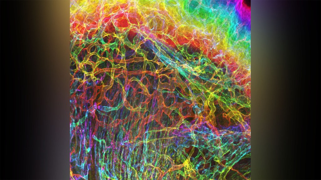
Credit: Sarah Lipp, Purdue University, and Sarah Calve, University of Colorado, Boulder
For experienced and aspiring shutterbugs alike, sometimes the best photo in the bunch turns out to be a practice shot. That’s also occasionally true in the lab when imaging cells and tissues, and it’s the story behind this spectacular image showing the interface of skin and muscle during mammalian development.
Here you see an area of the mouse forelimb located near a bone called the humerus. This particular sample was labeled for laminin, a protein found in the extracellular matrix (ECM) that undergirds cells and tissues to give them mechanical and biochemical support. Computer algorithms were used to convert the original 2D confocal scan into a 3D image, and colorization was added to bring the different layers of tissue into sharper relief.
Skin tissue (bright red and yellow) is located near the top of the image; blood vessels (paler red, orange, and yellow) are in the middle and branching downward; and muscle (green, blue, and purple) makes up the bottom layer.
The image was created by Sarah Lipp, a graduate student in the NIH-supported tissue engineering lab of Sarah Calve. The team focuses on tissue interfaces to better understand the ECM and help devise strategies to engineer musculoskeletal tissues, such as tendon and cartilage.
In February 2020, Lipp was playing around with some new software tools for tissue imaging. Before zeroing in on her main target—the mouse’s myotendinous junction, where muscle transfers its force to tendon, Lipp snapped this practice shot of skin meeting muscle. After processing the practice shot with a color-projecting macro in an image processing tool called Fiji, she immediately liked what she saw.
So, Lipp tweaked the color a bit more and entered the image in the 2020 BioArt Scientific Image & Video Competition, sponsored by the Federation of American Societies for Experimental Biology, Bethesda, MD. Last December, the grad student received the good news that her practice shot had snagged one of the prestigious contest’s top awards.
But she’s not stopping there. Lipp is continuing to pursue her research interests at the University of Colorado, Boulder, where the Calve lab recently moved from Purdue University, West Lafayette, IN. Here’s wishing her a career filled with more great images—and great science!
Links:
Muscle and Bone Diseases (National Institute of Arthritis and Musculoskeletal and Skin Diseases/NIH)
Musculoskeletal Extracellular Matrix Laboratory (University of Colorado, Boulder)
BioArt Scientific Image & Video Competition (Federation of American Societies for Experimental Biology, Bethesda, MD)
NIH Support: National Institute of Arthritis and Musculoskeletal and Skin Diseases
Posted In: Snapshots of Life
Tags: BioArt, developmental neurobiology, ECM, extracellular matrix, FASEB Bioart 2020, Fiji, humerus, imaging, laminin, mouse, muscle, myotendinous junction, skin, tendon, tissue engineering
Watch Flowers Spring to Life
Posted on April 25th, 2019 by Dr. Francis Collins
Spring has sprung! The famous Washington cherry blossoms have come and gone, and the tulips and azaleas are in full bloom. In this mesmerizing video, you’ll get a glimpse of the early steps in how some spring flowers bloom.
Floating into view are baby flowers, their cells outlined (red), at the tip of the stem of the mustard plant Arabidopsis thaliana. Stem cells that contain the gene STM (green) huddle in the center of this fast-growing region of the plant stem—these stem cells will later make all of the flower parts.
As the video pans out, slightly older flowers come into view. These contain organs called sepals (red, bumpy outer regions) that will grow into leafy support structures for the flower’s petals.
Movie credits go to Nathanaёl Prunet, an assistant professor at the University of California, Los Angeles, who shot this video while working in the NIH-supported lab of Elliot Meyerowitz at the California Institute of Technology, Pasadena. Prunet used confocal microscopy to display the different ages and stages of the developing flowers, generating a 3D data set of images. He then used software to produce a bird’s-eye view of those images and turned it into a cool movie. The video was one of the winners in the Federation of American Societies for Experimental Biology’s 2018 BioArt competition.
Beyond being cool, this video shows how a single gene, STM, plays a starring role in plant development. This gene acts like a molecular fountain of youth, keeping cells ever-young until it’s time to grow up and commit to making flowers and other plant parts.
Like humans, most plants begin life as a fertilized cell that divides over and over—first into a multi-cell embryo and then into mature parts, or organs. Because of its ease of use and low cost, Arabidopsis is a favorite model for scientists to learn the basic principles driving tissue growth and regrowth for humans as well as the beautiful plants outside your window. Happy Spring!
Links:
Meyerowitz Lab (California Institute of Technology, Pasadena)
Prunet Lab (University of California, Los Angeles)
The Arabidosis Information Resource (Phoenix Bioinformatics, Fremont, CA)
BioArt Scientific Image and Video Competition (Federation of American Societies for Experimental Biology, Bethesda, MD)
NIH Support: National Institute of General Medical Sciences
Posted In: Cool Videos
Tags: 2018 BioArt Scientific Image & Video Competition, Arabidopsis, Arabidopsis thaliana, BioArt, development, developmental biology, flowers, model organism, mustard plant, plants, stem cells, STM, video
Students Contribute to Research Through Ovarian Art
Posted on February 21st, 2019 by Dr. Francis Collins

Credit: Crystal D. Rogers and Mariano Loza-Coll, California State University, Northridge
Seeing the development of an organ under a microscope for the first time can be a truly unforgettable experience. But for a class taught by Crystal Rogers at California State University, Northridge, it can also be an award-winning moment.
This image, prepared during a biology lab course, was one of the winners in the 2018 BioArt Scientific Image & Video Competition, sponsored by the Federation of American Societies for Experimental Biology (FASEB). This colorful image shows the tip of an ovary from a fruit fly (Drosophila melanogaster), provided by Mariano Loza-Coll. You can see that the ovary is packed with oocytes (DNA stained blue). The orderly connective structure (pink) and signal-transmitting molecules like STAT (yellow) are common to egg maturation and reproductive processes in humans.
What makes this image unique among this year’s BioArt winners is that the prep work was done by undergraduate students just learning how to work in a lab. They did the tissue dissections, molecular labeling, and beautiful stainings in preparation for Rogers to “snap” the photo on her research lab’s optical-sectioning microscope.
What’s also fantastic is that many of Rogers’s students are from groups traditionally underrepresented in biomedicine. Many are considering careers in research and, from the looks of things, they are off to a beautiful start.
After teaching classes, Rogers also has an NIH-supported lab to run. She and her team study salamanders and chickens to determine how biological “glue” proteins, called cadherins, help to create neural crest cells, a critical cell type that arises very early in development [1].
For developmental biologists, it’s essential to understand what prompts these neural crest cells to migrate to locations throughout the body, from the heart to the skin to the cranium, or head. For example, cranial neural crest cells at first produce what appears to be the same generic, undifferentiated facial template in vertebrate species. And yet, neural crest cells and the surrounding ectodermal cells go on to generate craniofacial structures as distinct as the beak of a toucan, the tusk of a boar, or the horn of a rhinoceros.
But if the organ of interest is an ovary, the fruit fly has long been a go-to organism to learn more. Not only does the fruit fly open a window into ovarian development and health issues like infertility, it showcases the extraordinary beauty of biology.
Reference:
[1] A catenin-dependent balance between N-cadherin and E-cadherin controls neuroectodermal cell fate choices. Rogers CD, Sorrells LK, Bronner ME. Mech Dev. 2018 Aug;152:44-56.
Links:
Rogers Lab (California State University, Northridge)
BioArt Scientific Image & Video Competition (Federation of American Societies for Experimental Biology, Bethesda, MD)
NIH Support: Eunice Kennedy Shriver National Institute of Child Health and Human Development
Posted In: Snapshots of Life
Tags: BioArt, cadherin, development, developmental biology, Drosophila melanogaster, embryology, embryos, fruit fly, microscopy, model organism, neural crest cells, oocyte, ovary, STAT
Snapshots of Life: Finding Where HIV Hides
Posted on March 8th, 2018 by Dr. Francis Collins

Credit: Nadia Roan, University of California, San Francisco
Researchers have learned a tremendous amount about how the human immunodeficiency virus (HIV), which causes AIDS, infects immune cells. Much of that information comes from studying immune cells in the bloodstream of HIV-positive people. Less detailed is the picture of how HIV interacts with immune cells inside the lymph nodes, where the virus can hide.
In this image of lymph tissue taken from the neck of a person with uncontrolled HIV infection, you can see areas where HIV is replicating (red) amid a sea of immune cells (blue dots). Areas of greatest HIV replication are associated with a high density of a subtype of human CD4 T-cells (yellow circles) that have been found to be especially susceptible to HIV infection.
Tags: 2017 MIC Image Contest at the University of California Berkeley, AIDS, art, BioArt, CD127, CD4 T cells, cervical lymph node, HIV, HIV replication, human immunodeficiency virus, immunology, lymph node, retrovirus, virology
Snapshots of Life: Portrait of a Bacterial Biofilm
Posted on January 19th, 2017 by Dr. Francis Collins

Credit: Scott Chimileski and Roberto Kolter, Harvard Medical School, Boston
In nature, there is strength in numbers. Sometimes, those numbers also have their own unique beauty. That’s the story behind this image showing an intricate colony of millions of the single-celled bacterium Pseudomonas aeruginosa, a common culprit in the more than 700,000 hospital-acquired infections estimated to occur annually in the United States. [1]. The bacteria have self-organized into a sticky, mat-like colony called a biofilm, which allows them to cooperate with each other, adapt to changes in their environment, and ensure their survival.
In this image, the Pseudomonas biofilm has grown in a laboratory dish to about the size of a dime. Together, the millions of independent bacterial cells have created a tough extracellular matrix of secreted proteins, polysaccharide sugars, and even DNA that holds the biofilm together, stained in red. The darkened areas at the center come from the bacteria’s natural pigments.
Tags: antibacterial drugs, antibiotics, antimicrobial resistance, bacteria, BioArt, biofilm, extracellular matrix, Federation of American Societies for Experimental Biology’s 2016 BioArt, hospital acquired infections, microbes, microbiology, microbiome, Pseudomonas aeruginosa
Snapshots of Life: Tales from the (Intestinal) Crypt!
Posted on October 27th, 2016 by Dr. Francis Collins
Caption: This “spooky” video ends with a scientific image of intestinal crypts (blue and green) plus organoids made from cultured crypt stem cells (pink).
As Halloween approaches, some of you might be thinking about cueing up the old TV series “Tales from the Crypt” and diving into its Vault of Horror for a few hours. But today I’d like to share the story of a quite different and not nearly so scary kind of crypt: the crypts of Lieberkühn, more commonly called intestinal crypts.
This confocal micrograph depicts a row of such crypts (marked in blue and green) lining a mouse colon. In mice, as well as in humans, the intestines contain millions of crypts, each of which has about a half-dozen stem cells at its base that are capable of regenerating the various types of tissues that make up these tiny glands. What makes my tale of the crypt particularly interesting are the oval structures (pink), which are organoids that have been engineered from cultured crypt stem cells and then transplanted into a mouse model. If you look at the organoids closely, you’ll see Paneth cells (aqua blue), which are immune cells that support the stem cells and protect the intestines from bacterial invasion.
A winner in the 2016 “Image Awards” at the Koch Institute Public Galleries, Massachusetts Institute of Technology (MIT), Cambridge, this image was snapped by Jatin Roper, a physician-scientist in the lab of Omer Yilmaz, with the help of his MIT collaborator Tuomas Tammela. Roper and his colleagues have been making crypt organoids for a few years by placing the stem cells in a special 3D chamber, where they are bathed with the right protein growth factors at the right time to spur them to differentiate into the various types of cells found in a crypt.
Once the organoids are developmentally complete, Roper can inject them into mice and watch them take up residence. Then he can begin planning experiments.
For example, Roper’s group is now considering using the organoids to examine how high-fat and low-calorie diets affect intestinal function in mice. Another possibility is to use similar organoids to monitor the effect of aging on the colon or to test which of a wide array of targeted therapies might work best for a particular individual with colon cancer.
Links:
Video: Gut Reaction (Jatin Roper)
Jatin Roper (Tufts Medical Center, Boston)
Omer Yilmaz (Massachusetts Institute of Technology, Cambridge)
The Koch Institute Galleries (MIT)
NIH Support: National Cancer Institute; National Institute on Aging
Posted In: Health, Science, Uncategorized, Video
Tags: art, BioArt, colon, colon cancer, crypts, crypts of Lieberkühn, diet, high-fat diet, intestinal crypts, intestine, low-calorie diet, organoids, Paneth cells, stem cells, The Koch Institute Galleries
Snapshots of Life: A Flare for the Dramatic
Posted on September 29th, 2016 by Dr. Francis Collins

Credit: Valentin Romanov, University of Utah, Salt Lake City
Oil and water may not mix, but under the right conditions—like those in the photo above—it can sure produce some interesting science that resembles art. You’re looking at a water droplet suspended in an emulsion of olive oil (black and purple) and lipids, molecules that serve as the building blocks of cell membranes. Each lipid has been tagged with a red fluorescent marker, and what look like red and yellow flames are the markers reacting to a beam of UV light. Their glow shows the lipids sticking to the surface of the water droplet, which will soon engulf the droplet to form a single lipid bilayer, which can later be transformed into a lipid bilayer that closely resembles a cell membrane. Scientists use these bubbles, called liposomes, as artificial cells for a variety of research purposes.
In this case, the purpose is structural biology studies. Valentin Romanov, the graduate student at the University of Utah, Salt Lake City, who snapped the image, creates liposomes to study proteins that help cells multiply. By encapsulating and letting the proteins interact with lipids in the artificial cell membrane, Romanov and his colleagues in the NIH-supported labs of Bruce Gale at the University of Utah and Adam Frost at the University of California, San Francisco, can freeze and capture their changing 3D structures at various points in the cell division process with high-resolution imaging techniques. These snapshots will help the researchers to understand in finer detail how the proteins work and perhaps to design drugs to manipulate their functions.
Tags: BioArt, cell biology, cell membrane, cells, imaging, lipids, liposomes, membrane biology, microfluidics, Science as Art, structural biology, University of Utah, water droplet
Snapshots of Life: New Target for Herpes Treatment?
Posted on September 3rd, 2015 by Dr. Francis Collins
 Something about this image reminds me of that wacky and infectious old song: “It was a one-eyed, one-horned, flyin’ purple people eater …” Of course, this purple blob isn’t a people eater, but it does happen to be infectious. What you see here is a 3D rendering of a protein that the herpes simplex virus 1 (HSV-1)—one of two herpes viruses that cause genital herpes and cold sores—depends upon to infect human cells.
When a cell is infected with HSV-1, the virus inserts its DNA into human cells, periodically coming out of dormancy to make more copies of itself. However, errors sometimes occur when the DNA is replicated. When that happens, an HSV-1 protein, dubbed infected cell protein 8 (ICP8), stitches broken pieces of DNA back together. That’s what you see depicted in this schematic, which shows two single strands of DNA (red with multicolor bases) entering an ICP8 complex (purplish blue) to be reannealed into DNA’s familiar double-stranded helix (red).
Tags: 3D computational analysis, BioArt, BioArt 2014, cold sores, electron microscopy, genital herpes, herpes, herpes simplex virus 1, HSV-1, ICP8, infected cell protein 8, viral replication, virology, virus
Snapshots of Life: Reward Seeking, in Technicolor
Posted on March 13th, 2014 by Dr. Francis Collins

Credit: Saleem Nicola, Vincent B. McGinty, James J. Kim, and Sylvie Lardeux
Originally, this vibrant picture was just a set of black lines on a graph, charting the various paths of a laboratory rat as it made its way toward a lever to release a shot of sugar water. But Dr. Saleem Nicola, an NIH-funded researcher at Albert Einstein College of Medicine, Bronx, NY, wanted to pique the interest of his colleagues, so he decided to have a bit of fun with the image.
First, Dr. Nicola broadened the lines, giving them a noodle-like appearance. He then went on to use other information about the rat journeys to add rainbow hues, and, finally, he replaced the white background with black. The result is an eye-catching image that is among the winners of the Federation of American Societies for Experimental Biology’s 2013 BioArt competition.
Snapshots of Life: Mending Broken Hearts
Posted on February 13th, 2014 by Dr. Francis Collins

Caption: Micrograph of laboratory-grown rat heart muscle cells. Fluorescent labeling shows mitochondria (red), cytoskeleton (green), and nuclei (blue).
Credit: Credit: Douglas B. Cowan and James D. McCully, Harvard Medical School, Boston
This may not look like your average Valentine’s Day card, but it’s an image sure to warm the hearts of many doctors and patients. Why? This micrograph, a winner in the Federation of American Societies for Experimental Biology’s 2013 BioArt Competition, shows cells that have been specially engineered to repair the damage done by heart attacks—which strike more than 700,000 Americans every year.
Working with rat heart muscle cells grown in a lab dish, NIH-supported bioengineers at Harvard Medical School used transplant techniques to boost the number of tiny powerhouses, called mitochondria, within the cells. If you look closely at the image above, you’ll see the heart muscle cells are tagged in green, their nuclei in blue, and their mitochondria in red.